Hubbry Logo
search
logo
2002413

Process (anatomy)

logo
Community Hub0 Subscribers
Read side by side
from Wikipedia
Process
Details
Identifiers
Latinprocessus
TA98A02.0.00.028
TA2397
FMA75428
Anatomical terminology

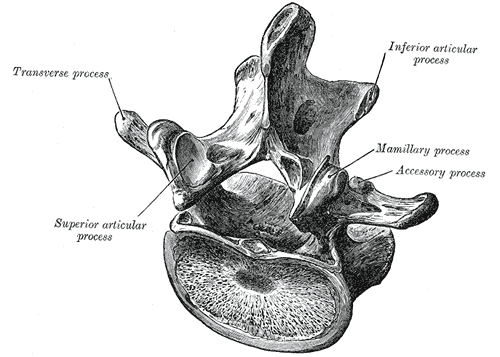
In anatomy, a process (Latin: processus) is a projection or outgrowth of tissue from a larger body.[1] For instance, in a vertebra, a process may serve for muscle attachment and leverage (as in the case of the transverse and spinous processes), or to fit (forming a synovial joint), with another vertebra (as in the case of the articular processes).[2] The word is also used at the microanatomic level, where cells can have processes such as cilia or pedicels. Depending on the tissue, processes may also be called by other terms, such as apophysis, tubercle, or protuberance.

Examples

[edit]

Examples of processes include:

See also

[edit]

Notes

[edit]

References

[edit]
Revisions and contributorsEdit on WikipediaRead on Wikipedia
from Grokipedia
In anatomy, a process (Latin: processus) is a projection or outgrowth of tissue from a larger body structure.[1] Bony processes are a common type, referring to projections or prominences extending from the surface of a bone, typically serving as attachment sites for muscles, ligaments, tendons, or contributing to the formation of joints.[2] These bony structures vary in shape, size, and function, arising during embryonic development through ossification processes influenced by genetic factors and mechanical stresses.[2] They are essential for skeletal stability, locomotion, and overall body support, with their prominence often adapted to specific biomechanical demands.[3] The term also applies to soft tissue and cellular processes, such as outgrowths in organs or neuronal extensions, as detailed in later sections. Common types of bony processes include the spinous process, a sharp, raised elevation found on vertebrae for muscle attachment and spinal articulation; the transverse process, a laterally projecting feature on vertebrae that anchors muscles and ligaments involved in torso movement; and the styloid process, a slender, pointed projection such as the one on the temporal bone that supports nearby muscles and ligaments.[2] Other notable examples encompass the acromial process of the scapula, which forms part of the shoulder joint by articulating with the clavicle, and the coracoid process, a beak-like hook on the scapula serving as an attachment for the pectoralis minor muscle and other structures.[2] These markings are classified under broader bone surface features, distinguishing them from depressions like fossae or foramina, and their study is fundamental to understanding skeletal morphology and pathology.[3]

Definition and Etymology

Definition

In anatomy, a process (Latin: processus, meaning "going forward" or "advance") is defined as a projection or outgrowth of tissue from a larger body or structure, functioning as an extension that can vary in size, shape, and purpose.[4] This term encompasses natural outgrowths, projections, or appendages arising from bone, cartilage, soft tissue, or other anatomical components. Unlike a general prominence, which denotes a broad elevation or swelling on a surface, a process refers to a more distinct, often elongated or sharp projection that is typically named and associated with particular structural roles, such as attachment sites for muscles or ligaments.[2] The concept of a process was first formalized in Renaissance anatomical literature as a descriptor for skeletal extensions, prominently featured in Andreas Vesalius' De humani corporis fabrica (1543), where terms like processus articularis detailed vertebral projections.[5] For instance, the spinous processes of vertebrae illustrate this usage in classical skeletal anatomy.[2]

Etymology

The term "process" in anatomical nomenclature originates from the Latin word processus, meaning "a going forward" or "advance," derived from the verb procedere, which combines pro- (forward) and cedere (to go).[6] This etymological root reflects a conceptual progression or extension, aptly describing anatomical projections as outgrowths extending from a main structure.[7] In the context of anatomy, the term processus gained prominence during the Renaissance, particularly through the work of Andreas Vesalius in his seminal text De Humani Corporis Fabrica (1543), where it was employed to denote bony protrusions and tissue extensions, marking a shift toward more precise Latin-based descriptions. This usage evolved from earlier Greek equivalents, such as apophysis—meaning "offshoot" from apo- (away) and physis (growth)—which Galen had applied broadly to bone processes in antiquity.[8] Vesalius's adoption helped standardize processus in Western anatomical literature, bridging classical Greek terminology with emerging modern dissection practices.[9] The Terminologia Anatomica (1998 and second edition 2019), published by the Federative Committee on Anatomical Terminology, formalized processus as the preferred Latin term for anatomical projections, such as the processus zygomaticus (zygomatic process) or processus mastoideus (mastoid process), alongside English equivalents to facilitate international medical communication. This bilingual approach in nomenclature underscores the term's enduring role in describing tissue outgrowths, consistent with its historical development.[10]

Anatomical Characteristics

Macroscopic Characteristics

Anatomical processes, as macroscopic projections on bones, exhibit significant variations in size and shape to accommodate diverse structural demands within the human skeleton. These projections range from small, rounded tubercles, typically measuring 2.5-3.5 cm in width, such as the greater tubercle of the humerus, to larger, elongated spines that can exceed 5 cm in length, exemplified by the spinous processes of thoracic vertebrae which average around 5 cm.[11][12] Shapes are often irregular, pointed, or blunt, with spines presenting as sharp, slender ridges and tubercles as compact eminences, reflecting adaptations to their positional roles in the skeleton.[13][2] In macroanatomy, processes are primarily composed of bone tissue, featuring an outer layer of dense cortical bone that provides rigidity and an inner network of trabecular bone for lightweight support, particularly in larger projections.[13][14] During developmental stages, these structures often originate from cartilage models through endochondral ossification, though some form via intramembranous ossification, where a cartilaginous covering is gradually replaced by bone.[15][16] Microscopic details of cellular organization within this composition are addressed elsewhere.[13] Processes integrate seamlessly with the parent bone, typically via a widened base or narrowed neck that ensures continuity and distributes mechanical loads effectively across the skeletal framework.[13] This attachment allows for stable extension from the main bone body, with the periosteum enveloping the junction to facilitate vascular and neural connections.[15]

Microscopic Characteristics

In bony processes, the cellular structure consists primarily of osteocytes, mature bone cells embedded within lacunae in the mineralized matrix, connected by cytoplasmic processes that extend through canaliculi to facilitate nutrient exchange and communication.[17] These osteocytes are derived from osteoblasts, which are cuboidal cells actively secreting osteoid, and are supported by osteoclasts, multinucleated cells responsible for bone resorption.[17] Histologically, bony processes exhibit a layered organization beginning with the outer periosteum, a fibrous connective tissue layer rich in collagen fibers and blood vessels that covers the external surface and contributes to appositional growth.[17] Internally, compact bone in these processes is structured around Haversian systems, or osteons, consisting of concentric lamellae surrounding central canals that house vascular and neural elements, with interstitial lamellae filling spaces between osteons for added strength.[17] Developmentally, bony processes form through ossification, where intramembranous ossification directly differentiates mesenchymal cells into osteoblasts that deposit a collagenous matrix mineralized with hydroxyapatite, or endochondral ossification replaces a hyaline cartilage template via chondrocyte hypertrophy and vascular invasion, leading to trabecular and cortical bone layers unique to the process's load-bearing role.[16] Matrix deposition in bone involves osteoid calcification for rigidity.[18]

Classification

Bony Processes

Bony processes, also known as osseous projections or prominences, are raised outgrowths extending from the surface of bones, forming part of the skeletal system's structural markings that facilitate attachments for muscles, ligaments, and tendons.[13] These projections are distinguished from other bone features like articulations or foramina and are classified primarily by their shape, size, and morphology rather than specific function, reflecting the mechanical stresses they endure.[19] Within this classification, subtypes include spines, which are sharp and elongated; tubercles, which are small and rounded; crests, which are linear elevations; and epicondyles, which are projections above condyles; among other variants that vary in prominence and contour.[13] Key subtypes of bony processes encompass the apophysis, defined as a non-articular outgrowth arising from a secondary ossification center, typically serving as a site for muscular or tendinous attachment without direct involvement in joint formation.[20] The trochanter represents a large, blunt, and irregular projection, often found on proximal long bones to accommodate robust muscle insertions.[13] Similarly, the styloid process is a slender, pointed, needle-like extension that varies in length, with the temporal styloid process typically measuring 2-3 cm, projecting from bone surfaces to provide precise attachment points.[21] These morphological distinctions align with standardized anatomical nomenclature, ensuring consistent identification across skeletal elements.[22] Anatomically, bony processes are distributed predominantly throughout the skull, vertebral column, and long bones of the appendicular skeleton, where they contribute to the overall architecture of the osseous framework as delineated in the Terminologia Anatomica. This prevalence underscores their role in regions subject to varied biomechanical demands, from cranial support to locomotor stability, though their precise forms adapt to local skeletal requirements.[13]

Functions

Structural and Mechanical Functions

In anatomy, bony processes primarily function as attachment sites for muscles and ligaments, providing anchors that increase the surface area for secure insertions and origins. These projections often feature roughened or textured surfaces, such as tuberosities or trochanters, which enhance frictional grip for tendons and ligaments, thereby distributing tensile forces across broader areas to prevent localized stress concentrations. For instance, the greater trochanter of the femur serves as a key attachment point for the gluteus medius and minimus muscles, as well as the iliofemoral ligament, enabling efficient force transmission during locomotion.[2][13] Elongated processes also contribute to mechanical leverage and overall skeletal stability by extending the moment arms of attached muscles, allowing for greater torque generation with minimal energy expenditure while dispersing mechanical loads across the skeleton. This design optimizes force distribution, reducing the risk of deformation under weight-bearing conditions; for example, the transverse processes of vertebrae provide leverage for paraspinal muscles and enhance spinal column rigidity by interlocking with adjacent structures. Such configurations support postural maintenance and load-bearing, as seen in the spinous processes of the thoracic vertebrae, which anchor erector spinae muscles to maintain upright stability.[2][23] Additionally, certain processes fulfill protective roles by forming ridges or projections that shield underlying soft tissues, vessels, and nerves from external trauma or compressive forces. These features create structural barriers that safeguard critical anatomy; the mastoid process of the temporal bone, for instance, protects the underlying middle and inner ear structures while also serving as an attachment site. Overall, these functions underscore the integral role of processes in maintaining skeletal integrity and biomechanical efficiency.[2][13]

Articulatory Functions

Articular processes, which are bony projections on vertebrae and other skeletal elements, play a critical role in forming joints by providing articular facets that enable precise interactions between bones. These facets create surfaces for synovial or fibrous joints, facilitating movements such as rotation and gliding while maintaining structural integrity. In synovial joints, the articular processes contribute to the formation of a joint cavity lined by synovial membrane, allowing for lubricated motion, whereas in fibrous joints, they support direct connective tissue linkages that permit limited flexibility.[24][25] In the vertebral column, superior and inferior articular processes specifically form zygapophyseal joints, which are plane synovial joints that guide spinal motion. The superior processes project upward to articulate with the downward-facing inferior processes of the adjacent vertebra above, creating paired facets that restrict excessive translation while permitting flexion, extension, and some rotation. This configuration is essential for the spine's overall articulatory function, as the orientation of these processes varies regionally to optimize movement—such as more coronal alignment in the lumbar region for stability during extension.[26][27][28] Evolutionary adaptations have shaped bony processes to enhance range of motion tailored to functional demands across skeletal regions. In the cranium, sutures adjacent to processes—such as those on the temporal and occipital bones—form fibrous joints that allow initial flexibility for brain growth during development, gradually ossifying to provide rigidity in adulthood. This adaptation reflects a balance between postnatal expansion and biomechanical stability, with suture complexity increasing in mammals to accommodate larger brains and diverse locomotor patterns. In the spine, articular processes evolved from primitive vertebral elements in early vertebrates to sophisticated facets that protect the spinal cord while enabling adaptive postures, as seen in hominins where spinal morphology adaptations supported bipedal locomotion.[29][30][31]

Examples

Processes in the Axial Skeleton

The axial skeleton includes the vertebral column, skull, and rib cage, each featuring distinct bony processes that contribute to structural integrity and interconnections. In the vertebral column, processes arise from the vertebral arch and body, providing key anatomical landmarks.[26] Vertebral processes are categorized into transverse, spinous, and articular types. Transverse processes project laterally from the junction of the pedicles and laminae, serving as attachment sites for paravertebral muscles and ligaments across cervical, thoracic, and lumbar regions.[32] In thoracic vertebrae, these processes specifically include costal facets for rib articulation.[33] Spinous processes extend posteriorly from the lamina junction, varying in length and orientation by spinal region—for instance, bifid in upper cervical vertebrae and short and hatchet-shaped in lumbar ones—to facilitate midline attachments.[27] Articular processes, consisting of superior and inferior pairs, form the zygapophyseal (facet) joints between adjacent vertebrae, with their oval-shaped facets oriented sagittally in the cervical spine and more coronally in lumbar regions.[34] In the skull, processes of the temporal bone exemplify cranial projections integral to the axial framework. The mastoid process is a conical projection inferior to the external acoustic meatus, arising from the petrous portion and providing anchorage for sternocleidomastoid and other neck muscles.[35] The styloid process, a slender, needle-like extension averaging 2-3 cm in length from the temporal bone's inferior surface, serves as an attachment point for the stylohyoid and stylomandibular ligaments.[21] Within the thoracic cage, rib-associated processes facilitate vertebral connections. Each rib features a tubercle—a posterolateral projection near the neck—that articulates with the costal facet on the transverse process of its corresponding thoracic vertebra, enabling precise alignment of the rib head and tubercle for costovertebral joint formation.[36] This tubercle includes both articular and non-articular surfaces, with the former engaging the vertebral transverse process.[37]

Processes in the Appendicular Skeleton

Processes in the appendicular skeleton are bony projections primarily on the pectoral and pelvic girdles as well as the long bones of the upper and lower limbs, serving as critical attachment sites for muscles that enable a wide range of movements, including arm elevation for manipulation and leg propulsion for locomotion. These structures enhance mechanical leverage, allowing precise control and force generation in the extremities. Unlike those in the axial skeleton, which emphasize core stability, appendicular processes prioritize dynamic mobility to support daily activities such as reaching, grasping, walking, and running. In the shoulder girdle, the acromion process extends laterally from the scapula's spine and articulates with the clavicle at the acromioclavicular joint, forming a stable yet mobile platform for upper limb actions. It provides the origin for the middle fibers of the deltoid muscle, which abducts the arm and stabilizes the shoulder during overhead movements essential for manipulation. The coracoid process, projecting anteriorly from the scapular neck, anchors several key structures for arm flexion and stabilization; the pectoralis minor inserts here to depress and protract the scapula, while the coracobrachialis and short head of the biceps brachii originate from its tip, facilitating elbow flexion and supination for precise hand positioning. Additionally, ligaments such as the coracoacromial and coracoclavicular attach to the coracoid, reinforcing the glenohumeral joint against dislocation during dynamic upper limb activities. The pelvic girdle features processes along the iliac crest and ischium that support lower limb mobility by attaching hip and pelvic floor muscles. The anterior superior iliac spine, a prominent projection at the anterior end of the iliac crest, serves as the origin for the sartorius muscle, which flexes, abducts, and laterally rotates the hip while flexing the knee, enabling actions like crossing the legs or kicking. Adjacent to this, the tensor fasciae latae originates from the anterior superior iliac spine and the outer lip of the iliac crest, tensioning the iliotibial tract to stabilize the knee and assist in hip abduction during walking and lateral movements. The ischial spine, located on the posterior ischium superior to the sciatic notch, provides attachment for the coccygeus muscle and the iliococcygeus component of the levator ani, which together support the pelvic floor and maintain stability during strenuous lower limb exertions like running or jumping. In the upper limb, the olecranon process forms the proximal, hook-like extension of the ulna, creating a pulley for the triceps brachii tendon. This muscle inserts on the olecranon, generating powerful elbow extension that is vital for pushing, throwing, and propelling the body during manipulative tasks. The process also articulates with the humerus in the olecranon fossa, locking the elbow in extension to enhance force transmission. Lower limb processes on the femur further amplify mobility, particularly at the hip joint. The greater trochanter, a large lateral projection just below the femoral neck, receives insertions from the gluteus medius and gluteus minimus muscles on its superoposterior and anterior facets, respectively; these abductors stabilize the pelvis during the swing phase of gait and prevent contralateral pelvic drop, ensuring efficient bipedal locomotion. The lesser trochanter, a smaller medial projection at the same level, is the insertion site for the iliopsoas muscle via its tendon, which flexes the hip to initiate leg swing and elevate the thigh in activities like climbing or running.

Clinical Significance

Injuries and Fractures

Injuries to anatomical processes often result from high-impact trauma or forceful muscular contractions, leading to fractures that can compromise structural integrity and associated functions. Bony processes, such as spinous and mastoid projections, are particularly vulnerable due to their protruding nature and attachment points for ligaments and muscles. These fractures typically occur in the context of motor vehicle accidents, falls, sports injuries, or occupational hazards, with avulsion and direct impact being the predominant mechanisms.[38][39] Avulsion fractures commonly affect spinous processes in the cervical or thoracic spine, where sudden, forceful muscle contractions—such as those from trapezius or rhomboid pulls during rapid flexion—detach a fragment at the ligamentous insertion site. A classic example is the clay-shoveler's fracture, an avulsion of the C7 spinous process tip, historically linked to repetitive shoveling but now more often seen in contact sports or trauma. In contrast, direct impact fractures involve the mastoid process of the temporal bone, resulting from lateral blunt force in assaults or falls, which can disrupt the air cell system and surrounding structures without necessarily involving the inner ear.[39][40][38] Diagnosis of process fractures relies on imaging to confirm the injury and evaluate its extent. Plain X-rays, particularly lateral views for spinal processes, reveal fracture lines or displaced fragments, while anteroposterior projections may show characteristic "double spinous process" shadows in avulsions. High-resolution CT scans serve as the gold standard, especially for mastoid or complex cranial processes, delineating displacement, involvement of adjacent bones, and soft tissue damage such as hematoma or ligament tears; MRI is adjunctive when neurological or extensive soft tissue involvement is suspected. Clinical assessment includes localized tenderness, bruising (e.g., Battle's sign over the mastoid), and functional deficits like reduced neck mobility or hearing impairment.[39][38][41] Treatment strategies prioritize stability and symptom relief, tailored to fracture displacement and functional impact. Stable, nondisplaced fractures—common in isolated spinous avulsions—are managed conservatively with immobilization using a cervical collar or brace for 4-12 weeks, alongside NSAIDs for pain and gradual physiotherapy to restore range of motion; high union rates are achieved with this approach. Displaced fractures affecting critical functions, such as those compromising spinal alignment or causing mastoid-related complications like cerebrospinal fluid leaks, require surgical intervention, including internal fixation with plates or excision of nonunited fragments to prevent chronic pain or instability. Observation for associated injuries, like facial nerve palsy in temporal bone cases, guides additional therapies such as steroids or tympanoplasty.[39][42][38]

Pathological Conditions

Pathological conditions affecting bony processes encompass a range of non-traumatic disorders, primarily involving developmental anomalies, degenerative changes, and infectious or inflammatory processes that alter the structure and function of these anatomical projections.[43] Developmental variations in processes often manifest as agenesis, hypoplasia, or fusion due to disruptions in embryonic somitogenesis and ossification. For instance, Klippel-Feil syndrome is characterized by congenital fusion of two or more cervical vertebrae, which can extend to the spinous and transverse processes, resulting in a shortened neck and restricted mobility.[43] Agenesis of specific processes is rarer but documented, such as the congenital absence of the posterior portion of the transverse process along with the superior articular facet in the seventh cervical vertebra, typically discovered incidentally and potentially leading to localized instability.[44] These anomalies may remain asymptomatic or contribute to chronic pain and neurological symptoms if they compromise adjacent neural structures.[45] Degenerative diseases, particularly osteoarthritis, frequently target the articular processes of the spine, known as facet joints. In spinal osteoarthritis, progressive cartilage erosion and subchondral bone remodeling in these synovial joints lead to osteophyte formation and hypertrophy of the articular processes, which narrows the spinal canal and foramina, precipitating lumbar spinal stenosis.[46] This condition is prevalent in older adults, with facet joint osteoarthritis contributing to 15–45% of chronic low back pain cases through mechanical irritation of surrounding tissues and nerve roots.[47] The resulting instability and inflammation exacerbate degenerative cascades, distinguishing this from acute trauma.[48] Infectious and inflammatory conditions can directly involve processes through bacterial invasion or extension from adjacent sites. Mastoiditis, an acute or chronic infection of the mastoid air cells within the mastoid process of the temporal bone, often arises as a complication of acute otitis media, causing mucosal inflammation, abscess formation, and potential erosion of the bony septa.[49] If untreated, it may lead to intracranial complications like meningitis due to the proximity of the process to vital structures.[50] Similarly, osteomyelitis—a hematogenous or contiguous bone infection—can affect processes in long bones, such as the greater trochanter of the femur, where subacute forms present with localized pain and swelling in pediatric patients, often requiring prolonged antibiotic therapy to prevent sequestrum formation.[51] These inflammatory processes highlight the vulnerability of vascularized bony projections to microbial spread.[52]

References

User Avatar
No comments yet.