



Community hub
Recent from talks
Knowledge base stats:
Talk channels stats:
Members stats:
Cardiac stress test
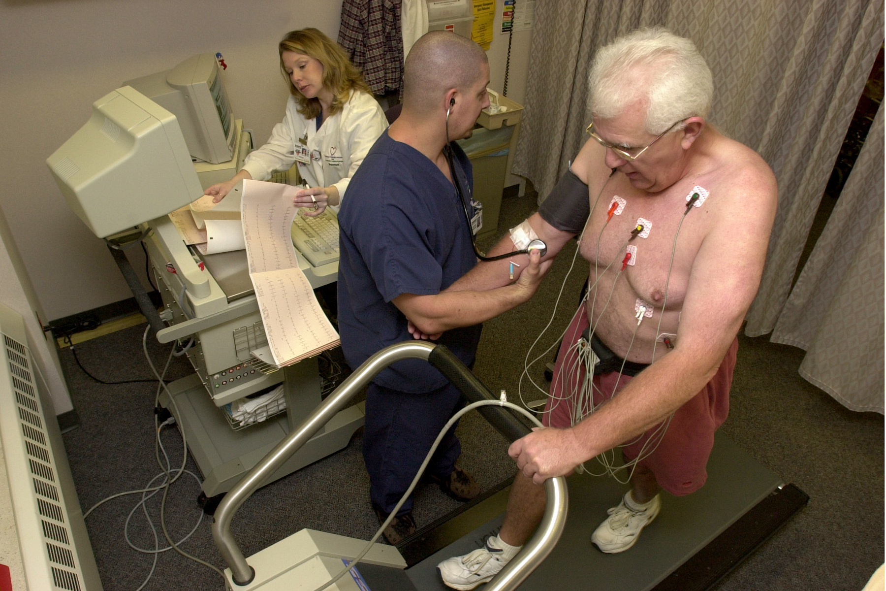
A cardiac stress test is a cardiological examination that evaluates the cardiovascular system's response to external stress within a controlled clinical setting. This stress response can be induced through physical exercise (usually a treadmill) or intravenous pharmacological stimulation of heart rate.
As the heart works progressively harder (stressed) it is monitored using an electrocardiogram (ECG) monitor. This measures the heart's electrical rhythms and broader electrophysiology. Pulse rate, blood pressure and symptoms such as chest discomfort or fatigue are simultaneously monitored by attending clinical staff. Clinical staff will question the patient throughout the procedure asking questions that relate to pain and perceived discomfort. Abnormalities in blood pressure, heart rate, ECG or worsening physical symptoms could be indicative of coronary artery disease.
Stress testing does not accurately diagnose all cases of coronary artery disease, and can often indicate that it exists in people who do not have the condition. The test can also detect heart abnormalities such as arrhythmias, and conditions affecting electrical conduction within the heart such as various types of fascicular blocks.
A "normal" stress test does not offer any substantial reassurance that a future unstable coronary plaque will not rupture and block an artery, inducing a heart attack. As with all medical diagnostic procedures, data is only from a moment in time. A primary reason stress testing is not perceived as a robust method of CAD detection — is that stress testing generally only detects arteries that are severely narrowed (~70% or more).
A stress test may be accompanied by echocardiography. The echocardiography is performed both before and after the exercise so that structural differences can be compared.
A resting echocardiogram is obtained prior to stress. The ultrasound images obtained are similar to the ones obtained during a full surface echocardiogram, commonly referred to as transthoracic echocardiogram. The patient is subjected to stress in the form of exercise or chemically (often dobutamine). After the target heart rate is achieved, 'stress' echocardiogram images are obtained. The two echocardiogram images are then compared to assess for any abnormalities in wall motion of the heart. This is used to detect obstructive coronary artery disease.
While also measuring breathing gases (e.g., oxygen saturation, maximal oxygen consumption), the test is often referred to as a cardiopulmonary exercise test. Common indications for a cardiopulmonary exercise test include evaluation of shortness of breath, workup before heart transplantation, and prognosis and risk assessment of heart failure patients.
The test is also common in sport science for measuring athletes' maximal oxygen consumption, V̇O2 max. In 2016, the American Heart Association published an official scientific statement advocating that cardiorespiratory fitness, quantifiable as V̇O2 max and measured during a cardiopulmonary exercise test, be categorized as a clinical vital sign and should be routinely assessed as part of clinical practice.
Hub AI
Cardiac stress test AI simulator
(@Cardiac stress test_simulator)
Cardiac stress test
A cardiac stress test is a cardiological examination that evaluates the cardiovascular system's response to external stress within a controlled clinical setting. This stress response can be induced through physical exercise (usually a treadmill) or intravenous pharmacological stimulation of heart rate.
As the heart works progressively harder (stressed) it is monitored using an electrocardiogram (ECG) monitor. This measures the heart's electrical rhythms and broader electrophysiology. Pulse rate, blood pressure and symptoms such as chest discomfort or fatigue are simultaneously monitored by attending clinical staff. Clinical staff will question the patient throughout the procedure asking questions that relate to pain and perceived discomfort. Abnormalities in blood pressure, heart rate, ECG or worsening physical symptoms could be indicative of coronary artery disease.
Stress testing does not accurately diagnose all cases of coronary artery disease, and can often indicate that it exists in people who do not have the condition. The test can also detect heart abnormalities such as arrhythmias, and conditions affecting electrical conduction within the heart such as various types of fascicular blocks.
A "normal" stress test does not offer any substantial reassurance that a future unstable coronary plaque will not rupture and block an artery, inducing a heart attack. As with all medical diagnostic procedures, data is only from a moment in time. A primary reason stress testing is not perceived as a robust method of CAD detection — is that stress testing generally only detects arteries that are severely narrowed (~70% or more).
A stress test may be accompanied by echocardiography. The echocardiography is performed both before and after the exercise so that structural differences can be compared.
A resting echocardiogram is obtained prior to stress. The ultrasound images obtained are similar to the ones obtained during a full surface echocardiogram, commonly referred to as transthoracic echocardiogram. The patient is subjected to stress in the form of exercise or chemically (often dobutamine). After the target heart rate is achieved, 'stress' echocardiogram images are obtained. The two echocardiogram images are then compared to assess for any abnormalities in wall motion of the heart. This is used to detect obstructive coronary artery disease.
While also measuring breathing gases (e.g., oxygen saturation, maximal oxygen consumption), the test is often referred to as a cardiopulmonary exercise test. Common indications for a cardiopulmonary exercise test include evaluation of shortness of breath, workup before heart transplantation, and prognosis and risk assessment of heart failure patients.
The test is also common in sport science for measuring athletes' maximal oxygen consumption, V̇O2 max. In 2016, the American Heart Association published an official scientific statement advocating that cardiorespiratory fitness, quantifiable as V̇O2 max and measured during a cardiopulmonary exercise test, be categorized as a clinical vital sign and should be routinely assessed as part of clinical practice.