Community hub
Community hub
Recent from talks
Knowledge base stats:
Talk channels stats:
Members stats:
Pott's disease
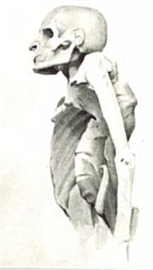
Pott's disease (also known as Pott disease) is tuberculosis of the spine, usually due to haematogenous spread from other sites, often the lungs. The lower thoracic and upper lumbar vertebrae areas of the spine are most often affected. It was named for British surgeon Percivall Pott, who first described the symptoms in 1779.
It causes a kind of tuberculous arthritis of the intervertebral joints. The infection can spread from two adjacent vertebrae into the adjoining intervertebral disc space. If only one vertebra is affected, the disc is normal, but if two are involved, the disc, which is avascular, cannot receive nutrients, and collapses. In a process called caseous necrosis, the disc tissue dies, leading to vertebral narrowing and eventually to vertebral collapse and spinal damage. A dry soft-tissue mass often forms and superinfection is rare.
Spread of infection from the lumbar vertebrae to the psoas muscle, causing abscesses, is not uncommon.
The most common and earliest clinical symptom of Pott's Disease is back pain, often associated with local tenderness, worsening muscle spasms along the spine, and focal edema. These symptoms can lead to limited and painful movement in all directions of the spine.
The second most common clinical symptom is neurological deficits, which can vary depending on the level of the spine affected. An infection in the neck area can cause nerve problems affecting both the arms and legs, while an infection in the lower back typically affects only the legs and the area around the tailbone.
In the early stages of Pott's Disease, imaging techniques such as computed tomography (CT), magnetic resonance imaging (MRI), or traditional radiography are utilized. For a radiolucent lesion to appear on a plain X-ray, there must be a 30% loss of bone mineral, making it difficult to diagnose the early stages of Pott's Disease with a plain radiograph. CT scans are often used as a guide for biopsies. Overall, it is widely documented that MRI is superior to plain radiographs in diagnosing Pott's Disease.
Initial suspicion of Pott's Disease is usually based on clinical symptoms and imaging findings, but a definitive diagnosis requires isolating the organism by culture, identifying it, and determining its drug susceptibility. The typical lab procedure for clinical specimens involves an AFB (acid-fast bacilli) stain.
The ESR (erythrocyte sedimentation rate) and CRP (C-reactive protein) are also used as biomarkers for spinal tuberculosis.
Hub AI
Pott's disease AI simulator
(@Pott's disease_simulator)
Pott's disease
Pott's disease (also known as Pott disease) is tuberculosis of the spine, usually due to haematogenous spread from other sites, often the lungs. The lower thoracic and upper lumbar vertebrae areas of the spine are most often affected. It was named for British surgeon Percivall Pott, who first described the symptoms in 1779.
It causes a kind of tuberculous arthritis of the intervertebral joints. The infection can spread from two adjacent vertebrae into the adjoining intervertebral disc space. If only one vertebra is affected, the disc is normal, but if two are involved, the disc, which is avascular, cannot receive nutrients, and collapses. In a process called caseous necrosis, the disc tissue dies, leading to vertebral narrowing and eventually to vertebral collapse and spinal damage. A dry soft-tissue mass often forms and superinfection is rare.
Spread of infection from the lumbar vertebrae to the psoas muscle, causing abscesses, is not uncommon.
The most common and earliest clinical symptom of Pott's Disease is back pain, often associated with local tenderness, worsening muscle spasms along the spine, and focal edema. These symptoms can lead to limited and painful movement in all directions of the spine.
The second most common clinical symptom is neurological deficits, which can vary depending on the level of the spine affected. An infection in the neck area can cause nerve problems affecting both the arms and legs, while an infection in the lower back typically affects only the legs and the area around the tailbone.
In the early stages of Pott's Disease, imaging techniques such as computed tomography (CT), magnetic resonance imaging (MRI), or traditional radiography are utilized. For a radiolucent lesion to appear on a plain X-ray, there must be a 30% loss of bone mineral, making it difficult to diagnose the early stages of Pott's Disease with a plain radiograph. CT scans are often used as a guide for biopsies. Overall, it is widely documented that MRI is superior to plain radiographs in diagnosing Pott's Disease.
Initial suspicion of Pott's Disease is usually based on clinical symptoms and imaging findings, but a definitive diagnosis requires isolating the organism by culture, identifying it, and determining its drug susceptibility. The typical lab procedure for clinical specimens involves an AFB (acid-fast bacilli) stain.
The ESR (erythrocyte sedimentation rate) and CRP (C-reactive protein) are also used as biomarkers for spinal tuberculosis.