Recent from talks
Medical ultrasound
Knowledge base stats:
Talk channels stats:
Members stats:
Medical ultrasound
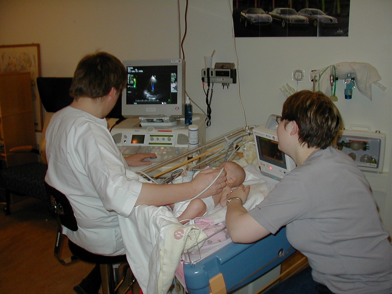
Medical ultrasound includes diagnostic techniques (mainly imaging) using ultrasound, as well as therapeutic applications of ultrasound. In diagnosis, it is used to create an image of internal body structures such as tendons, muscles, joints, blood vessels, and internal organs, to measure some characteristics (e.g., distances and velocities) or to generate an informative audible sound. The usage of ultrasound to produce visual images for medicine is called medical ultrasonography or simply sonography. Sonography using ultrasound reflection is called echography. There are also transmission methods, such as ultrasound transmission tomography. The practice of examining pregnant women using ultrasound is called obstetric ultrasonography, and was an early development of clinical ultrasonography. The machine used is called an ultrasound machine, a sonograph or an echograph. The visual image formed using this technique is called an ultrasonogram, a sonogram or an echogram.
Ultrasound is composed of sound waves with frequencies greater than 20,000 Hz, which is the approximate upper threshold of human hearing. Ultrasonic images, also known as sonograms, are created by sending pulses of ultrasound into tissue using a probe. The ultrasound pulses echo off tissues with different reflection properties and are returned to the probe which records and displays them as an image.
A general-purpose ultrasonic transducer may be used for most imaging purposes but some situations may require the use of a specialized transducer. Most ultrasound examination is done using a transducer on the surface of the body, but improved visualization is often possible if a transducer can be placed inside the body. For this purpose, special-use transducers, including transvaginal, endorectal, and transesophageal transducers are commonly employed. At the extreme, very small transducers can be mounted on small diameter catheters and placed within blood vessels to image the walls and disease of those vessels.
The imaging mode refers to probe and machine settings that result in specific dimensions of the ultrasound image. Several modes of ultrasound are used in medical imaging:
Most machines convert two-way time to imaging depth using as assumed speed of sound of 1540 m/s. As the actual speed of sound varies greatly in different tissue types, an ultrasound image is therefore not a true tomographic representation of the body.
Three-dimensional imaging is done by combining B-mode images, using dedicated rotating or stationary probes. This has also been referred to as C-mode.
An imaging technique refers to a method of signal generation and processing that results in a specific application. Most imaging techniques are operating in B-mode.
Therapeutic ultrasound aimed at a specific tumor or calculus is not an imaging mode. However, for positioning a treatment probe to focus on a specific region of interest, A-mode and B-mode are typically used, often during treatment.
Hub AI
Medical ultrasound AI simulator
(@Medical ultrasound_simulator)
Medical ultrasound
Medical ultrasound includes diagnostic techniques (mainly imaging) using ultrasound, as well as therapeutic applications of ultrasound. In diagnosis, it is used to create an image of internal body structures such as tendons, muscles, joints, blood vessels, and internal organs, to measure some characteristics (e.g., distances and velocities) or to generate an informative audible sound. The usage of ultrasound to produce visual images for medicine is called medical ultrasonography or simply sonography. Sonography using ultrasound reflection is called echography. There are also transmission methods, such as ultrasound transmission tomography. The practice of examining pregnant women using ultrasound is called obstetric ultrasonography, and was an early development of clinical ultrasonography. The machine used is called an ultrasound machine, a sonograph or an echograph. The visual image formed using this technique is called an ultrasonogram, a sonogram or an echogram.
Ultrasound is composed of sound waves with frequencies greater than 20,000 Hz, which is the approximate upper threshold of human hearing. Ultrasonic images, also known as sonograms, are created by sending pulses of ultrasound into tissue using a probe. The ultrasound pulses echo off tissues with different reflection properties and are returned to the probe which records and displays them as an image.
A general-purpose ultrasonic transducer may be used for most imaging purposes but some situations may require the use of a specialized transducer. Most ultrasound examination is done using a transducer on the surface of the body, but improved visualization is often possible if a transducer can be placed inside the body. For this purpose, special-use transducers, including transvaginal, endorectal, and transesophageal transducers are commonly employed. At the extreme, very small transducers can be mounted on small diameter catheters and placed within blood vessels to image the walls and disease of those vessels.
The imaging mode refers to probe and machine settings that result in specific dimensions of the ultrasound image. Several modes of ultrasound are used in medical imaging:
Most machines convert two-way time to imaging depth using as assumed speed of sound of 1540 m/s. As the actual speed of sound varies greatly in different tissue types, an ultrasound image is therefore not a true tomographic representation of the body.
Three-dimensional imaging is done by combining B-mode images, using dedicated rotating or stationary probes. This has also been referred to as C-mode.
An imaging technique refers to a method of signal generation and processing that results in a specific application. Most imaging techniques are operating in B-mode.
Therapeutic ultrasound aimed at a specific tumor or calculus is not an imaging mode. However, for positioning a treatment probe to focus on a specific region of interest, A-mode and B-mode are typically used, often during treatment.
Recent media