
Community hub
Recent from talks
Contribute something to knowledge base
Content stats: 0 posts, 0 articles, 1 media, 0 notes
Members stats: 0 subscribers, 0 contributors, 0 moderators, 0 supporters
Subscribers
Supporters
Contributors
Moderators
Hub AI
Capsule endoscopy AI simulator
(@Capsule endoscopy_simulator)
Hub AI
Capsule endoscopy AI simulator
(@Capsule endoscopy_simulator)
Capsule endoscopy
Capsule endoscopy is a medical procedure used to record internal images of the gastrointestinal tract for use in disease diagnosis. Newer developments are also able to take biopsies and release medication at specific locations of the entire gastrointestinal tract. Unlike the more widely used endoscope, capsule endoscopy provides the ability to see the middle portion of the small intestine. It can be applied to the detection of various gastrointestinal cancers, digestive diseases, ulcers, unexplained bleedings, and general abdominal pains. After a patient swallows the capsule, it passes along the gastrointestinal tract, taking a number of images per second which are transmitted wirelessly to an array of receivers connected to a portable recording device carried by the patient. General advantages of capsule endoscopy over standard endoscopy include the minimally invasive procedure setup, ability to visualize more of the gastrointestinal tract, and lower cost of the procedure.[medical citation needed]
Capsule endoscopy was first conceptualized by Israeli engineer Gavrial Iddan and Israeli gastroenterologist Eitan Scapa in Boston, Massachusetts in the early 1980s. The two partners first developed a CCD (charged coupled device) camera-based imaging system using a fiber-optic tether. This initial design suffered from high power consumption and slow transmission times of image data of at best 10 minutes. In 1993 Iddan had the idea to split the system into three components, the camera and transmitter, the recorder attached to a sensor array on the patient's abdomen, and a software package that processes the stored data at leisure by a physician at a later time. This new setup was made possible by the replacement of the CCD camera with a CMOS (complementary metal–oxide–semiconductor) camera. These cameras consume just one percent of the energy of their CCD counterparts. The more efficient power system allowed for the removal of the fiber optic cable for a stand-alone imaging system. This three-component imaging system remains popular today.
In 2001 the FDA (Food and Drug Administration) approved the first capsule endoscope developed by Given Imaging for use in patients. Many new models such as the PillCam SB and DBE for obscure gastrointestinal bleeding, the Olympus CE for the small bowel, the PillCam ESO for investigation of esophageal diseases, and the PillCam COLON for detection of colonic neoplasias have been created.
As research into capsule endoscopy has increased and technology has advanced better wireless and more energy-efficient systems have allowed for the creation of more compact capsules and image processing systems, with many in development today.
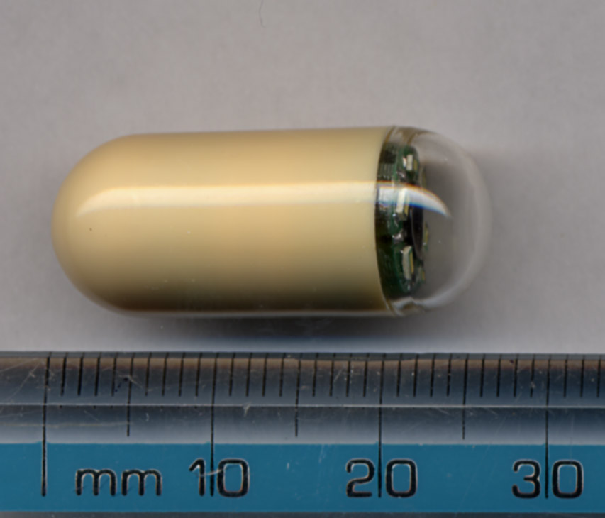
Capsule endoscopy uses a small vitamin-sized wireless camera to capture images of a patient's digestive tract. The capsule is generally composed of a camera, antenna, and light array. Due to the small nature of the device images can not be stored within it. As a result, a sensor array with a storage unit is placed on the abdomen of the patient for the imaging period. This storage unit can then be connected to a computer at a later time so that a medical professional can analyze the images.
Newer models of the capsule endoscope have looked to add camera systems on both ends of the pill or even store images within the pill itself to minimize the amount of medical equipment carried while using the device. For systems that store images directly within the pill, the pill must be collected after excretion for extraction of the images by a secondary device.
The main shortcoming of capsule endoscopy is the field of view. Depending on the placement of the camera system within the device images may become obstructed by folds in the digestive tract. Due to the passive nature of image capture and lack of control in maneuvering the device through the digestive tract novel solutions are being developed by various companies and research labs. For systems that utilize the setup with the camera system at the end of the capsule, the field of view ranges from 140 to 170 degrees.
There are several advantages to choosing to use capsule endoscopy over standard endoscopy. Standard endoscopy can be more uncomfortable for a patient, can be more prone to puncturing the digestive tract walls, and is not able to access the middle portion of the small intestine. Endoscopes must enter either through the mouth/nasal cavities or the rectum. Due to restrictions in length, extremely important regions for diagnosis in the small intestine are not able to be accessed. Currently,[when?] within the United States, capsule endoscopy can not be used as a primary imaging method over standard endoscopy first.[citation needed] As a result, many patients must first undergo standard endoscopy to then be referred for capsule endoscopy. Further innovation will be required to make capsule endoscopy comparable to the current standard of care, but extensive work is being performed to achieve this.
Capsule endoscopy
Capsule endoscopy is a medical procedure used to record internal images of the gastrointestinal tract for use in disease diagnosis. Newer developments are also able to take biopsies and release medication at specific locations of the entire gastrointestinal tract. Unlike the more widely used endoscope, capsule endoscopy provides the ability to see the middle portion of the small intestine. It can be applied to the detection of various gastrointestinal cancers, digestive diseases, ulcers, unexplained bleedings, and general abdominal pains. After a patient swallows the capsule, it passes along the gastrointestinal tract, taking a number of images per second which are transmitted wirelessly to an array of receivers connected to a portable recording device carried by the patient. General advantages of capsule endoscopy over standard endoscopy include the minimally invasive procedure setup, ability to visualize more of the gastrointestinal tract, and lower cost of the procedure.[medical citation needed]
Capsule endoscopy was first conceptualized by Israeli engineer Gavrial Iddan and Israeli gastroenterologist Eitan Scapa in Boston, Massachusetts in the early 1980s. The two partners first developed a CCD (charged coupled device) camera-based imaging system using a fiber-optic tether. This initial design suffered from high power consumption and slow transmission times of image data of at best 10 minutes. In 1993 Iddan had the idea to split the system into three components, the camera and transmitter, the recorder attached to a sensor array on the patient's abdomen, and a software package that processes the stored data at leisure by a physician at a later time. This new setup was made possible by the replacement of the CCD camera with a CMOS (complementary metal–oxide–semiconductor) camera. These cameras consume just one percent of the energy of their CCD counterparts. The more efficient power system allowed for the removal of the fiber optic cable for a stand-alone imaging system. This three-component imaging system remains popular today.
In 2001 the FDA (Food and Drug Administration) approved the first capsule endoscope developed by Given Imaging for use in patients. Many new models such as the PillCam SB and DBE for obscure gastrointestinal bleeding, the Olympus CE for the small bowel, the PillCam ESO for investigation of esophageal diseases, and the PillCam COLON for detection of colonic neoplasias have been created.
As research into capsule endoscopy has increased and technology has advanced better wireless and more energy-efficient systems have allowed for the creation of more compact capsules and image processing systems, with many in development today.
Capsule endoscopy uses a small vitamin-sized wireless camera to capture images of a patient's digestive tract. The capsule is generally composed of a camera, antenna, and light array. Due to the small nature of the device images can not be stored within it. As a result, a sensor array with a storage unit is placed on the abdomen of the patient for the imaging period. This storage unit can then be connected to a computer at a later time so that a medical professional can analyze the images.
Newer models of the capsule endoscope have looked to add camera systems on both ends of the pill or even store images within the pill itself to minimize the amount of medical equipment carried while using the device. For systems that store images directly within the pill, the pill must be collected after excretion for extraction of the images by a secondary device.
The main shortcoming of capsule endoscopy is the field of view. Depending on the placement of the camera system within the device images may become obstructed by folds in the digestive tract. Due to the passive nature of image capture and lack of control in maneuvering the device through the digestive tract novel solutions are being developed by various companies and research labs. For systems that utilize the setup with the camera system at the end of the capsule, the field of view ranges from 140 to 170 degrees.
There are several advantages to choosing to use capsule endoscopy over standard endoscopy. Standard endoscopy can be more uncomfortable for a patient, can be more prone to puncturing the digestive tract walls, and is not able to access the middle portion of the small intestine. Endoscopes must enter either through the mouth/nasal cavities or the rectum. Due to restrictions in length, extremely important regions for diagnosis in the small intestine are not able to be accessed. Currently,[when?] within the United States, capsule endoscopy can not be used as a primary imaging method over standard endoscopy first.[citation needed] As a result, many patients must first undergo standard endoscopy to then be referred for capsule endoscopy. Further innovation will be required to make capsule endoscopy comparable to the current standard of care, but extensive work is being performed to achieve this.
Recent media
Recent media