
Community hub
Recent from talks
Contribute something to knowledge base
Content stats: 0 posts, 0 articles, 1 media, 0 notes
Members stats: 0 subscribers, 0 contributors, 0 moderators, 0 supporters
Subscribers
Supporters
Contributors
Moderators
Hub AI
Cadherin-1 AI simulator
(@Cadherin-1_simulator)
Hub AI
Cadherin-1 AI simulator
(@Cadherin-1_simulator)
Cadherin-1
Cadherin-1 or Epithelial cadherin (E-cadherin), is a protein that in humans is encoded by the CDH1 gene (not to be confused with the APC/C activator protein CDH1). Mutations are correlated with gastric, breast, colorectal, thyroid, and ovarian cancers. CDH1 has also been designated as CD324 (cluster of differentiation 324). It is a tumor suppressor gene.
The discovery of cadherin cell-cell adhesion proteins is attributed to Masatoshi Takeichi, whose experience with adhering epithelial cells began in 1966. His work originally began by studying lens differentiation in chicken embryos at Nagoya University, where he explored how retinal cells regulate lens fiber differentiation. To do this, Takeichi initially collected media that had previously cultured neural retina cells (CM) and suspended lens epithelial cells in it. He observed that cells suspended in the CM media had delayed attachment compared to cells in his regular medium. His interest in cell adherence was sparked, and he moved on to examine attachment in other conditions such as in the presence of protein, magnesium, and calcium. At this point in 1970s, little was understood about the specific roles these ions played. Therefore, Takeichi's work in discovering calcium's role in cell-cell adhesion was highly transformative.
Takeichi went on to discover the existence of multiple cadherins, beginning with E-cadherin. Using rats immunized with F9 cells, he worked with an undergraduate student in the Okada laboratory, Noboru Suzuki, to generate mouse antibodies called ECCD1. This antibody blocked cell-adhesion ability and showed a calcium-dependent interaction with its antigen, E-cadherin. They went on to find that ECCD1 reacted to a variety of epithelial cells when comparing antibody distributions. The delay Takeichi experienced in specifically discovering E-cadherin was most likely due to the model he used to initially investigate cell adherence. The chinese hamster V79 cells apparently did not express E-cadherin, but instead 20 other subtypes that have since been discovered.
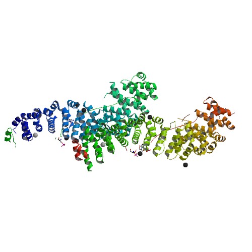
Cadherin-1 is a classical member of the cadherin superfamily. The encoded protein is a calcium-dependent cell–cell adhesion glycoprotein composed of five extracellular cadherin repeats, a transmembrane region, and a highly conserved cytoplasmic tail. Mutations in this gene are correlated with gastric, breast, colorectal, thyroid, and ovarian cancers. Loss of function is thought to contribute to progression in cancer by increasing proliferation, invasion, and/or metastasis. The ectodomain of this protein mediates bacterial adhesion to mammalian cells, and the cytoplasmic domain is required for internalization. Identified transcript variants arise from mutation at consensus splice sites.
E-cadherin (epithelial) is the most well-studied member of the cadherin family and is an essential transmembrane protein within adherens junctions. In addition to E-cadherin, adherens junctions are composed of the intracellular components, p120-catenin, beta-catenin, and alpha-catenin. Together, these proteins stabilize epithelial tissues and regulate intercellular exchange. The structure of E-cadherin consists of 5 cadherin repeats (EC1 ~ EC5) in the extracellular domain, one transmembrane domain, and a highly-phosphorylated intracellular domain. This region is vital to beta-catenin binding and, therefore, to E-cadherin function. Beta-catenin can also bind to alpha-catenin. Alpha-catenin participates in regulation of actin-containing cytoskeletal filaments. In epithelial cells, E-cadherin-containing cell-to-cell junctions are often adjacent to actin-containing filaments of the cytoskeleton.
E-cadherin is first expressed in the 2-cell stage of mammalian development, and becomes phosphorylated by the 8-cell stage, where it causes compaction. In adult tissues, E-cadherin is expressed in epithelial tissues, where it is constantly regenerated with a 5-hour half-life on the cell surface. [citation needed] Cell–cell interactions mediated by E-cadherin are crucial to blastula formation in many animals.
E-cadherin has been known to mediate adhesion-dependent proliferation inhibition by triggering cell cycle exit via contact inhibition of proliferation (CIP) and recruitment of the Hippo pathway. E-cadherin adhesions inhibit growth signals, which initiates a kinase cascade that excludes the transcription factor YAP from the nucleus. Conversely, decreasing cell density (decreasing cell-cell adhesion) or applying mechanical stretch to place E-cadherins under increased tension promotes cell cycle entry and YAP nuclear localization.
E-cadherin has been found to have a role in epithelial morphogenesis and branching, such as during the formation of epithelial buds. Physiologically, branching is an important feature that allows tissues, such as salivary glands and pancreatic buds, to maximize functional surface areas. It has been discovered that the application of appropriate growth factors and extracellular matrix can induce branching in tissue, but the mechanisms of branching appear to differ between single-layered and stratified epithelium.
Cadherin-1
Cadherin-1 or Epithelial cadherin (E-cadherin), is a protein that in humans is encoded by the CDH1 gene (not to be confused with the APC/C activator protein CDH1). Mutations are correlated with gastric, breast, colorectal, thyroid, and ovarian cancers. CDH1 has also been designated as CD324 (cluster of differentiation 324). It is a tumor suppressor gene.
The discovery of cadherin cell-cell adhesion proteins is attributed to Masatoshi Takeichi, whose experience with adhering epithelial cells began in 1966. His work originally began by studying lens differentiation in chicken embryos at Nagoya University, where he explored how retinal cells regulate lens fiber differentiation. To do this, Takeichi initially collected media that had previously cultured neural retina cells (CM) and suspended lens epithelial cells in it. He observed that cells suspended in the CM media had delayed attachment compared to cells in his regular medium. His interest in cell adherence was sparked, and he moved on to examine attachment in other conditions such as in the presence of protein, magnesium, and calcium. At this point in 1970s, little was understood about the specific roles these ions played. Therefore, Takeichi's work in discovering calcium's role in cell-cell adhesion was highly transformative.
Takeichi went on to discover the existence of multiple cadherins, beginning with E-cadherin. Using rats immunized with F9 cells, he worked with an undergraduate student in the Okada laboratory, Noboru Suzuki, to generate mouse antibodies called ECCD1. This antibody blocked cell-adhesion ability and showed a calcium-dependent interaction with its antigen, E-cadherin. They went on to find that ECCD1 reacted to a variety of epithelial cells when comparing antibody distributions. The delay Takeichi experienced in specifically discovering E-cadherin was most likely due to the model he used to initially investigate cell adherence. The chinese hamster V79 cells apparently did not express E-cadherin, but instead 20 other subtypes that have since been discovered.
Cadherin-1 is a classical member of the cadherin superfamily. The encoded protein is a calcium-dependent cell–cell adhesion glycoprotein composed of five extracellular cadherin repeats, a transmembrane region, and a highly conserved cytoplasmic tail. Mutations in this gene are correlated with gastric, breast, colorectal, thyroid, and ovarian cancers. Loss of function is thought to contribute to progression in cancer by increasing proliferation, invasion, and/or metastasis. The ectodomain of this protein mediates bacterial adhesion to mammalian cells, and the cytoplasmic domain is required for internalization. Identified transcript variants arise from mutation at consensus splice sites.
E-cadherin (epithelial) is the most well-studied member of the cadherin family and is an essential transmembrane protein within adherens junctions. In addition to E-cadherin, adherens junctions are composed of the intracellular components, p120-catenin, beta-catenin, and alpha-catenin. Together, these proteins stabilize epithelial tissues and regulate intercellular exchange. The structure of E-cadherin consists of 5 cadherin repeats (EC1 ~ EC5) in the extracellular domain, one transmembrane domain, and a highly-phosphorylated intracellular domain. This region is vital to beta-catenin binding and, therefore, to E-cadherin function. Beta-catenin can also bind to alpha-catenin. Alpha-catenin participates in regulation of actin-containing cytoskeletal filaments. In epithelial cells, E-cadherin-containing cell-to-cell junctions are often adjacent to actin-containing filaments of the cytoskeleton.
E-cadherin is first expressed in the 2-cell stage of mammalian development, and becomes phosphorylated by the 8-cell stage, where it causes compaction. In adult tissues, E-cadherin is expressed in epithelial tissues, where it is constantly regenerated with a 5-hour half-life on the cell surface. [citation needed] Cell–cell interactions mediated by E-cadherin are crucial to blastula formation in many animals.
E-cadherin has been known to mediate adhesion-dependent proliferation inhibition by triggering cell cycle exit via contact inhibition of proliferation (CIP) and recruitment of the Hippo pathway. E-cadherin adhesions inhibit growth signals, which initiates a kinase cascade that excludes the transcription factor YAP from the nucleus. Conversely, decreasing cell density (decreasing cell-cell adhesion) or applying mechanical stretch to place E-cadherins under increased tension promotes cell cycle entry and YAP nuclear localization.
E-cadherin has been found to have a role in epithelial morphogenesis and branching, such as during the formation of epithelial buds. Physiologically, branching is an important feature that allows tissues, such as salivary glands and pancreatic buds, to maximize functional surface areas. It has been discovered that the application of appropriate growth factors and extracellular matrix can induce branching in tissue, but the mechanisms of branching appear to differ between single-layered and stratified epithelium.
Recent media
Recent media