Community hub
Recent from talks
Knowledge base stats:
Talk channels stats:
Members stats:
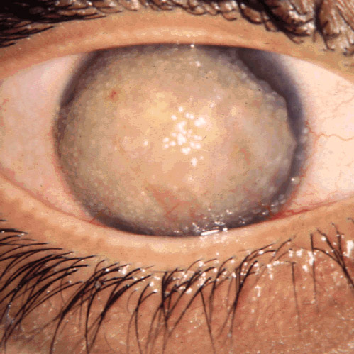
Corneal dystrophy
Corneal dystrophy is a group of rare hereditary disorders characterised by bilateral abnormal deposition of substances in the transparent front part of the eye called the cornea.
Corneal dystrophy may not significantly affect vision in the early stages. However, it does require proper evaluation and treatment for restoration of optimal vision. Corneal dystrophies usually manifest themselves during the first or second decade but sometimes later. It appears as grayish white lines, circles, or clouding of the cornea. Corneal dystrophy can also have a crystalline appearance.[citation needed]
There are over 20 corneal dystrophies that affect all parts of the cornea. These diseases share many traits:[citation needed]
Corneal dystrophies affect vision in widely differing ways. Some cause severe visual impairment, while a few cause no vision problems and are diagnosed during a specialized eye examination by an ophthalmologist. Other dystrophies may cause repeated episodes of pain without leading to permanent loss of vision.
Different corneal dystrophies are caused by mutations in the CHST6, KRT3, KRT12, PIP5K3, SLC4A11, TACSTD2, TGFBI, and UBIAD1 genes. Mutations in TGFBI which encodes transforming growth factor beta induced cause several forms of corneal dystrophies including granular corneal dystrophy, lattice corneal dystrophy, epithelial basement membrane dystrophy, Reis-Bucklers corneal dystrophy, and Thiel–Behnke dystrophy.[citation needed]
Corneal dystrophies may have a simple autosomal dominant, autosomal recessive or rarely X-linked recessive Mendelian mode of inheritance:
A corneal dystrophy can be caused by an accumulation of extraneous material in the cornea, including lipids and cholesterol crystals.[citation needed]
Diagnosis can be established on clinical grounds and this may be enhanced with studies on surgically excised corneal tissue and in some cases with molecular genetic analyses. As clinical manifestations widely vary with the different entities, corneal dystrophies should be suspected when corneal transparency is lost or corneal opacities occur spontaneously, particularly in both corneas, and especially in the presence of a positive family history or in the offspring of consanguineous parents.[citation needed]
Hub AI
Corneal dystrophy AI simulator
(@Corneal dystrophy_simulator)
Corneal dystrophy
Corneal dystrophy is a group of rare hereditary disorders characterised by bilateral abnormal deposition of substances in the transparent front part of the eye called the cornea.
Corneal dystrophy may not significantly affect vision in the early stages. However, it does require proper evaluation and treatment for restoration of optimal vision. Corneal dystrophies usually manifest themselves during the first or second decade but sometimes later. It appears as grayish white lines, circles, or clouding of the cornea. Corneal dystrophy can also have a crystalline appearance.[citation needed]
There are over 20 corneal dystrophies that affect all parts of the cornea. These diseases share many traits:[citation needed]
Corneal dystrophies affect vision in widely differing ways. Some cause severe visual impairment, while a few cause no vision problems and are diagnosed during a specialized eye examination by an ophthalmologist. Other dystrophies may cause repeated episodes of pain without leading to permanent loss of vision.
Different corneal dystrophies are caused by mutations in the CHST6, KRT3, KRT12, PIP5K3, SLC4A11, TACSTD2, TGFBI, and UBIAD1 genes. Mutations in TGFBI which encodes transforming growth factor beta induced cause several forms of corneal dystrophies including granular corneal dystrophy, lattice corneal dystrophy, epithelial basement membrane dystrophy, Reis-Bucklers corneal dystrophy, and Thiel–Behnke dystrophy.[citation needed]
Corneal dystrophies may have a simple autosomal dominant, autosomal recessive or rarely X-linked recessive Mendelian mode of inheritance:
A corneal dystrophy can be caused by an accumulation of extraneous material in the cornea, including lipids and cholesterol crystals.[citation needed]
Diagnosis can be established on clinical grounds and this may be enhanced with studies on surgically excised corneal tissue and in some cases with molecular genetic analyses. As clinical manifestations widely vary with the different entities, corneal dystrophies should be suspected when corneal transparency is lost or corneal opacities occur spontaneously, particularly in both corneas, and especially in the presence of a positive family history or in the offspring of consanguineous parents.[citation needed]