
Community hub
Recent from talks
Contribute something to knowledge base
Content stats: 0 posts, 0 articles, 1 media, 0 notes
Members stats: 0 subscribers, 0 contributors, 0 moderators, 0 supporters
Subscribers
Supporters
Contributors
Moderators
Hub AI
Golgi tendon organ AI simulator
(@Golgi tendon organ_simulator)
Hub AI
Golgi tendon organ AI simulator
(@Golgi tendon organ_simulator)
Golgi tendon organ
The Golgi tendon organ (GTO) (also known as Golgi organ, tendon organ, neurotendinous organ or neurotendinous spindle) is a skeletal muscle stretch receptor proprioceptor. It is situated at the interface between a muscle and its tendon known as the musculotendinous junction. It senses muscle tension (whereas muscle spindles are responsible for detecting muscle length and changes in muscle length). It is innervated by type Ib sensory nerve fibers.
It represents the sensory leg of the Golgi tendon reflex arc.
The Golgi tendon organ is one of several eponymous terms named after the Italian physician Camillo Golgi.
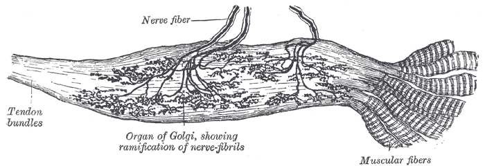
The body of the Golgi tendon organ is made up of braided strands of collagen (intrafusal fasciculi) that are less compact than elsewhere in the tendon and are encapsulated. The capsule is connected in series (along a single path) with a group of muscle fibers (10-20 fibers) at one end, and merge into the tendon proper at the other. Each capsule is about 1 mm long, has a diameter of about 0.1 mm.
One or more fast-conducting type Ib sensory nerve fibers penetrate the capsule and lose their medullary[clarification needed] sheaths, branch, intertwine with the collagen fibers, and terminate as flattened leaf-like endings between the collagen strands (see figure).
When the muscle generates force, the sensory terminals of the Ib afferent axons are compressed and become deformed which causes the opening of stretch-sensitive cation channels, depolarizing the axon and causing it to fire nerve impulses. The action potential frequency encodes the force being developed by the muscle fibers associated with the Golgi tendon organ. The average level of activity in a tendon organ population is representative of the whole muscle force.
The Ib sensory feedback generates stretch reflexes and supraspinal responses which control muscle contraction. Ib afferents synapse with interneurons in the spinal cord that also project to the brain cerebellum and cerebral cortex. The Golgi tendon reflex assists in regulating muscle contraction force. It is associated with the Ib. Tendon organs signal muscle force through the entire physiological range, not only at high strain.
During locomotion, Ib input excites rather than inhibits motoneurons of the receptor-bearing muscles, and it affects the timing of the transitions between the stance and swing phases of locomotion. The switch to autogenic excitation is a form of positive feedback.
Golgi tendon organ
The Golgi tendon organ (GTO) (also known as Golgi organ, tendon organ, neurotendinous organ or neurotendinous spindle) is a skeletal muscle stretch receptor proprioceptor. It is situated at the interface between a muscle and its tendon known as the musculotendinous junction. It senses muscle tension (whereas muscle spindles are responsible for detecting muscle length and changes in muscle length). It is innervated by type Ib sensory nerve fibers.
It represents the sensory leg of the Golgi tendon reflex arc.
The Golgi tendon organ is one of several eponymous terms named after the Italian physician Camillo Golgi.
The body of the Golgi tendon organ is made up of braided strands of collagen (intrafusal fasciculi) that are less compact than elsewhere in the tendon and are encapsulated. The capsule is connected in series (along a single path) with a group of muscle fibers (10-20 fibers) at one end, and merge into the tendon proper at the other. Each capsule is about 1 mm long, has a diameter of about 0.1 mm.
One or more fast-conducting type Ib sensory nerve fibers penetrate the capsule and lose their medullary[clarification needed] sheaths, branch, intertwine with the collagen fibers, and terminate as flattened leaf-like endings between the collagen strands (see figure).
When the muscle generates force, the sensory terminals of the Ib afferent axons are compressed and become deformed which causes the opening of stretch-sensitive cation channels, depolarizing the axon and causing it to fire nerve impulses. The action potential frequency encodes the force being developed by the muscle fibers associated with the Golgi tendon organ. The average level of activity in a tendon organ population is representative of the whole muscle force.
The Ib sensory feedback generates stretch reflexes and supraspinal responses which control muscle contraction. Ib afferents synapse with interneurons in the spinal cord that also project to the brain cerebellum and cerebral cortex. The Golgi tendon reflex assists in regulating muscle contraction force. It is associated with the Ib. Tendon organs signal muscle force through the entire physiological range, not only at high strain.
During locomotion, Ib input excites rather than inhibits motoneurons of the receptor-bearing muscles, and it affects the timing of the transitions between the stance and swing phases of locomotion. The switch to autogenic excitation is a form of positive feedback.
Recent media
Recent media