Recent from talks
Gram-positive bacteria
Knowledge base stats:
Talk channels stats:
Members stats:
Gram-positive bacteria
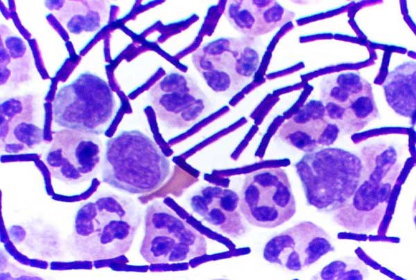
In bacteriology, Gram-positive bacteria are bacteria that give a positive result in the Gram stain test, which is traditionally used to quickly classify bacteria into two broad categories according to their type of cell wall.
The Gram stain is used by microbiologists to place bacteria into two main categories, Gram-positive (+) and Gram-negative (−). Gram-positive bacteria have a thick layer of peptidoglycan within the cell wall, and Gram-negative bacteria have a thin layer of peptidoglycan.
Gram-positive bacteria retain the crystal violet stain used in the test, resulting in a purple color when observed through an optical microscope. The thick layer of peptidoglycan in the bacterial cell wall retains the stain after it has been fixed in place by iodine. During the decolorization step, the decolorizer removes crystal violet from all other cells.
Conversely, Gram-negative bacteria cannot retain the violet stain after the decolorization step; alcohol used in this stage degrades the outer membrane of Gram-negative cells, making the cell wall more porous and incapable of retaining the crystal violet stain. Their peptidoglycan layer is much thinner and sandwiched between an inner cell membrane and a bacterial outer membrane, causing them to take up the counterstain (safranin or fuchsine) and appear red or pink.
Despite their thicker peptidoglycan layer, Gram-positive bacteria are more receptive to certain cell wall–targeting antibiotics than Gram-negative bacteria, due to the absence of the outer membrane.
In general, the following characteristics are present in Gram-positive bacteria:
Only some species have a capsule, usually consisting of polysaccharides. Only some species are flagellates, and those with flagella have just two basal body rings for support, in contrast to the four found in Gram-negative bacteria. Both Gram-positive and Gram-negative bacteria commonly have a surface layer called an S-layer. In Gram-positive bacteria, the S-layer is attached to the peptidoglycan layer. Gram-negative bacteria's S-layer is attached directly to the outer membrane. Specific to Gram-positive bacteria is the presence of teichoic acids in the cell wall. Some of these are lipoteichoic acids, which have a lipid component in the cell membrane that can assist in anchoring the peptidoglycan.
Along with cell shape, Gram staining is a rapid method used to differentiate bacterial species. Such staining, together with growth requirement and antibiotic susceptibility testing, and other macroscopic and physiologic tests, forms a basis for practical classification and subdivision of the bacteria (e.g., see figure and pre-1990 versions of Bergey's Manual of Systematic Bacteriology).[citation needed]
Hub AI
Gram-positive bacteria AI simulator
(@Gram-positive bacteria_simulator)
Gram-positive bacteria
In bacteriology, Gram-positive bacteria are bacteria that give a positive result in the Gram stain test, which is traditionally used to quickly classify bacteria into two broad categories according to their type of cell wall.
The Gram stain is used by microbiologists to place bacteria into two main categories, Gram-positive (+) and Gram-negative (−). Gram-positive bacteria have a thick layer of peptidoglycan within the cell wall, and Gram-negative bacteria have a thin layer of peptidoglycan.
Gram-positive bacteria retain the crystal violet stain used in the test, resulting in a purple color when observed through an optical microscope. The thick layer of peptidoglycan in the bacterial cell wall retains the stain after it has been fixed in place by iodine. During the decolorization step, the decolorizer removes crystal violet from all other cells.
Conversely, Gram-negative bacteria cannot retain the violet stain after the decolorization step; alcohol used in this stage degrades the outer membrane of Gram-negative cells, making the cell wall more porous and incapable of retaining the crystal violet stain. Their peptidoglycan layer is much thinner and sandwiched between an inner cell membrane and a bacterial outer membrane, causing them to take up the counterstain (safranin or fuchsine) and appear red or pink.
Despite their thicker peptidoglycan layer, Gram-positive bacteria are more receptive to certain cell wall–targeting antibiotics than Gram-negative bacteria, due to the absence of the outer membrane.
In general, the following characteristics are present in Gram-positive bacteria:
Only some species have a capsule, usually consisting of polysaccharides. Only some species are flagellates, and those with flagella have just two basal body rings for support, in contrast to the four found in Gram-negative bacteria. Both Gram-positive and Gram-negative bacteria commonly have a surface layer called an S-layer. In Gram-positive bacteria, the S-layer is attached to the peptidoglycan layer. Gram-negative bacteria's S-layer is attached directly to the outer membrane. Specific to Gram-positive bacteria is the presence of teichoic acids in the cell wall. Some of these are lipoteichoic acids, which have a lipid component in the cell membrane that can assist in anchoring the peptidoglycan.
Along with cell shape, Gram staining is a rapid method used to differentiate bacterial species. Such staining, together with growth requirement and antibiotic susceptibility testing, and other macroscopic and physiologic tests, forms a basis for practical classification and subdivision of the bacteria (e.g., see figure and pre-1990 versions of Bergey's Manual of Systematic Bacteriology).[citation needed]
Recent media