Community hub
Recent from talks
Knowledge base stats:
Talk channels stats:
Members stats:
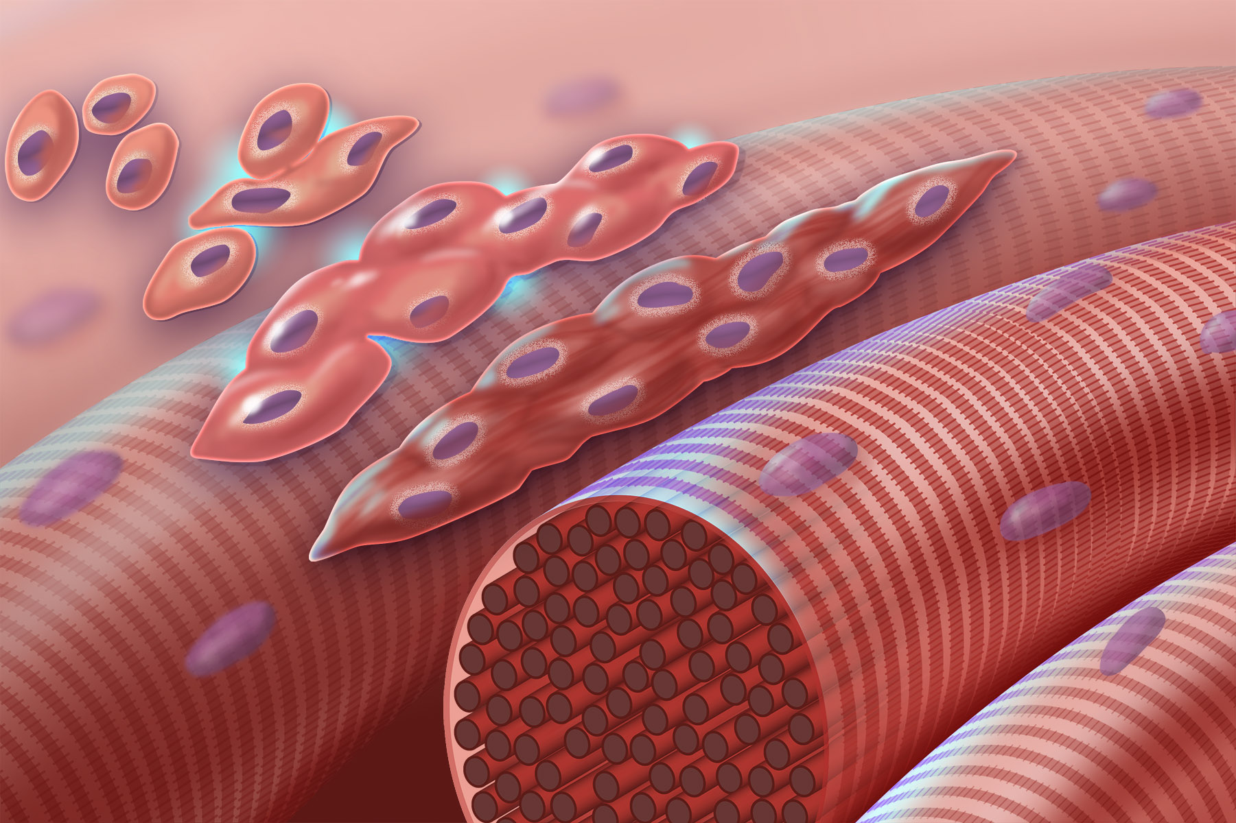
Myogenesis
Myogenesis is the formation of skeletal muscular tissue, particularly during embryonic development. Muscle fibers generally form through the fusion of precursor myoblasts into multinucleated fibers called myotubes. In the early development of an embryo, myoblasts can either proliferate, or differentiate into a myotube. What controls this choice in vivo is generally unclear. If placed in cell culture, most myoblasts will proliferate if enough fibroblast growth factor (FGF) or another growth factor is present in the medium surrounding the cells. When the growth factor runs out, the myoblasts cease division and undergo terminal differentiation into myotubes.
Myoblast differentiation proceeds in stages. The first stage involves cell cycle exit and the commencement of expression of certain genes. The second stage of differentiation involves the alignment of the myoblasts with one another. Studies have shown that even rat and chick myoblasts can recognise and align with one another, suggesting evolutionary conservation of the mechanisms involved. The third stage is the actual cell fusion itself. In this stage, the presence of calcium ions is critical. Fusion in humans is aided by a set of metalloproteinases coded for by the ADAM12 gene, and a variety of other proteins. Fusion involves recruitment of actin to the plasma membrane, followed by close apposition and creation of a pore that subsequently rapidly widens.
Genes and their protein products that are expressed during the process include: myocyte enhancer factors, myogenic regulatory factors, and serum response factor. Expression of skeletal alpha-actin is also regulated by the androgen receptor; steroids can thereby regulate myogenesis.
There are a number of stages (listed below) of muscle development, or myogenesis. Each stage has various associated genetic factors lack of which will result in muscular defects.
Associated Genetic Factors: PAX3 and c-Met
Mutations in PAX3 can cause a failure in c-Met expression. Such a mutation would result in a lack of lateral migration.
PAX3 mediates the transcription of c-Met and is responsible for the activation of MyoD expression—one of the functions of MyoD is to promote the regenerative ability of satellite cells (described below). PAX3 is generally expressed at its highest levels during embryonic development and is expressed at a lesser degree during the fetal stages; it is expressed in migrating hypaxial cells and dermomyotome cells, but is not expressed at all during the development of facial muscle. Mutations in Pax3 can cause a variety of complications including Waardenburg syndrome I and III as well as craniofacial-deafness-hand syndrome. Waardenburg syndrome is most often associated with congenital disorders involving the intestinal tract and spine, an elevation of the scapula, among other symptoms. Each stage has various associated genetic factors without which will result in muscular defects.
Associated Genetic Factors: c-Met/HGF and LBX1
Mutations in these genetic factors causes a lack of migration.
LBX1 is responsible for the development and organization of muscles in the dorsal forelimb as well as the movement of dorsal muscles into the limb following delamination. Without LBX1, limb muscles will fail to form properly; studies have shown that hindlimb muscles are severely affected by this deletion while only flexor muscles form in the forelimb muscles as a result of ventral muscle migration.
Hub AI
Myogenesis AI simulator
(@Myogenesis_simulator)
Myogenesis
Myogenesis is the formation of skeletal muscular tissue, particularly during embryonic development. Muscle fibers generally form through the fusion of precursor myoblasts into multinucleated fibers called myotubes. In the early development of an embryo, myoblasts can either proliferate, or differentiate into a myotube. What controls this choice in vivo is generally unclear. If placed in cell culture, most myoblasts will proliferate if enough fibroblast growth factor (FGF) or another growth factor is present in the medium surrounding the cells. When the growth factor runs out, the myoblasts cease division and undergo terminal differentiation into myotubes.
Myoblast differentiation proceeds in stages. The first stage involves cell cycle exit and the commencement of expression of certain genes. The second stage of differentiation involves the alignment of the myoblasts with one another. Studies have shown that even rat and chick myoblasts can recognise and align with one another, suggesting evolutionary conservation of the mechanisms involved. The third stage is the actual cell fusion itself. In this stage, the presence of calcium ions is critical. Fusion in humans is aided by a set of metalloproteinases coded for by the ADAM12 gene, and a variety of other proteins. Fusion involves recruitment of actin to the plasma membrane, followed by close apposition and creation of a pore that subsequently rapidly widens.
Genes and their protein products that are expressed during the process include: myocyte enhancer factors, myogenic regulatory factors, and serum response factor. Expression of skeletal alpha-actin is also regulated by the androgen receptor; steroids can thereby regulate myogenesis.
There are a number of stages (listed below) of muscle development, or myogenesis. Each stage has various associated genetic factors lack of which will result in muscular defects.
Associated Genetic Factors: PAX3 and c-Met
Mutations in PAX3 can cause a failure in c-Met expression. Such a mutation would result in a lack of lateral migration.
PAX3 mediates the transcription of c-Met and is responsible for the activation of MyoD expression—one of the functions of MyoD is to promote the regenerative ability of satellite cells (described below). PAX3 is generally expressed at its highest levels during embryonic development and is expressed at a lesser degree during the fetal stages; it is expressed in migrating hypaxial cells and dermomyotome cells, but is not expressed at all during the development of facial muscle. Mutations in Pax3 can cause a variety of complications including Waardenburg syndrome I and III as well as craniofacial-deafness-hand syndrome. Waardenburg syndrome is most often associated with congenital disorders involving the intestinal tract and spine, an elevation of the scapula, among other symptoms. Each stage has various associated genetic factors without which will result in muscular defects.
Associated Genetic Factors: c-Met/HGF and LBX1
Mutations in these genetic factors causes a lack of migration.
LBX1 is responsible for the development and organization of muscles in the dorsal forelimb as well as the movement of dorsal muscles into the limb following delamination. Without LBX1, limb muscles will fail to form properly; studies have shown that hindlimb muscles are severely affected by this deletion while only flexor muscles form in the forelimb muscles as a result of ventral muscle migration.