Hubbry Logo
MyogenesisMyogenesisMain
Open search
Myogenesis
Community hub
Myogenesis
logo
8 pages, 0 posts
0 subscribers
Be the first to start a discussion here.
Myogenesis
Myogenesis
from Wikipedia
Myoblasts (cells with a single nucleus, represented in violet) fusing together to form muscle fibers (multinucleated muscle cells) during myogenesis

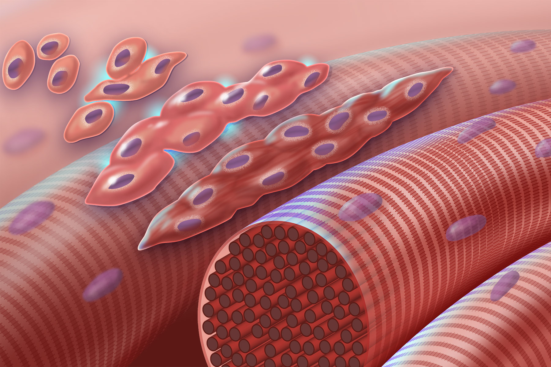
Myogenesis is the formation of skeletal muscular tissue, particularly during embryonic development. Muscle fibers generally form through the fusion of precursor myoblasts into multinucleated fibers called myotubes. In the early development of an embryo, myoblasts can either proliferate, or differentiate into a myotube. What controls this choice in vivo is generally unclear. If placed in cell culture, most myoblasts will proliferate if enough fibroblast growth factor (FGF) or another growth factor is present in the medium surrounding the cells. When the growth factor runs out, the myoblasts cease division and undergo terminal differentiation into myotubes.

Myoblast differentiation proceeds in stages. The first stage involves cell cycle exit and the commencement of expression of certain genes. The second stage of differentiation involves the alignment of the myoblasts with one another. Studies have shown that even rat and chick myoblasts can recognise and align with one another, suggesting evolutionary conservation of the mechanisms involved.[1] The third stage is the actual cell fusion itself. In this stage, the presence of calcium ions is critical. Fusion in humans is aided by a set of metalloproteinases coded for by the ADAM12 gene, and a variety of other proteins. Fusion involves recruitment of actin to the plasma membrane, followed by close apposition and creation of a pore that subsequently rapidly widens.

Genes and their protein products that are expressed during the process include: myocyte enhancer factors, myogenic regulatory factors, and serum response factor. Expression of skeletal alpha-actin is also regulated by the androgen receptor; steroids can thereby regulate myogenesis.[2]

Overview

[edit]

There are a number of stages (listed below) of muscle development, or myogenesis.[3] Each stage has various associated genetic factors lack of which will result in muscular defects.

Stages

[edit]
Stage Associated Genetic Factors
Delamination PAX3, c-Met
Migration c-met/HGF, LBX1
Proliferation PAX3, c-Met, Mox2, MSX1, Six1/4, Myf5, MyoD
Determination Myf5 and MyoD
Differentiation Myogenin, MCF2, Six1/4, MyoD, Myf6
Specific Muscle Formation Lbx1, Meox2
Satellite Cells PAX7

Delamination

[edit]
Patient with Waardenburg Syndrom III (Waardenburg-Klein Syndrome)
Patient with Waardenburg syndrome III (Waardenburg Klein Syndrome) with wide-set eyes.

Associated Genetic Factors: PAX3 and c-Met
Mutations in PAX3 can cause a failure in c-Met expression. Such a mutation would result in a lack of lateral migration.

PAX3 mediates the transcription of c-Met and is responsible for the activation of MyoD expression—one of the functions of MyoD is to promote the regenerative ability of satellite cells (described below).[3] PAX3 is generally expressed at its highest levels during embryonic development and is expressed at a lesser degree during the fetal stages; it is expressed in migrating hypaxial cells and dermomyotome cells, but is not expressed at all during the development of facial muscle.[3] Mutations in Pax3 can cause a variety of complications including Waardenburg syndrome I and III as well as craniofacial-deafness-hand syndrome.[3] Waardenburg syndrome is most often associated with congenital disorders involving the intestinal tract and spine, an elevation of the scapula, among other symptoms. Each stage has various associated genetic factors without which will result in muscular defects.[3]

Migration

[edit]

Associated Genetic Factors: c-Met/HGF and LBX1
Mutations in these genetic factors causes a lack of migration.

LBX1 is responsible for the development and organization of muscles in the dorsal forelimb as well as the movement of dorsal muscles into the limb following delamination.[3] Without LBX1, limb muscles will fail to form properly; studies have shown that hindlimb muscles are severely affected by this deletion while only flexor muscles form in the forelimb muscles as a result of ventral muscle migration.[3]

c-Met is a tyrosine kinase receptor that is required for the survival and proliferation of migrating myoblasts. A lack of c-Met disrupts secondary myogenesis and—as in LBX1—prevents the formation of limb musculature.[3] It is clear that c-Met plays an important role in delamination and proliferation in addition to migration. PAX3 is needed for the transcription of c-Met.[3]

Proliferation

[edit]

Associated Genetic Factors: PAX3, c-Met, Mox2, MSX1, Six, Myf5, and MyoD

Mox2 (also referred to as MEOX-2) plays an important role in the induction of mesoderm and regional specification.[3] Impairing the function of Mox2 will prevent the proliferation of myogenic precursors and will cause abnormal patterning of limb muscles.[4] Specifically, studies have shown that hindlimbs are severely reduced in size while specific forelimb muscles will fail to form.[3]

Myf5 is required for proper myoblast proliferation.[3] Studies have shown that mice muscle development in the intercostal and paraspinal regions can be delayed by inactivating Myf-5.[3] Myf5 is considered to be the earliest expressed regulatory factor gene in myogenesis. If Myf-5 and MyoD are both inactivated, there will be a complete absence of skeletal muscle.[3] These consequences further reveal the complexity of myogenesis and the importance of each genetic factor in proper muscle development.

MyoD1 (MYF3)
MyoD1 (MYF3).

Determination

[edit]

Associated Genetic Factors: Myf5 and MyoD
One of the most important stages in myogenesis determination requires both Myf5 and MyoD to function properly in order for myogenic cells to progress normally. Mutations in either associated genetic factor will cause the cells to adopt non-muscular phenotypes.[3]

As stated earlier, the combination of Myf5 and MyoD is crucial to the success of myogenesis. Both MyoD and Myf5 are members of the myogenic bHLH (basic helix-loop-helix) proteins transcription factor family.[5] Cells that make myogenic bHLH transcription factors (including MyoD or Myf5) are committed to development as a muscle cell.[6] Consequently, the simultaneous deletion of Myf5 and MyoD also results in a complete lack of skeletal muscle formation.[6] Research has shown that MyoD directly activates its own gene; this means that the protein made binds the myoD gene and continues a cycle of MyoD protein production.[6] Meanwhile, Myf5 expression is regulated by Sonic hedgehog, Wnt1, and MyoD itself.[3] By noting the role of MyoD in regulating Myf5, the crucial interconnectedness of the two genetic factors becomes clear.[3]

Serum response factor is needed for myogenesis and muscle development.[7] Interaction of SRF with other proteins, such as steroid hormone receptors, may contribute to regulation of muscle growth by steroids.[8]

Differentiation

[edit]

Associated genetic factors: Myogenin, Mcf2, Six, MyoD, and Myf6
Mutations in these associated genetic factors will prevent myocytes from advancing and maturing.

Muscular Dystrophy Histopathology
Muscular Dystrophy Histopathology.

Myogenin (also known as Myf4) is required for the fusion of myogenic precursor cells to either new or previously existing fibers.[3] In general, myogenin is associated with amplifying expression of genes that are already being expressed in the organism. Deleting myogenin results in nearly complete loss of differentiated muscle fibers and severe loss of skeletal muscle mass in the lateral/ventral body wall.[3]

Gowers's sign
Depiction of man exhibiting the Gowers's sign: common symptom of centronuclear myopathy that results from the weakness of lower limb muscles.

Myf-6 (also known as MRF4 or Herculin) is important to myotube differentiation and is specific to skeletal muscle.[3] Mutations in Myf-6 can provoke disorders including centronuclear myopathy and Becker muscular dystrophy.[3]

Specific muscle formation

[edit]

Associated genetic factors: LBX1 and Mox2
In specific muscle formation, mutations in associated genetic factors begin to affect specific muscular regions. Because of its large responsibility in the movement of dorsal muscles into the limb following delamination, mutation or deletion of Lbx1 results in defects in extensor and hindlimb muscles.[3] As stated in the Proliferation section, Mox2 deletion or mutation causes abnormal patterning of limb muscles. The consequences of this abnormal patterning include severe reduction in size of hindlimbs and complete absence of forelimb muscles.[3]

Satellite cells

[edit]

Associated genetic factors: PAX7
Mutations in Pax7 will prevent the formation of satellite cells and, in turn, prevent postnatal muscle growth.[3]

Satellite cells are described as quiescent myoblasts and neighbor muscle fiber sarcolemma.[3] They are crucial for the repair of muscle, but have a very limited ability to replicate. Activated by stimuli such as injury or high mechanical load, satellite cells are required for muscle regeneration in adult organisms.[3] In addition, satellite cells have the capability to also differentiate into bone or fat. In this way, satellite cells have an important role in not only muscle development, but in the maintenance of muscle through adulthood.[3]

Skeletal muscle

[edit]

During embryogenesis, the dermomyotome and/or myotome in the somites contain the myogenic progenitor cells that will evolve into the prospective skeletal muscle.[9] The determination of dermomyotome and myotome is regulated by a gene regulatory network that includes a member of the T-box family, tbx6, ripply1, and mesp-ba.[10] Skeletal myogenesis depends on the strict regulation of various gene subsets in order to differentiate the myogenic progenitors into myofibers. Basic helix-loop-helix (bHLH) transcription factors, MyoD, Myf5, myogenin, and MRF4 are critical to its formation. MyoD and Myf5 enable the differentiation of myogenic progenitors into myoblasts, followed by myogenin, which differentiates the myoblast into myotubes.[9] MRF4 is important for blocking the transcription of muscle-specific promoters, enabling skeletal muscle progenitors to grow and proliferate before differentiating.

Basic helix–loop–helix
Basic helix–loop–helix.

There are a number of events that occur in order to propel the specification of muscle cells in the somite. For both the lateral and medial regions of the somite, paracrine factors induce myotome cells to produce MyoD protein—thereby causing them to develop as muscle cells.[11] A transcription factor (TCF4) of connective tissue fibroblasts is involved in the regulation of myogenesis. Specifically, it regulates the type of muscle fiber developed and its maturations.[3] Low levels of TCF4 promote both slow and fast myogenesis, overall promoting the maturation of muscle fiber type. Thereby this shows the close relationship of muscle with connective tissue during the embryonic development.[12]

Regulation of myogenic differentiation is controlled by two pathways: the phosphatidylinositol 3-kinase/Akt pathway and the Notch/Hes pathway, which work in a collaborative manner to suppress MyoD transcription.[5] The O subfamily of the forkhead proteins (FOXO) play a critical role in regulation of myogenic differentiation as they stabilize Notch/Hes binding. Research has shown that knockout of FOXO1 in mice increases MyoD expression, altering the distribution of fast-twitch and slow-twitch fibers.[5]

Muscle fusion

[edit]

Primary muscle fibers originate from primary myoblasts and tend to develop into slow muscle fibers.[3] Secondary muscle fibers then form around the primary fibers near the time of innervation. These muscle fibers form from secondary myoblasts and usually develop as fast muscle fibers. Finally, the muscle fibers that form later arise from satellite cells.[3]

Two genes significant in muscle fusion are Mef2 and the twist transcription factor. Studies have shown knockouts for Mef2C in mice lead to muscle defects in cardiac and smooth muscle development, particularly in fusion.[13] The twist gene plays a role in muscle differentiation.

The SIX1 gene plays a critical role in hypaxial muscle differentiation in myogenesis. In mice lacking this gene, severe muscle hypoplasia affected most of the body muscles, specifically hypaxial muscles.[14]

In myoblasts, PtdIns5P, produced by the lipid phosphatase MTM1, is rapidly metabolized by PI5P 4-kinase α into PI(4,5)P2, which accumulates at the plasma membrane. This accumulation facilitates the formation of podosome-like protrusions, where the fusogen Myomaker is localized, playing a crucial role in the spatiotemporal regulation of myoblast fusion.[15]

Protein synthesis and actin heterogeneity

[edit]

There are 3 types of proteins produced during myogenesis.[4] Class A proteins are the most abundant and are synthesized continuously throughout myogenesis. Class B proteins are proteins that are initiated during myogenesis and continued throughout development. Class C proteins are those synthesized at specific times during development. Also 3 different forms of actin were identified during myogenesis.

Sim2, a BHLH-Pas transcription factor, inhibits transcription by active repression and displays enhanced expression in ventral limb muscle masses during chick and mouse embryonic development. It accomplishes this by repressing MyoD transcription by binding to the enhancer region, and prevents premature myogenesis.[16]

Delta1 expression in neural crest cells is necessary for muscle differentiation of the somites, through the Notch signaling pathway. Gain and loss of this ligand in neural crest cells results in delayed or premature myogenesis.[17]

Techniques

[edit]

The significance of alternative splicing was elucidated using microarrary analysis of differentiating C2C12 myoblasts.[18] 95 alternative splicing events occur during C2C12 differentiation in myogenesis. Therefore, alternative splicing is necessary in myogenesis.

Systems approach

[edit]

Systems approach is a method used to study myogenesis, which manipulates a number of different techniques like high-throughput screening technologies, genome wide cell-based assays, and bioinformatics, to identify different factors of a system.[9] This has been specifically used in the investigation of skeletal muscle development and the identification of its regulatory network.

Systems approach using high-throughput sequencing and ChIP-chip analysis has been essential in elucidating the targets of myogenic regulatory factors like MyoD and myogenin, their inter-related targets, and how MyoD acts to alter the epigenome in myoblasts and myotubes.[9] This has also revealed the significance of PAX3 in myogenesis, and that it ensures the survival of myogenic progenitors.[9]

This approach, using cell based high-throughput transfection assay and whole-mount in situ hybridization, was used in identifying the myogenetic regulator RP58, and the tendon differentiation gene, Mohawk homeobox.[9]

References

[edit]
[edit]
Revisions and contributorsEdit on WikipediaRead on Wikipedia
from Grokipedia
Myogenesis is the multistep responsible for the formation and development of tissue, beginning with the specification and proliferation of mesenchymal precursor cells derived from the , followed by their differentiation into myoblasts, fusion into multinucleated myotubes, and maturation into functional myofibers. This process is essential for enabling movement, , and metabolic functions, and it occurs in distinct phases throughout development and adulthood. Embryonic myogenesis initiates in the somites, where paraxial segments give rise to myogenic progenitor cells (MPCs) expressing paired box transcription factors Pax3 and Pax7, which migrate to form the primary myotomes and limb muscle masses. These progenitors then commit to the myogenic lineage under the control of myogenic regulatory factors (MRFs), a family of basic helix-loop-helix transcription factors including Myf5, , myogenin, and MRF4, which activate muscle-specific and drive exit. The process unfolds in waves: primary myotubes form first along axial and limb templates, followed by secondary myotubes during the fetal stage to expand muscle mass, all regulated by extrinsic signals such as Wnt and Sonic hedgehog pathways. In postnatal life, myogenesis continues through the activation of satellite cells—quiescent stem cells marked by Pax7 expression and located beneath the of myofibers—which proliferate in response to or growth demands, differentiate into myoblasts, and fuse with existing fibers to facilitate repair and . This adult regenerative phase shares core molecular mechanisms with embryonic myogenesis, particularly the hierarchical action of MRFs, where and Myf5 promote commitment and proliferation, while myogenin and MRF4 oversee terminal differentiation and fusion. Disruptions in myogenesis can lead to congenital muscle disorders or impaired regeneration in conditions like , underscoring its clinical significance.

Introduction

Definition and Scope

Myogenesis refers to the by which tissue forms from precursor cells, primarily through the commitment, proliferation, differentiation, and fusion of myoblasts into multinucleated myofibers. This process is fundamental to generating functional muscle capable of contraction and force production across various tissue types. The scope of myogenesis encompasses both embryonic development and postnatal regeneration, with distinct mechanisms for the three primary muscle types: , , and . myogenesis, the most extensively studied, originates from precursors in the somites during embryogenesis and continues postnatally via cells for growth and repair. forms from cardiogenic early in development, resulting in striated, involuntary fibers connected by intercalated discs, while arises from diverse origins including and neural crest-derived cells (), yielding non-striated, involuntary cells specialized for organ-specific functions like vascular tone. Although the core stages of myogenesis—such as , proliferation, and fusion—are referenced here, they are elaborated in subsequent sections on skeletal-specific processes. Myogenesis plays a critical role in organismal development, enabling locomotion through s that comprise over 600 distinct units in humans, supporting organ function via cardiac and smooth muscles, and facilitating tissue repair after injury, particularly in . Disruptions in this process contribute to debilitating conditions, such as muscular dystrophies, where impaired regeneration leads to progressive muscle weakness and degeneration.

Historical Development

The study of myogenesis began with early microscopic observations in the , where researchers identified mononucleated cells, later termed myoblasts, as precursors to multinucleated muscle fibers during embryonic development. These observations, based on light microscopy of and avian embryos, laid the groundwork for understanding muscle formation through , though the precise mechanisms remained unclear until advanced imaging techniques emerged in the . In the mid-20th century, embryological research advanced significantly with the identification of satellite cells as quiescent muscle stem cells capable of contributing to postnatal myogenesis. Alexander Mauro's 1961 electron microscopy study of frog revealed these mononucleated cells positioned between the and plasma membrane of muscle fibers, proposing them as reserve cells for regeneration. Concurrently, 1980s studies by Wachtler and Christ using quail-chick chimeras demonstrated that precursors originate from somites, the segmental blocks of paraxial flanking the , with myogenic cells delaminating from the dermomyotome to populate limb and body wall muscles. The molecular era of myogenesis research was ushered in by the 1987 discovery of , a helix-loop-helix identified by Weintraub and colleagues through subtractive hybridization of cDNA libraries from myoblasts and fibroblasts. This seminal work showed that of could convert non-muscle cells into myoblasts, establishing it as a master regulator of determination. In the , advancements highlighted the roles of various populations in myogenesis, including demonstrations that embryonic stem cells could differentiate into skeletal myoblasts under defined culture conditions, opening avenues for regenerative therapies. The 2010s and 2020s shifted focus to and cellular heterogeneity, with reviews elucidating how histone modifications and orchestrate myogenic gene in satellite cells and progenitors. Single-cell sequencing studies further revealed transcriptional diversity among muscle stem cells, identifying subpopulations with distinct regenerative potentials and highlighting heterogeneity in myogenic commitment during development and repair. In the 2020s, research has advanced with investigations into epitranscriptomic modifications, such as m6A methylation, in regulating myogenic differentiation and optimized in vitro models, including organoids, for studying and bovine myogenesis as of 2025.

Embryonic Origins

Mesodermal Specification

Myogenesis begins with the specification of the during in embryos, where epiblast cells ingress through the to form a trilaminar structure comprising , , and . The paraxial , positioned bilaterally along the , emerges as a key compartment destined to give rise to somites, which are transient segmental structures essential for development. This presomitic maintains a mesenchymal state before undergoing segmentation, setting the stage for myogenic commitment. Commitment of the paraxial mesoderm to the myogenic lineage is driven by inductive signals from adjacent axial structures, particularly the and dorsal . (BMP) antagonists, such as noggin and chordin, secreted by these tissues, play a critical role by inhibiting ventralizing BMP signaling, thereby promoting dorsal somitic fates including myogenesis. Noggin, expressed in the node, , and early , is essential for proper somitogenesis and the patterning of myogenic regulators like in the medial somite. Similarly, chordin contributes to anterior somite specification by inducing early myogenic markers such as Myf5, ensuring the medial-lateral patterning necessary for formation. Hox genes establish rostro-caudal identity in the paraxial mesoderm, influencing the regional specificity of myogenic differentiation along the embryonic axis. These transcription factors are expressed in collinear domains within the presomitic mesoderm, with specific paralog groups (e.g., and ) regulating the activation of myogenic factors like Myf6 in hypaxial muscle progenitors. Disruption of Hox expression alters vertebral and somitic identities, underscoring their role in coordinating myogenic programs with axial patterning. Early molecular markers of myogenic specification in the presomitic include the transcription factors and Msx1, which are upregulated in response to these commitment signals. expression initiates in the presomitic mesoderm and marks committed progenitors prior to formation, playing a pivotal role in early myogenic lineage specification. Msx1, expressed concurrently in paraxial mesoderm derivatives, supports progenitor maintenance by modulating interactions with , preventing premature differentiation while promoting migration competence.

Somitogenesis and Myotome Formation

Somitogenesis is the process by which the unsegmented paraxial , positioned lateral to the and , undergoes periodic segmentation to form in a sequential manner from anterior to posterior along the embryonic axis. This segmentation establishes the metameric pattern essential for the development of the vertebral column, , and skeletal muscles. In embryos, the first somite pair appears around embryonic day 8 (E8), with subsequent pairs forming at a rate that initially averages about 90 minutes per pair, slowing to approximately 120 minutes as development progresses, resulting in roughly 50-65 somite pairs by E13. The presomitic mesoderm (PSM) continuously elongates from the , providing the tissue that will be partitioned into somites. The clock-and-wavefront model provides the primary framework for understanding the spatiotemporal regulation of somitogenesis. The "wavefront" refers to a posterior-to-anterior of maturation competence in the PSM, established by opposing signaling gradients: (FGF) and Wnt signaling emanate from the tailbud to maintain PSM progenitors in an undifferentiated state, while produced by nascent somites promotes anterior maturation and defines the determination front. The "clock" consists of oscillatory cycles within PSM cells, with a period of approximately 120 minutes in mice, that synchronize to trigger somite boundary formation when cells at the determination front phase-lock in their oscillations. Key components of the clock include the Notch/Delta pathway, where cyclic expression of ligands like Delta-like 1 (Dll1) and targets such as Lunatic fringe () and Hairy and enhancer of split 7 (Hes7) generate feedback loops that propagate oscillations cell-autonomously and synchronize neighboring cells via . Wnt and FGF signaling further modulate these oscillations, with Wnt3a driving cyclic expression of clock genes in the posterior PSM. Following formation, the epithelial differentiates into distinct compartments, with the emerging as the primary myogenic domain from the medial and lateral edges of the dermomyotome. The comprises two subdivisions: the epaxial , located dorsally and innervated by the dorsal ramus of the spinal nerves to form extensor and deep back muscles, and the hypaxial , positioned ventrally and innervated by the to contribute to intercostal, abdominal, and limb muscles. Initial myoblasts within the express the paired-box throughout the progenitor population and the myogenic regulatory factor Myf5, which is activated first in the epaxial domain around E8 in mice, marking the onset of myogenic specification. Myf5 expression expands to the hypaxial by E9.5, preceding overt differentiation. By E10.5, cells begin to populate the myogenic lineage, establishing the foundational structure for development.

Stages of Skeletal Myogenesis

Delamination and Migration

During myogenesis, delamination marks the initial detachment of myogenic progenitor cells from the dermomyotome, a key step in forming skeletal muscle precursors. Pax3-expressing cells, located in the hypaxial domain of the dermomyotome, undergo an epithelial-to-mesenchymal transition (EMT) to exit the epithelial structure and adopt a migratory mesenchymal phenotype. This process is tightly regulated by the c-Met receptor tyrosine kinase, which is transcriptionally activated by Pax3 and essential for initiating cell emigration from the dermomyotome. Without c-Met signaling, these progenitors remain trapped in the somite, preventing their dispersal. Migration of these delaminated myoblasts is driven by chemotactic cues that direct them to specific destinations in the . Hepatocyte growth factor (HGF), also known as scatter factor (SF), serves as the primary for c-Met, produced by surrounding mesenchymal tissues to induce directed and through extracellular matrices. This HGF/c-Met interaction promotes , enabling long-range migration along predefined paths while maintaining cell survival and preventing premature differentiation. The transcription factor LBX1 plays a crucial role in specifying and executing this migration, particularly for limb-bound progenitors, by regulating genes involved in pathfinding and responsiveness to guidance signals such as /SDF1. LBX1 is co-expressed with in hypaxial precursors and is required for their lateral progression, though it does not affect initial . Delaminated myoblasts, primarily from the hypaxial domain, follow trajectories to populate limb buds, diaphragm, and other ventral structures. Epaxial progenitors contribute to back muscles with minimal migration, differentiating locally. In the limbs, LBX1 ensures proper entry via a lateral pathway, contributing to the formation of both proximal and distal muscle masses. Disruptions in these processes, such as in c-Met, HGF, or LBX1 mutants, result in severe defects including failed limb colonization and subsequent muscle , with Pax3-positive cells accumulating ectopically or undergoing . For instance, LBX1-deficient mice exhibit near-complete loss of hindlimb muscles and reduced forelimb extensors due to misguided or stalled migration.

Proliferation and Determination

During embryonic skeletal myogenesis, myoblasts delaminate from the myotome and undergo extensive proliferation to expand the progenitor pool prior to commitment. This expansion primarily occurs through symmetric cell divisions, which are regulated by the transcription factor Pax3 that promotes cell cycle progression while maintaining low levels of the myogenic regulatory factors (MRFs) Myf5 and MyoD to prevent premature differentiation. Low Myf5 and MyoD expression in these proliferating progenitors ensures continued division without initiating the myogenic program, as higher levels would trigger lineage commitment. The cell cycle in these myoblasts is driven by cyclin D1 in complex with cyclin-dependent kinases CDK4 and CDK6, which facilitate G1/S phase transition and sustain proliferation by interacting with MyoD to sequester it in the cytoplasm, thereby inhibiting its transcriptional activity. Myoblast determination marks the irreversible commitment to the myogenic lineage, often triggered by asymmetric cell divisions or environmental cues such as signaling from the niche, which upregulate Myf5 and expression to lock cells into a myoblast fate. In asymmetric divisions, one cell retains stem-like properties while the other expresses elevated Myf5 and , committing to myogenesis; environmental factors like withdrawal of mitogens or activation of pathways such as Wnt further promote this upregulation. Commitment is reinforced by withdrawal, mediated by p21 (encoded by Cdkn1a), a inhibitor induced by that blocks CDK4/6 activity and halts proliferation to prepare cells for subsequent maturation. In mouse embryogenesis, distinguish primary and secondary myoblasts during this phase. Recent studies have revealed early lineage segregation of primary myotubes from secondary progenitors, occurring prior to overt differentiation waves. Primary myoblasts, derived from the initial progenitors, proliferate predominantly between embryonic day (E) 11.5 and E14.5 to generate the first wave of muscle fibers, expanding the pool through Pax3-dependent symmetric divisions under low MRF conditions. Secondary myoblasts emerge later, around E14.5, from a distinct subset often marked by Pax7, and their proliferation contributes to additional fiber formation, with determination similarly involving Myf5/ upregulation and p21-mediated cycle exit to balance pool maintenance and commitment. This temporal segregation ensures sufficient cell numbers for trunk and limb muscle development without overlapping with later differentiation events.

Differentiation and Fusion

During myoblast differentiation, committed myogenic cells exit the and initiate terminal maturation into myocytes, marked by the upregulation of transcription factors such as myogenin and MEF2 family members. Myogenin, a basic helix-loop-helix , plays a pivotal role in activating the expression of muscle-specific structural genes, including heavy chain (MyHC), which is essential for assembly. Similarly, MEF2 proteins cooperate with myogenin to drive via recruitment of the Brg1, thereby enabling the transcription of late-stage muscle genes like MyHC. This cooperative regulation ensures the precise temporal activation of contractile proteins, with MEF2 also directly controlling expression to support calcium-handling capabilities in nascent muscle cells. The fusion of differentiated myocytes into multinucleated myotubes represents a critical step in formation, involving orchestrated cell alignment and membrane merger. Initial myoblast alignment is facilitated by cell adhesion molecules, including M-cadherin, a calcium-dependent that mediates homophilic interactions at contact sites and is essential for fusion competence. ADAM12, a disintegrin and metalloprotease, further supports this process by binding to α-actinin-2 and promoting cytoskeletal rearrangements that stabilize myoblast interactions prior to fusion. The actual membrane fusion is calcium-dependent and driven by specialized fusogenic proteins: myomaker, a multipass that initiates and pore formation, and myomerger (also known as myomixer), which independently regulates subsequent hemifusion-to-pore transition steps through exposure on the outer membrane leaflet. These proteins act in concert to remodel membranes without relying on traditional viral fusogens, ensuring efficient formation. Intracellular calcium transients, coordinated with these molecular events, further synchronize fusion dynamics across myoblasts. Following fusion, myotubes undergo maturation, developing functional contractility through organization and excitation-contraction coupling. Contractile ability emerges as MyHC and integrate into organized myofibrils, allowing myotubes to generate force in response to , a process refined by biomechanical cues. Concurrently, motor neurons extend axons to innervate myotubes, forming neuromuscular junctions (NMJs) that stabilize myofiber maturation and enhance contractile properties by modulating and trophic signaling. This innervation is indispensable for achieving adult-like twitch characteristics and preventing during late embryonic development.

Molecular Regulation

Myogenic Regulatory Factors

The myogenic regulatory factors (MRFs) constitute a family of four basic helix-loop-helix (bHLH) transcription factors—Myf5, MyoD, myogenin, and MRF4—that orchestrate skeletal muscle commitment, differentiation, and maturation during development. These proteins share a conserved bHLH domain, enabling them to bind DNA and regulate muscle-specific gene expression, with their sequential activation ensuring precise control over myogenesis. Myf5, the earliest expressed MRF, initiates myogenic commitment in somitic precursors around embryonic day 8 in mice, promoting epaxial myotome formation and exhibiting functional redundancy with MyoD to ensure progenitor survival. MyoD, a pivotal bHLH protein discovered in 1987, drives myoblast determination and induces expression of downstream factors like myogenin, converting non-muscle cells into myogenic lineages. Myogenin, identified in 1989, functions primarily as a differentiation driver, essential for myoblast fusion and fiber assembly starting around embryonic day 8.5. MRF4 plays a maturation role, with biphasic expression—first at embryonic day 9 and reappearing postnatally—supporting late-stage muscle gene regulation and maintenance in adult tissue. MRFs exert their effects through heterodimerization with ubiquitous E-proteins (such as E12 or E47) via the HLH domain, forming complexes that bind E-box motifs with the consensus sequence CANNTG in the regulatory regions of target genes. This binding recruits chromatin-remodeling complexes, including SWI/SNF and histone acetyltransferases, to open chromatin structure and activate muscle-specific transcription, as exemplified by MyoD's role in remodeling at loci like myogenin. Myf5 similarly modifies chromatin but shows weaker intrinsic activation potential compared to MyoD or myogenin. Genetic studies reveal significant redundancy among MRFs; single knockouts of Myf5 or MyoD yield viable mice with normal musculature, but double knockouts (Myf5^{-/-}; MyoD^{-/-}) result in complete absence of myoblasts and skeletal muscle at birth, with no desmin-positive progenitors detectable, underscoring their essential, overlapping roles in myoblast specification and propagation. The MRF family exhibits strong evolutionary conservation across vertebrates, arising from duplications of a single ancestral bHLH gene during early chordate evolution, with the four members (Myf5, MyoD, myogenin, MRF4/Myf6) present in jawed vertebrates. The bHLH domains, particularly those involved in DNA binding, remain highly invariant, while activation domains show evidence of positive selection driving functional divergence, such as in differentiation-specific roles for myogenin and MRF4. This conservation highlights the MRFs' fundamental role in vertebrate myogenesis, with orthologs in species from fish to mammals maintaining the core myogenic code.

Signaling Pathways

Myogenesis is tightly regulated by a network of extracellular and intracellular signaling pathways that orchestrate the timing of progenitor proliferation, determination, differentiation, and fusion. These signals integrate inputs from the , neighboring tissues, and growth factors to ensure precise myogenic fate decisions during embryonic development and adult regeneration. Key pathways such as Wnt, Notch, and PI3K/Akt play pivotal roles in balancing self-renewal against terminal differentiation, while growth factors like IGF-1 and HGF/SF provide context-specific cues for migration and enhancement of myogenic programs. The exhibits dual functionality in myogenesis, with its branch promoting progenitor proliferation and the non-canonical branch facilitating myoblast fusion. Wnt/β-catenin signaling, activated by ligands such as Wnt1 and Wnt3a binding to receptors, stabilizes β-catenin to drive transcription of target genes that maintain myogenic precursors in a proliferative state, preventing premature differentiation. In contrast, non-canonical Wnt signaling, particularly via Wnt7a through -7 and Rac1, enhances myoblast alignment and membrane protrusion essential for fusion, thereby supporting multinucleated myofiber formation during later stages. Notch signaling acts primarily as an inhibitor of myogenic differentiation, maintaining progenitors in an undifferentiated pool through mechanisms. Upon ligand binding (e.g., Delta or ) to Notch receptors on myoblasts, the intracellular domain translocates to the nucleus and activates transcription factors like Hes1, which repress myogenic regulatory factor (MRF) expression and block exit. This inhibitory role is crucial during early somitogenesis to expand the population, with downregulation of Notch allowing progression to differentiation. The PI3K/Akt pathway, downstream of receptor kinases, promotes postnatal by enhancing protein synthesis and inhibiting programs, particularly in response to mechanical loading or . Activation of PI3K generates PIP3 to recruit and phosphorylate Akt, which in turn phosphorylates targets like to drive myofiber growth without directly initiating embryonic myogenesis. Growth factors further modulate these processes: insulin-like growth factor-1 (IGF-1) enhances MyoD transcriptional activity and myoblast proliferation by activating the IGF-1 receptor and downstream PI3K/Akt, thereby amplifying commitment to the myogenic lineage. Hepatocyte growth factor/scatter factor (HGF/SF), secreted by mesenchymal cells, stimulates myoblast migration during from the via c-Met receptor activation and ERK/MAPK signaling, facilitating limb muscle patterning. Conversely, members of the TGF-β family, including TGF-β1 and , inhibit differentiation by repressing MRF expression through Smad2/3-mediated transcription, ensuring temporal control and preventing ectopic myotube formation. Crosstalk between pathways refines myogenic outcomes; for instance, Sonic hedgehog (Shh) from the and floor plate sustains Myf5 expression in epaxial somites via transcription factors, establishing the initial myogenic field. Additionally, feedback loops integrate these signals with MRFs, where Wnt and IGF-1 pathways amplify and Myf5 activity to coordinate proliferation with differentiation entry.

Epigenetic Control

Epigenetic modifications play a crucial role in regulating myogenic during development and regeneration by establishing stable states that influence the accessibility of myogenic loci. These include histone modifications, patterns, and the actions of non-coding RNAs, which collectively fine-tune the transition from proliferation to differentiation without altering the underlying DNA sequence. Such mechanisms ensure precise temporal control, allowing myoblasts to respond to developmental cues while suppressing alternative cell fates. Histone modifications, particularly and , activate key myogenic transcription factors. Trimethylation of at 4 () on the Myf5 promoter is mediated by the MLL1 complex, which promotes Myf5 expression essential for myoblast proliferation and differentiation. This modification enhances accessibility at myogenic regulatory factor (MRF) loci, facilitating their activation during early myogenesis. Similarly, acetylation of H3K27 by the p300/CBP acetyltransferases enriches active enhancers associated with myogenin (MYOG) promoters, driving the upregulation of MYOG and supporting late-stage differentiation in primary myoblasts. These marks create a permissive environment for MRF binding to epigenetically modified . DNA methylation dynamics further refine myogenic commitment by silencing non-myogenic genes and enabling differentiation-specific expression. Hypermethylation occurs at promoters of and T-box genes, which are non-myogenic developmental regulators, thereby repressing alternative lineages during myoblast specification. In contrast, active demethylation by ten-eleven translocation (TET) enzymes, particularly TET2, targets enhancers of differentiation genes like myogenin, reducing CpG methylation to increase openness and recruitment. TET2 deficiency impairs myoblast fusion and muscle regeneration by elevating methylation at these sites, highlighting its role in transitioning quiescent cells to active progenitors. Non-coding RNAs provide an additional layer of epigenetic control, modulating modifiers and promoting myogenic progression. MicroRNAs miR-1 and miR-206 enhance differentiation by directly targeting 4 (HDAC4), a of muscle , thereby alleviating HDAC4-mediated inhibition of myogenic transcription factors. Long non-coding RNAs (lncRNAs), such as the YY1-associated muscle lincRNA (Yam-1), regulate cell activation during regeneration; Yam-1 peaks early post-injury, activates miR-715 to suppress Wnt7b, and inhibits premature differentiation, ensuring coordinated muscle repair.

Myogenesis in Cardiac and Smooth Muscle

Cardiac Muscle Formation

Cardiac muscle formation originates from mesodermal precursors in the early , specifically the splanchnic , which differentiates into the first heart field to form the primitive heart tube comprising the left ventricle and parts of the atria. The second heart field, derived from pharyngeal splanchnic , contributes additional progenitors that populate the outflow tract, right ventricle, and atrial septum through progressive addition to the heart tube. Early specification of these cardiac progenitors is marked by the expression of transcription factors such as Gata4 and Nkx2.5 in the precardiac , which are essential for initiating cardiogenic programs and lineage commitment. The process of cardiomyocyte development involves proliferation of cells followed by differentiation into contractile cardiomyocytes, distinct from skeletal myogenesis as cardiac precursors do not undergo myoblast fusion to form multinucleated fibers; instead, they interconnect via intercalated discs to create a functional . In the second heart field, the Hand2 regulates outflow tract by promoting proliferation and specifying myocardial identity, with its disruption leading to defects such as . This differentiation is orchestrated by signaling pathways including BMP, FGF, and Wnt, which balance proliferation and maturation without reliance on fusion events. Maturation of cardiac muscle entails trabeculation, where subsets of ventricular cardiomyocytes delaminate from the outer layer and invade the cardiac jelly to form trabeculae, enhancing oxygen in the avascular myocardium through endocardial-myocardial via Neuregulin-ErbB signaling. Subsequently, the compact layer develops through cardiomyocyte proliferation driven by epicardial-derived factors like FGF9, culminating in a stratified myocardium that supports efficient pumping. In contrast to , which relies on cells for robust regeneration, cardiac muscle lacks such a population and exhibits limited postnatal regenerative potential, with injury typically resulting in fibrotic scarring rather than functional repair.

Smooth Muscle Differentiation

Smooth muscle cells (SMCs) originate primarily from the , including its splanchnic component, which contributes to the smooth muscle layers of major arteries like the dorsal aorta and visceral organs. Additionally, cells serve as progenitors for SMCs in the arteries and outflow tract vessels, highlighting the diverse embryonic origins of smooth muscle lineages. These progenitors differentiate under the control of key transcription factors, notably serum response factor (SRF) and its coactivator myocardin, which form a complex that binds CArG box motifs in the promoters of SMC-specific genes to drive differentiation. Unlike , which involves myoblast fusion into multinucleated fibers, smooth muscle differentiation proceeds without , resulting in mononucleated cells that assemble into continuous sheets or concentric layers in vessel walls and visceral structures. This process is marked by the progressive expression of contractile proteins, including α- actin (SMA) as an early indicator and calponin as a later marker of maturation, enabling the cells to acquire contractile properties essential for vascular tone and organ motility. The mononucleated nature allows SMCs to retain high proliferative and migratory capacity during development and repair, forming interconnected networks rather than discrete fibers. Smooth muscle exhibits two main types: vascular SMCs, which form the media of blood vessels and exhibit a multi-unit organization with dense innervation, and visceral SMCs, which constitute the walls of hollow organs like the gut and in a single-unit configuration with gap junctions for coordinated contraction. While myocardin is indispensable for visceral SMC development, it is dispensable for vascular SMCs, reflecting subtype-specific regulatory dependencies. Differentiation in both types is potently induced by transforming growth factor-β (TGF-β), which activates the MRTF-A/SRF pathway to upregulate SMC marker genes such as SMA and calponin, ensuring proper contractile acquisition. This mechanism parallels aspects of formation, where SRF also plays a central role, but emphasizes non-striated, sheet-like architecture.

Postnatal Processes

Satellite Cells and Regeneration

Satellite cells are quiescent muscle stem cells characterized by the expression of the Pax7 and located in a specialized niche beneath the of adult fibers. These cells remain dormant under homeostatic conditions but serve as the primary source for postnatal muscle repair and growth following injury or stress. Upon muscle injury, satellite cells exit quiescence and become activated through signaling pathways, including the downregulation of Notch signaling, which normally maintains their dormant state. Activated satellite cells then proliferate, with a subset expressing low levels of Myf5 during this phase to support expansion while preserving stem cell potential. From the proliferating pool, some cells return to quiescence by upregulating Pax7 to replenish the stem cell reserve, while others commit to differentiation by increasing expression, leading to myoblast formation. Differentiated myoblasts subsequently fuse with existing damaged myofibers or form new ones, restoring muscle architecture and function in a cyclic process that balances regeneration and self-renewal. Recent advances in single-cell RNA sequencing (scRNA-seq) during the 2020s have uncovered significant heterogeneity within cell populations, revealing subpopulations with distinct transcriptional profiles that influence their regenerative contributions. For instance, scRNA-seq studies have highlighted interactions between cells and fibro-adipogenic progenitors (FAPs), where FAPs support myogenesis through secreted factors like FGF7, which binds FGFR2 on cells to enhance proliferation and repair efficiency. This heterogeneity underscores the niche's role in modulating cell fate and improving outcomes in muscle regeneration models. Therapeutic strategies targeting satellite cell dysfunction hold promise for treating (DMD), a condition marked by deficiency and impaired regeneration. Induced pluripotent stem cells (iPSCs) derived from DMD patients can be reprogrammed into satellite-like cells, offering a renewable source for transplantation to restore muscle repair capacity without immune rejection risks. Preclinical studies in the 2020s demonstrate that iPSC-derived myogenic progenitors integrate into the host niche, express Pax7, and contribute to myofiber formation in DMD mouse models, paving the way for personalized cell therapies. However, challenges such as low engraftment efficiency, immune rejection in allogeneic approaches, and risks of tumorigenicity from iPSCs persist, alongside controversies over ethical aspects of chimeric cell strategies, as reviewed in late 2025.

Muscle Adaptation and Hypertrophy

Muscle adaptation and hypertrophy represent key postnatal processes in skeletal muscle, enabling tissues to remodel in response to mechanical loading, disuse, or aging. These mechanisms involve coordinated changes in protein synthesis, fiber composition, and cellular contributions, primarily driven by satellite cells and intracellular signaling pathways. Unlike injury-induced regeneration, adaptation focuses on physiological responses to exercise or inactivity, promoting either growth or maintenance of muscle mass. Hypertrophy, the increase in muscle size, is prominently induced by resistance training, particularly eccentric contractions that generate high mechanical tension. Eccentric exercise activates the Akt/ signaling pathway, which enhances protein synthesis and accretion by phosphorylating downstream targets like p70S6K, leading to ribosomal biogenesis and myofibrillar assembly. This pathway's upregulation is essential for load-induced , as inhibition by rapamycin blocks muscle growth in experimental models. Additionally, sustained often requires myonuclear addition, where activated cells fuse with existing myofibers to provide transcriptional capacity for expanded cytoplasmic volume, ensuring the myonuclear domain remains viable. Studies in humans undergoing resistance training demonstrate that myonuclear accretion correlates positively with cross-sectional area gains, particularly in type II fibers. Muscle fibers also adapt by altering their metabolic and contractile properties, such as switching from fast-twitch (glycolytic) to slow-twitch (oxidative) types in response to endurance activities. This fiber type transition is mediated by peroxisome proliferator-activated receptor gamma coactivator 1-alpha (PGC-1α), a transcriptional coactivator that promotes and upregulates slow-fiber genes like those for heavy chain I. Overexpression of PGC-1α in shifts fast fibers toward a slow , enhancing resistance and oxidative capacity. In contrast, denervation or disuse triggers through FOXO upregulation, which induces E3 ligases like atrogin-1/MAFbx and MuRF1, promoting proteasomal degradation and autophagic flux. FOXO activation in denervated muscle leads to rapid fiber wasting, with genetic ablation of FOXO isoforms preventing in rodent models. Aging profoundly impacts these adaptive processes, culminating in —a progressive loss of muscle mass and function. impairs satellite cell function through reduced proliferative capacity and altered niche signaling, such as diminished Notch pathway activity, limiting regenerative potential and responses. This age-related decline correlates with lower myonuclear addition and fiber type shifts, exacerbating susceptibility. Resistance training interventions counteract these effects by augmenting satellite cell content and activation, thereby restoring protein synthesis and muscle size in older adults. For instance, progressive resistance programs increase type II fiber and satellite cell numbers, mitigating sarcopenic progression even in frail populations.

Advanced Mechanisms

Protein Synthesis Dynamics

During myogenesis, protein synthesis is tightly regulated to support the assembly of sarcomeres, the fundamental contractile units of muscle fibers. A key mechanism involves the mammalian target of rapamycin complex 1 (mTORC1), which promotes cap-dependent translation by phosphorylating eukaryotic translation initiation factor 4E-binding protein 1 (4E-BP1). This phosphorylation releases 4E-BP1 from eIF4E, enabling the formation of the eIF4F initiation complex and facilitating the translation of mRNAs encoding myofibrillar proteins essential for sarcomere formation. In myogenic cells, mTORC1 activation during differentiation sustains 4E-BP1 hyperphosphorylation, driving increased translation rates necessary for myotube maturation. The synthesis of core sarcomeric proteins follows a precise temporal sequence to ensure ordered assembly. , particularly the α-skeletal muscle isoform (ACTA1), is synthesized early, forming thin filaments that provide the initial scaffold for myofibrillogenesis. , the largest sarcomeric protein, is also produced concurrently with actin, spanning the sarcomere from Z-disk to M-line and stabilizing nascent structures. heavy chain (MYH) isoforms, components of thick filaments, are synthesized subsequently, integrating into the pre-existing actin-titin framework to complete thick filament assembly. This sequential order—actin and titin preceding —ensures structural integrity during sarcomere elongation and alignment in developing myofibrils. Regulation of protein synthesis in myogenesis is further modulated by growth factors and post-transcriptional mechanisms. (IGF-1) activates the PI3K/Akt pathway, which stimulates ribosomal biogenesis through enhanced transcription of ribosomal proteins and rRNA, thereby increasing translational capacity for myofibrillar components. MicroRNAs (miRNAs) also contribute to this regulation; for example, miR-21-5p has been shown to promote the proliferation and differentiation of satellite cells, influencing myogenic progression. These regulatory layers ensure that protein synthesis aligns with the demands of myogenic progression, preventing imbalances that could disrupt organization.

Actin and Cytoskeletal Heterogeneity

In myogenesis, exists in multiple isoforms that exhibit tissue-specific and stage-specific expression patterns, contributing to the dynamic cytoskeletal architecture required for development. The α-skeletal isoform, encoded by the ACTA1 , serves as the primary component of the thin filaments in mature sarcomeres, enabling contractile function. In contrast, the α-cardiac isoform, encoded by ACTC1, predominates in but can also appear in contexts, such as during regeneration, where it supports maintenance and exhibits functional redundancy with ACTA1. Cytoplasmic γ-, encoded by ACTG1, is highly expressed in proliferating myoblasts during early myogenesis, facilitating non-contractile cytoskeletal roles before its downregulation upon differentiation into myocytes. Actin heterogeneity in myogenic cells manifests as distinct sarcomeric and non-sarcomeric pools, which underpin specialized functions throughout myogenesis. Sarcomeric , primarily α-skeletal, assembles into highly ordered thin filaments within the , forming the structural basis for and force transmission. Non-sarcomeric pools, including cytoplasmic β- and γ-s, localize to the extra-sarcomeric and persist at low levels in mature muscle, supporting dynamic processes like cell motility and maintenance of myofiber integrity. This compartmentalization allows for differential regulation, with non-sarcomeric actin enabling rapid remodeling in response to developmental cues. Polymerization dynamics further highlight cytoskeletal heterogeneity, particularly through the , which nucleates branched networks essential for myoblast migration. During migration, Arp2/3-mediated drives the formation of lamellipodia at the , promoting protrusive activity and directed movement of myogenic progenitors toward fusion sites. In fusion events, actin remodeling is orchestrated by Wiskott-Aldrich syndrome protein (WASp), which activates Arp2/3 to generate dense F-actin foci at the plasma membrane, facilitating membrane protrusion and pore expansion between fusing myoblasts. Disruptions in actin organization, particularly in sarcomeric pools, underlie pathological conditions such as , a congenital disorder characterized by and rod-like structures in myofibers. Mutations in ACTA1, accounting for approximately 20-50% of cases depending on severity, impair α-skeletal and stability, leading to defective thin filament assembly and nemaline body formation. These defects highlight the critical role of actin isoform fidelity in maintaining cytoskeletal integrity during and after myogenesis.

Research Methods

Experimental Models

In vitro models have been instrumental in dissecting the molecular and cellular mechanisms of myogenesis, particularly through the use of immortalized cell lines and primary cultures. The mouse myoblast cell line, derived from normal adult mouse satellite cells, serves as a widely adopted system for studying myogenic differentiation. Upon serum withdrawal or exposure to differentiation media, cells exit the proliferative phase, express myogenic regulatory factors, and fuse into multinucleated myotubes, recapitulating key aspects of formation. This model has facilitated assays for fusion efficiency, contractile protein expression, and responses to growth factors, though it exhibits limitations such as altered metabolic profiles compared to primary cells. Primary human myoblasts, isolated from skeletal muscle biopsies, provide a more physiologically relevant alternative for myogenesis studies, especially in the context of human disease modeling. These cells, obtained via enzymatic dissociation and expansion in growth media, differentiate into myotubes upon switching to low-serum conditions, enabling investigations into species-specific and age-related changes. Protocols for culturing myoblasts from adult donors have optimized yields to support functional assays, including migration, fusion, and assembly, while minimizing donor variability through standardized biopsy sites like the vastus lateralis. In vivo models offer dynamic insights into myogenesis within intact organisms, leveraging genetic tractability and imaging capabilities. Zebrafish embryos, with their optical transparency, enable live imaging of myoblast migration, fusion, and fiber type specification during early development. Transgenic lines expressing fluorescent reporters in myogenic cells allow real-time visualization of somite-derived progenitors, revealing rapid myofibrillogenesis and the role of hedgehog signaling in fast-twitch muscle formation. Chick-quail chimeras have been pivotal for in avian myogenesis, exploiting species-specific nuclear markers to trace somitic contributions to limb and axial musculature. By grafting quail into chick hosts, researchers demonstrated that medial somite halves give rise to epaxial muscles, while lateral halves contribute to hypaxial and limb muscles, establishing distinct myogenic lineages. This approach has clarified progenitor migrations and segmental origins, influencing mammalian models. Mouse knockout models, such as -null (-/-) mice, reveal the non-redundant roles of myogenic regulatory factors . Despite viable birth and normal morphology, -/- mice exhibit delayed myoblast differentiation and compensatory upregulation of Myf5, highlighting MyoD's essential function in postnatal regeneration and fiber type maintenance. These mutants have been crossed with dystrophic strains to study impaired satellite cell function, underscoring MyoD's role in adult myogenesis. Advancements in the have introduced three-dimensional organoids derived from pluripotent stem cells or primary muscle stem cells, providing sophisticated platforms for studying myoblast fusion and tissue morphogenesis. These self-organizing structures mimic fetal myogenesis by generating aligned myofibers with functional neuromuscular junctions, allowing quantitative assessment of fusion indices and maturation. Organoids integrate cues to enhance myotube diameter and contractility, bridging gaps between 2D cultures and complexity.

Omics and Systems Approaches

Omics approaches have revolutionized the study of myogenesis by providing high-resolution profiles of and regulatory landscapes during muscle development and regeneration. RNA sequencing () techniques, particularly single-cell RNA-seq (scRNA-seq), have been instrumental in dissecting the dynamics of myogenic progenitors. For instance, scRNA-seq applied to freshly isolated mouse skeletal muscle cells revealed transcriptional diversity, identifying distinct subpopulations with varying commitments to self-renewal or differentiation based on markers like Pax7 and Myf5. In cells from resting muscle, scRNA-seq uncovered functional heterogeneity, with clusters exhibiting differential expression of genes involved in quiescence, activation, and inflammatory responses, highlighting how these cells adapt to regenerative cues. Reviews of scRNA-seq in further emphasize its role in mapping lineage trajectories from embryonic progenitors to mature myofibers, revealing temporal shifts in transcriptomic states driven by myogenic regulatory factors (MRFs). Chromatin immunoprecipitation followed by sequencing (ChIP-seq) has mapped the genome-wide binding sites of MRFs, such as , to elucidate their regulatory mechanisms in myogenesis. Early genome-wide ChIP-seq studies in cells identified tens of thousands of MyoD binding sites in myoblasts and myotubes, with developmentally regulated occupancy linked to enhancer and muscle-specific . In myoblasts, comparative ChIP-seq showed that MyoD binding patterns overlap significantly with those in mouse cells but exhibit cell-type-specific differences, influencing the efficiency of myogenic . These analyses revealed that MyoD collaborates with co-factors like E proteins at shared sites, promoting sequential during differentiation, as demonstrated in time-course ChIP-seq experiments. Such mappings have pinpointed core enhancers that integrate signals from pathways like Wnt to drive MRF-dependent transcription. Systems biology approaches, including network modeling, integrate data to model interactions among key signaling pathways and MRFs in myogenesis. Computational models of regulatory networks (GRNs) have delineated how upstream regulators like and Pax7 activate MRF expression, with feedback loops ensuring lineage commitment from somites to myofibers. Specifically, models incorporating Wnt and Notch signaling illustrate a temporal switch: Notch maintains satellite cell quiescence and proliferation via Hes/Hey repressors, while Wnt promotes differentiation by stabilizing β-catenin and enhancing MRF activity, as reconstructed from multi-omics datasets in models. These networks predict that disruptions in Wnt-Notch-MRF crosstalk, such as in aging muscle, lead to impaired regeneration, validated through dynamic simulations of pathway interactions during postnatal myogenesis. The advent of CRISPR-based screens post-2015 has accelerated the discovery of novel regulators of myogenesis by enabling genome-wide . Pooled CRISPR knockout screens in myoblast lines identified RNA-binding proteins like Eef1a1 as critical for myogenic differentiation, with loss-of-function revealing defects in translation of MRF targets. In bovine mesenchymal s, CRISPR screens targeting stem cell regulators uncovered genes enhancing proliferation for applications, including those modulating myogenic commitment. Similarly, genome-wide CRISPR/Cas9 screens in mouse myoblasts pinpointed transcription factors like Zfp607b as essential for myotube formation, with knockouts disrupting MRF downstream cascades. These high-throughput efforts, booming since CRISPR's maturation, have prioritized over 100 candidates influencing cell fate, providing a rich for systems-level validation. Recent advancements in have illuminated gradients and compartmentalization in formation during embryonic myogenesis. profiling of embryonic mouse diaphragm muscle at E14.5 revealed regional domains, with gradients of MRF transcripts delineating primary versus secondary myotube precursors along the dorsoventral axis. In embryos, integrative single-cell and of somites and identified spatially restricted accessibility at enhancers, correlating with migratory waves of myogenic progenitors and Wnt-driven patterning. These techniques uncover subdomains where Notch gradients suppress precocious differentiation, ensuring timed expansion, as seen in high-resolution maps of developmental transcripts. In the 2020s, AI-driven models have enhanced the prediction of myogenic enhancers by analyzing sequence features and epigenomic data. Seminal approaches using phylogenetic profiling classified cell-type-specific transcriptional regulators of myogenesis, identifying motifs in enhancers that drive MyoD-dependent expression in progenitors. More recent frameworks, such as those decoding enhancer complexity from sequences, have been applied to predict tissue-specific regulatory elements, including myogenic ones active in cells and during differentiation. These models integrate ChIP-seq and inputs to forecast enhancer activity with high accuracy, prioritizing candidates for functional validation in muscle regeneration contexts.

References

Add your contribution
Related Hubs
User Avatar
No comments yet.