Community hub
Recent from talks
Knowledge base stats:
Talk channels stats:
Members stats:
Laminitis
Laminitis is a disease of the feet of ungulates, found mostly in horses and cattle involving inflammation of the laminae. Clinical signs include foot tenderness progressing to inability to walk, increased digital pulses, and increased temperature in the hooves. Severe cases with outwardly visible clinical signs are known by the colloquial term founder, and progression of the disease will lead to perforation of the coffin bone through the sole of the hoof or being unable to stand up, often requiring euthanasia.
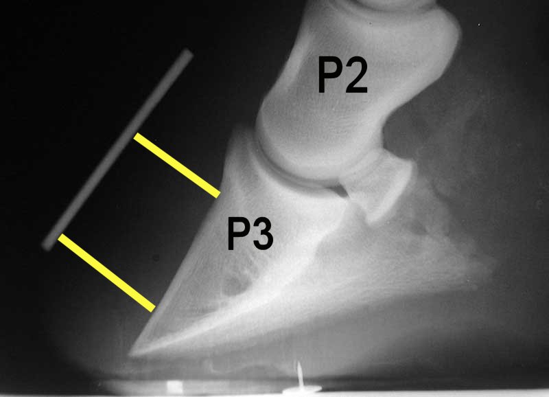
The bones of the hoof are suspended within the axial hooves of ungulates by layers of modified skin cells, known as laminae or lamellae, which suspend the bony column from the hoof wall, contributing to shock absorption during locomotion. In horses, there are about 550–600 pairs of primary epidermal laminae, each with 150–200 secondary laminae projecting from their surface. These interdigitate with equivalent structures attached to the surface of the coffin bone (PIII, P3, the third phalanx, pedal bone, or distal phalanx), known as dermal laminae. The secondary laminae contain basal cells which attach via hemidesmosomes to the basement membrane. The basement membrane is then attached to the coffin bone via the connective tissue of the dermis.
Laminitis literally means inflammation of the laminae, and while it remains controversial whether this is the primary mechanism of disease, evidence of inflammation occurs very early in some instances of the disease. A severe inflammatory event is thought to damage the basal epithelial cells, resulting in dysfunction of the hemidesmosomes and subsequent reduction in adherence between the epithelial cells and the basement membrane. Normal forces placed on the hoof are then strong enough to tear the remaining laminae, resulting in a failure of the interdigitation of the epidermal and dermal laminae between the hoof wall and the coffin bone. When severe enough, this results in displacement of the coffin bone within the hoof capsule. Most cases of laminitis occur in both front feet, but laminitis may be seen in all four feet, both hind feet, or in cases of support limb laminitis, in a single foot.
The mechanism remains unclear and is the subject of much research. Three conditions are thought to cause secondary laminitis:
Inflammatory events that are associated with laminitis include sepsis, endotoxemia, retained placenta, carbohydrate overload (excessive grain or pasture), enterocolitis, pleuropneumonia, and contact with black walnut shavings. In these cases, there is an increase in blood flow to the hoof, bringing in damaging substances and inflammatory cells into the hoof.
Endocrinopathy is usually the result of improper insulin regulation, and is most commonly seen with pituitary pars intermedia dysfunction (also called equine Cushing's syndrome) and equine metabolic syndrome (EMS), as well as obesity and glucocorticoid administration. In cases of EMS, most episodes occur in the spring when the grass is lush.
Mechanical laminitis starts when the hoof wall is pulled away from the bone or lost, as a result of external influences. Mechanical laminitis can occur when a horse habitually paws, is ridden or driven on hard surfaces ("road founder"), or in cases of excessive weight-bearing due to compensation for the opposing limb, a process called support limb laminitis. Support limb laminitis is most common in horses suffering from severe injury to one limb, such as fracture, resulting in a non-weight bearing state that forces them to take excessive load on the opposing limb. This causes decreased blood flow to the cells, decreasing oxygen and nutrient delivery, and thus altering their metabolism which results in laminitis.
One of the newest theories for the molecular basis of laminitis involves matrix metalloproteinases (MMPs). Metalloproteinases are enzymes that can degrade collagen, growth factors, and cytokines to remodel the extracellular matrix of tissues. To prevent tissue damage, they are regulated by tissue inhibitors of metalloproteinases (TIMPs). In cases of laminitis, an underlying cause is thought to cause an imbalance of MMPs and TIMPs, favoring MMPs, so that they may cleave substances within the extracellular matrix and therefore break down the basement membrane. Since the basement membrane is the main link between the hoof wall and the connective tissue of P3, it is thought that its destruction results in their separation. MMP-2 and MMP-9 are the primary enzymes thought to be linked to laminitis.
Hub AI
Laminitis AI simulator
(@Laminitis_simulator)
Laminitis
Laminitis is a disease of the feet of ungulates, found mostly in horses and cattle involving inflammation of the laminae. Clinical signs include foot tenderness progressing to inability to walk, increased digital pulses, and increased temperature in the hooves. Severe cases with outwardly visible clinical signs are known by the colloquial term founder, and progression of the disease will lead to perforation of the coffin bone through the sole of the hoof or being unable to stand up, often requiring euthanasia.
The bones of the hoof are suspended within the axial hooves of ungulates by layers of modified skin cells, known as laminae or lamellae, which suspend the bony column from the hoof wall, contributing to shock absorption during locomotion. In horses, there are about 550–600 pairs of primary epidermal laminae, each with 150–200 secondary laminae projecting from their surface. These interdigitate with equivalent structures attached to the surface of the coffin bone (PIII, P3, the third phalanx, pedal bone, or distal phalanx), known as dermal laminae. The secondary laminae contain basal cells which attach via hemidesmosomes to the basement membrane. The basement membrane is then attached to the coffin bone via the connective tissue of the dermis.
Laminitis literally means inflammation of the laminae, and while it remains controversial whether this is the primary mechanism of disease, evidence of inflammation occurs very early in some instances of the disease. A severe inflammatory event is thought to damage the basal epithelial cells, resulting in dysfunction of the hemidesmosomes and subsequent reduction in adherence between the epithelial cells and the basement membrane. Normal forces placed on the hoof are then strong enough to tear the remaining laminae, resulting in a failure of the interdigitation of the epidermal and dermal laminae between the hoof wall and the coffin bone. When severe enough, this results in displacement of the coffin bone within the hoof capsule. Most cases of laminitis occur in both front feet, but laminitis may be seen in all four feet, both hind feet, or in cases of support limb laminitis, in a single foot.
The mechanism remains unclear and is the subject of much research. Three conditions are thought to cause secondary laminitis:
Inflammatory events that are associated with laminitis include sepsis, endotoxemia, retained placenta, carbohydrate overload (excessive grain or pasture), enterocolitis, pleuropneumonia, and contact with black walnut shavings. In these cases, there is an increase in blood flow to the hoof, bringing in damaging substances and inflammatory cells into the hoof.
Endocrinopathy is usually the result of improper insulin regulation, and is most commonly seen with pituitary pars intermedia dysfunction (also called equine Cushing's syndrome) and equine metabolic syndrome (EMS), as well as obesity and glucocorticoid administration. In cases of EMS, most episodes occur in the spring when the grass is lush.
Mechanical laminitis starts when the hoof wall is pulled away from the bone or lost, as a result of external influences. Mechanical laminitis can occur when a horse habitually paws, is ridden or driven on hard surfaces ("road founder"), or in cases of excessive weight-bearing due to compensation for the opposing limb, a process called support limb laminitis. Support limb laminitis is most common in horses suffering from severe injury to one limb, such as fracture, resulting in a non-weight bearing state that forces them to take excessive load on the opposing limb. This causes decreased blood flow to the cells, decreasing oxygen and nutrient delivery, and thus altering their metabolism which results in laminitis.
One of the newest theories for the molecular basis of laminitis involves matrix metalloproteinases (MMPs). Metalloproteinases are enzymes that can degrade collagen, growth factors, and cytokines to remodel the extracellular matrix of tissues. To prevent tissue damage, they are regulated by tissue inhibitors of metalloproteinases (TIMPs). In cases of laminitis, an underlying cause is thought to cause an imbalance of MMPs and TIMPs, favoring MMPs, so that they may cleave substances within the extracellular matrix and therefore break down the basement membrane. Since the basement membrane is the main link between the hoof wall and the connective tissue of P3, it is thought that its destruction results in their separation. MMP-2 and MMP-9 are the primary enzymes thought to be linked to laminitis.