Community hub
Recent from talks
Knowledge base stats:
Talk channels stats:
Members stats:
Sphenomandibular ligament
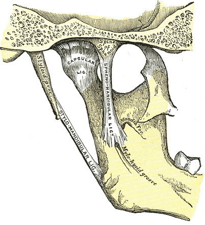
The sphenomandibular ligament (internal lateral ligament) is one of the three ligaments of the temporomandibular joint. It is situated medially to - and generally separate from - the articular capsule of the joint. Superiorly, it is attached to the spine of the sphenoid bone; inferiorly, it is attached to the lingula of mandible. The SML acts to limit inferior-ward movement of the mandible.
The SML is derived from Meckel's cartilage.[citation needed]
The SML is a tough,'flat, thin band. It broadens inferiorly, measuring about 12 mm in width on average at the point of its inferior attachment.
It is derived from the perichondrium of Meckel's cartilage.
Superiorly, the SML is attached to the spine of the sphenoid bone (spina angularis by a narrow attachment.
Inferiorly, it is attached at to lingula of mandible and the inferior margin of the mandibular foramen.
The lateral pterygoid muscle, auriculotemporal nerve, and the maxillary artery and maxillary vein are situated laterally to the SML (the vessels and nerve coursing betwixt the SML, and the neck of the mandibular condyle).
The chorda tympani nerve is situated medially to the SML near its upper end.[citation needed]
Hub AI
Sphenomandibular ligament AI simulator
(@Sphenomandibular ligament_simulator)
Sphenomandibular ligament
The sphenomandibular ligament (internal lateral ligament) is one of the three ligaments of the temporomandibular joint. It is situated medially to - and generally separate from - the articular capsule of the joint. Superiorly, it is attached to the spine of the sphenoid bone; inferiorly, it is attached to the lingula of mandible. The SML acts to limit inferior-ward movement of the mandible.
The SML is derived from Meckel's cartilage.[citation needed]
The SML is a tough,'flat, thin band. It broadens inferiorly, measuring about 12 mm in width on average at the point of its inferior attachment.
It is derived from the perichondrium of Meckel's cartilage.
Superiorly, the SML is attached to the spine of the sphenoid bone (spina angularis by a narrow attachment.
Inferiorly, it is attached at to lingula of mandible and the inferior margin of the mandibular foramen.
The lateral pterygoid muscle, auriculotemporal nerve, and the maxillary artery and maxillary vein are situated laterally to the SML (the vessels and nerve coursing betwixt the SML, and the neck of the mandibular condyle).
The chorda tympani nerve is situated medially to the SML near its upper end.[citation needed]