Community hub
Recent from talks
Knowledge base stats:
Talk channels stats:
Members stats:
Syndesmosis procedure
Syndesmosis procedure is one of the more than twenty bunion surgeries currently being performed. While the majority of bunion surgeries involve the breaking and shifting of bones (osteotomy procedures), syndesmosis procedure is one of few surgical techniques that use a soft-tissue or non-osteotomy (non-bone-breaking) approach to afford the same correction. More than 130 different surgical techniques have been described for correction of one single condition of the foot: the bunion deformity.
Bunion (hallux valgus) deformity is actually part of a complex of anatomical derangements of protruding mass (bunion), buckling of big toe (hallux valgus) and the bone behind it (metatarsus primus varus), displaced sesamoid bones (detrimental to the important walking function of big toe), collapsed metatarsal arch and several other secondary changes that are the domino effects of metatarsal primus varus. Thus, metatarsus primus varus correction has become the primary objective of all bunion surgeries.[citation needed]
Primus varus deformity is the leaning of the first metatarsal bone away from the second metatarsal and towards the opposite foot (Fig. 1). As it leans over, its head sticks out to form the bunion bump and it also widens the forefoot to cause shoes feeling too tight. Thus when bunion pain becomes unmanageable, surgical correction is to narrow the forefoot by repositioning of the first metatarsal head back to its normal position. This can be done by osteotomy (bone-breaking), soft tissue (non-osteotomy) or fusion techniques.[citation needed]
What causes the metatarsus primus varus deformity?
A first metatarsal bone would lean towards one side is because “the medial hood ligament is stretched” and as it becomes weak and incompetent the first metatarsal would lose its support and then gradually shift out of place to form the metatarsus primus varus deformity.
What causes ligaments to fail?
Female hormone estrogen and genetic inheritance are the two primary factors behind the weak ligaments of bunion feet. Bunion deformity was found more common in people wearing shoes by a study in Hong Kong and increased in Japan due to changing in footwear to Western style. Aging, degeneration and trauma have also been attributed to weakening of the responsible ligaments.[citation needed]
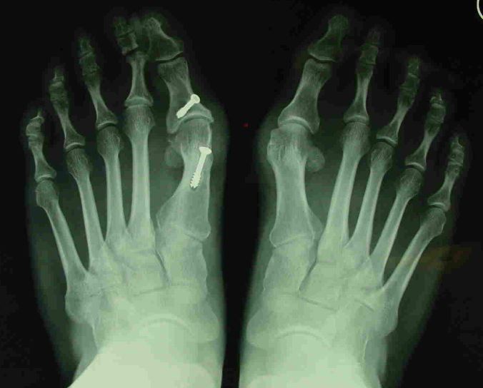
Syndesmosis procedure addresses specifically the two fundamental problems of metatarsus primus varus deformity that gives rise to the bunion deformity. They are leaning and instability of the first metatarsal bone . Syndesmosis procedure uprights the leaning first metatarsal bone with strong binding sutures between it and the second metatarsal bone (Fig. 2) and then also stabilizes it uniquely by creating a fibrous connecting bridge between these two bones (Fig. 3, 4). First metatarsal bone can be readily realigned because by definition of the metatarsus primus varus deformity its first metatarsal is abnormally loose and mobile.[citation needed]
Hub AI
Syndesmosis procedure AI simulator
(@Syndesmosis procedure_simulator)
Syndesmosis procedure
Syndesmosis procedure is one of the more than twenty bunion surgeries currently being performed. While the majority of bunion surgeries involve the breaking and shifting of bones (osteotomy procedures), syndesmosis procedure is one of few surgical techniques that use a soft-tissue or non-osteotomy (non-bone-breaking) approach to afford the same correction. More than 130 different surgical techniques have been described for correction of one single condition of the foot: the bunion deformity.
Bunion (hallux valgus) deformity is actually part of a complex of anatomical derangements of protruding mass (bunion), buckling of big toe (hallux valgus) and the bone behind it (metatarsus primus varus), displaced sesamoid bones (detrimental to the important walking function of big toe), collapsed metatarsal arch and several other secondary changes that are the domino effects of metatarsal primus varus. Thus, metatarsus primus varus correction has become the primary objective of all bunion surgeries.[citation needed]
Primus varus deformity is the leaning of the first metatarsal bone away from the second metatarsal and towards the opposite foot (Fig. 1). As it leans over, its head sticks out to form the bunion bump and it also widens the forefoot to cause shoes feeling too tight. Thus when bunion pain becomes unmanageable, surgical correction is to narrow the forefoot by repositioning of the first metatarsal head back to its normal position. This can be done by osteotomy (bone-breaking), soft tissue (non-osteotomy) or fusion techniques.[citation needed]
What causes the metatarsus primus varus deformity?
A first metatarsal bone would lean towards one side is because “the medial hood ligament is stretched” and as it becomes weak and incompetent the first metatarsal would lose its support and then gradually shift out of place to form the metatarsus primus varus deformity.
What causes ligaments to fail?
Female hormone estrogen and genetic inheritance are the two primary factors behind the weak ligaments of bunion feet. Bunion deformity was found more common in people wearing shoes by a study in Hong Kong and increased in Japan due to changing in footwear to Western style. Aging, degeneration and trauma have also been attributed to weakening of the responsible ligaments.[citation needed]
Syndesmosis procedure addresses specifically the two fundamental problems of metatarsus primus varus deformity that gives rise to the bunion deformity. They are leaning and instability of the first metatarsal bone . Syndesmosis procedure uprights the leaning first metatarsal bone with strong binding sutures between it and the second metatarsal bone (Fig. 2) and then also stabilizes it uniquely by creating a fibrous connecting bridge between these two bones (Fig. 3, 4). First metatarsal bone can be readily realigned because by definition of the metatarsus primus varus deformity its first metatarsal is abnormally loose and mobile.[citation needed]