Hubbry Logo
Ball-and-socket jointBall-and-socket jointMain
Open search
Ball-and-socket joint
Community hub
Ball-and-socket joint
logo
8 pages, 0 posts
0 subscribers
Be the first to start a discussion here.
Ball-and-socket joint
from Wikipedia
Ball and socket joint
1: Ball and socket joint; 2: Condyloid joint (Ellipsoid); 3: Saddle joint; 4 Hinge joint; 5: Pivot joint
Capsule of shoulder-joint (distended). Anterior aspect.
Identifiers
TA98A03.0.00.050
TA21562
FMA75301
Anatomical terminology

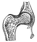
The ball-and-socket joint (or spheroid joint) is a type of synovial joint in which the ball-shaped surface of one rounded bone fits into the cup-like depression of another bone. The distal bone is capable of motion around an indefinite number of axes, which have one common center. This enables the joint to move in many directions.

An enarthrosis is a special kind of spheroidal joint in which the socket covers the sphere beyond its equator.[1]

Examples of joints

[edit]

Examples of this form of articulation are found in the hip, where the round head of the femur (ball) rests in the cup-like acetabulum (socket) of the pelvis; and in the shoulder joint, where the rounded upper extremity of the humerus (ball) rests in the cup-like glenoid fossa (socket) of the shoulder blade.[2] (The shoulder also includes a sternoclavicular joint.)

Ball and Socket Joint Animation

[edit]
Ball and Socket joint animation

Diagrams

[edit]

References

[edit]
[edit]
Revisions and contributorsEdit on WikipediaRead on Wikipedia
from Grokipedia
A ball-and-socket joint is a type of characterized by the spherical head of one fitting into a cup-like depression on another , enabling multiaxial movement in three or more planes for a broad . These joints are among the most mobile in the , allowing actions such as flexion, extension, abduction, adduction, and both internal and external . The only ball-and-socket joints in the are the glenohumeral (, where the head of the articulates with the of the , and the hip joint, where the fits into the of the . Structurally, ball-and-socket joints are enclosed within a fibrous capsule reinforced by ligaments and lined by a that produces lubricating fluid to minimize friction during movement. In the , additional stability comes from the muscles and tendons, though this joint's shallow socket makes it prone to despite its exceptional mobility, which supports eight distinct movements. The hip joint, by contrast, features a deeper socket augmented by a fibrocartilaginous labrum, providing greater stability for activities while still permitting six key movements. These joints are classified as polyaxial synovial joints, facilitating complex, three-dimensional motions essential for everyday activities like reaching, walking, and rotating the limbs. Ball-and-socket joints play a critical role in human locomotion and upper body function, but their design also predisposes them to conditions such as , dislocations, and labral tears, particularly under repetitive stress or injury. Their backward, forward, sideways, and rotational capabilities distinguish them from less versatile joint types, underscoring their importance in enabling fluid, adaptive movement throughout the skeletal system.

Anatomy

Structure

A ball-and-socket joint is a type of characterized by the articulation of a spherical or ball-shaped head of one fitting into a cup-like or concave socket of another . This configuration forms a joint cavity enclosed by an articular capsule. The primary articular surfaces consist of the rounded and the receiving socket, both covered by a layer of that provides a smooth, low-friction interface. , a type of avascular , is nourished by from the surrounding and helps distribute loads across the joint surfaces. The , composed of dense fibrous externally and reinforced internally by a , encases these structures and defines the joint boundaries. The lines the inner aspect of the and secretes , a viscous that fills the cavity and reduces during articulation. Socket depth can vary across ball-and-socket joints, with shallower sockets allowing broader contact dynamics and deeper ones providing enhanced enclosure of the . Ligaments associated with the capsule contribute to overall integrity (see Supporting Structures).

Supporting Structures

The supporting structures of ball-and-socket joints encompass ligaments, muscles, the labrum, bursae, tendons, and neurovascular components that collectively enhance joint stability while accommodating multiaxial mobility. These elements reinforce the relatively shallow socket, preventing dislocations and distributing loads effectively across the articulation. Ligaments form the primary static stabilizers, integrating with the joint capsule to restrict excessive translation and rotation. In the hip joint, the iliofemoral ligament—the strongest ligament in the body—spans from the anterior inferior iliac spine to the intertrochanteric line of the femur, limiting extension and external rotation to maintain upright posture. The pubofemoral ligament, extending from the superior pubic ramus to the intertrochanteric fossa, restricts abduction and extension, while the ischiofemoral ligament, attaching from the posterior acetabular rim to the greater trochanter, limits internal rotation and adduction during flexion. In the shoulder (glenohumeral) joint, the superior, middle, and inferior glenohumeral ligaments reinforce the anterior capsule, becoming taut at different degrees of abduction and rotation to prevent anterior dislocation of the humeral head. The acetabular labrum in the hip and the glenoid labrum in the shoulder, both fibrocartilaginous rings, deepen their respective sockets—the acetabulum by approximately 21% and the glenoid by 50%—increasing articular surface area and congruence to bolster stability against shear forces. Muscles provide dynamic stabilization through compression and proprioceptive feedback, counterbalancing the joint's inherent laxity. In the shoulder, the muscles—supraspinatus, infraspinatus, teres minor, and subscapularis—encase the humeral head, with the supraspinatus initiating abduction, infraspinatus and teres minor facilitating external , and subscapularis enabling internal ; together, they depress and center the humeral head within the . For the hip, the (gluteus maximus for extension and external , medius and minimus for abduction) stabilize the during , while the muscle, a primary flexor originating from the lumbar spine and , contributes to anterior stability by countering posterior forces. Tendons of these muscles blend with the capsule, further augmenting tensile strength. Bursae, fluid-filled sacs, minimize friction in these high-motion areas; notably, the in the shoulder cushions the tendons against the , preventing impingement during overhead activities. In the hip, analogous structures like the iliopsoas bursa protect the from compression against the . Blood supply and innervation support the metabolic demands and sensory-motor control of these structures, ensuring coordinated function. The shoulder joint receives arterial supply primarily from the anterior (34%) and posterior (64%) , branches of the , which form an anastomotic network around the humeral head to sustain capsular and ligamentous integrity. Innervation arises from the (from the posterior cord), which innervates the deltoid and teres minor while providing sensory branches to the , aiding in and stability reflexes. In the hip, the medial femoral dominates the supply to the and capsule post-infancy, with contributions from the lateral and obturator arteries forming a retinacular network critical for prevention. Sensory and motor innervation derives from the femoral, obturator, and sciatic nerves (including the nerve to quadratus femoris), which supply the surrounding muscles and capsule to facilitate dynamic stabilization and pain feedback.

Function

Movements

Ball-and-socket joints, also known as spheroidal joints, permit a wide array of movements due to their multiaxial design, where the rounded head of one fits into the cup-like of another. The primary movements include flexion and extension, which involve bending and straightening the in the ; abduction and adduction, which move the limb away from or toward the body's midline in the frontal plane; and internal and external , which twist the limb around its longitudinal axis. Additionally, circumduction—a combining flexion, extension, abduction, adduction, and —allows for conical tracing of the limb's distal end without actual of the itself. These joints exhibit of freedom, enabling triaxial around three perpendicular axes: mediolateral for flexion/extension, anteroposterior for abduction/adduction, and longitudinal for . This configuration provides greater mobility than any other type, though the exact range varies by joint and individual factors such as and . For instance, the hip joint typically allows up to 120° of flexion and 45° of abduction, while the permits up to 180° of abduction and 180° of flexion. Kinematically, during motion, the ball-shaped femoral or humeral head both rolls and slides within the socket to maintain congruent contact and minimize friction, facilitated by lubrication. Stabilizing ligaments, such as the in the , contribute to controlled movement by limiting excessive translation.

Biomechanics

Ball-and-socket joints exemplify a fundamental trade-off between stability and mobility in design, where deeper acetabular coverage of the in the enhances congruence and resistance to at the expense of reduced , contrasting with the shallower in the that permits greater multidirectional excursion but increases instability risk. This architectural variation aligns with functional demands: the prioritizes load-bearing stability during weight transfer, while the facilitates overhead reaching and arm positioning. Mechanical forces in ball-and-socket joints primarily manifest as compressive loads perpendicular to the articular surfaces, which are distributed across to minimize peak pressures on underlying subchondral bone, and shear forces parallel to the surfaces that arise during rotational movements and can strain the matrix. , comprising 60-80% water, 10-20% , and 4-7% proteoglycans, absorbs these compressive forces through pressurization and deformation, while fibers resist shear by anchoring the matrix. contributes to load-bearing by facilitating boundary lubrication and a "weeping" effect, where joint compression expels from to reduce and support nutrient under sustained loads. Torque in ball-and-socket joints is governed by the equation τ=F×d\tau = F \times d where τ\tau is , FF is the applied , and dd is the moment arm defined as the perpendicular distance from the joint center to the action; this quantifies rotational tendencies around the fixed geometric center of the spherical articulation. The instantaneous center of (ICR) represents the point of zero during motion, coinciding with the joint center in ideal ball-and-socket to enable pure without translation, though real motions may shift the ICR due to surface incongruities. Excessive can precipitate by overcoming congruence and soft-tissue restraints, as seen in posterior dislocations where high-impact flexion and adduction exceed resistive capacities. Muscle co-contraction enhances dynamic stability by generating compressive forces that seat the deeper into the socket, thereby increasing stiffness and damping without net production, a mechanism critical during rapid directional changes. This co-activation trades some efficiency for reduced translation under load, underscoring the joint's reliance on neuromuscular control for .

Examples

Hip Joint

The hip joint, also known as the coxal joint, is a multiaxial ball-and-socket formed by the articulation of the rounded with the cup-shaped of the . The , which constitutes approximately one-third of a , fits deeply into the , providing inherent stability due to the socket's depth and the joint's congruence. The , a fibrocartilaginous ring attached to the acetabular rim, deepens the socket further, enhances joint stability, and helps distribute compressive forces across the articular surfaces. Stability is further reinforced by a robust capsular ligament complex, including the strong Y-shaped anteriorly, which limits hyperextension; the pubofemoral ligament anteroinferiorly, which restricts abduction and extension; and the ischiofemoral ligament posteriorly, which limits internal rotation and adduction, particularly with flexion. These ligaments, along with surrounding muscles like the gluteals and , enable the hip to withstand significant loads while permitting essential movements. In terms of function, the hip joint plays a critical role in locomotion by supporting body weight and facilitating the cycle, where it alternates between stance and swing phases. During walking, the hip absorbs and transfers forces up to several times body weight, primarily through extension and slight abduction to maintain pelvic stability and forward progression. Its is more limited than that of the to prioritize stability for , allowing approximately 120° of flexion, 30° of extension, 45° of abduction, 30° of adduction, 40° of internal rotation, and 45° of external rotation. This constrained mobility supports efficient bipedal , including movements like flexion and extension referenced in general ball-and-socket joint . Evolutionarily, the human hip joint exhibits adaptations for , such as a shortened, broadened with a laterally flared ilium that repositions the for improved abductor leverage to stabilize the during single-leg stance. These changes, evident in early hominins like , shifted the 's orientation for upright posture, reducing energy expenditure in walking compared to quadrupedal locomotion in . A unique feature of the hip's vascular anatomy is its reliance on retinacular arteries, branches of the medial femoral circumflex artery, which supply the via an extracapsular route; disruption of these vessels, often from trauma or , can lead to due to the bone's limited collateral circulation.

Shoulder Joint

The shoulder joint, also known as the glenohumeral joint, exemplifies a ball-and-socket articulation where the convex humeral head fits into the shallow of the , allowing multiaxial movement while relying heavily on surrounding soft tissues for stability. The is a shallow, pear-shaped depression on the lateral aspect of the , deepened only slightly by the fibrocartilaginous , which covers less than half of the humeral head's surface area. This configuration contrasts with more enclosed sockets in other synovial joints, emphasizing the shoulder's design for mobility over inherent bony constraint. Key supporting structures include the , a musculotendinous complex comprising the supraspinatus, infraspinatus, teres minor, and subscapularis muscles, which envelop the humeral head and compress it against the glenoid to enhance stability during motion. The , originating from the of the and blending with the rotator cuff tendons, reinforces the superior aspect of the , limiting excessive external rotation and inferior translation of the humeral head. Due to the glenoid fossa's shallow depth, the shoulder is inherently more susceptible to instability and dislocations compared to the hip joint, where the provides greater osseous containment. Functionally, the shoulder enables a wide range of activities, including overhead reaching and throwing, through its extensive motion capabilities: approximately 180° of flexion, 150° of abduction, and up to 90° of external . These movements are coordinated by the muscles, which initiate and fine-tune actions like abduction (supraspinatus) and external (infraspinatus and teres minor). Innervation arises primarily from branches of the , notably the (from C5-C6 roots), which supplies the supraspinatus and infraspinatus muscles critical for joint stabilization. Vascularization is provided by the (a branch of the ) and the anterior and posterior circumflex humeral arteries (branches of the ), ensuring nutrient delivery to the capsule, labrum, and tendons.

Clinical Significance

Disorders

Ball-and-socket joints, such as the and , are prone to a range of disorders that can impair their multidirectional mobility and stability. These conditions often stem from degenerative processes, trauma, , or congenital anomalies, leading to symptoms like , , , and reduced . While the specific manifestations vary by joint and underlying cause, early recognition is crucial for managing long-term joint health. Degenerative disorders primarily involve osteoarthritis (OA), characterized by the gradual breakdown of due to mechanical over time. In the , a major weight-bearing ball-and-socket joint, OA is particularly prevalent, affecting up to 27% of older adults and resulting from repetitive loading that erodes the protective cartilage layer between the and . Symptoms typically include deep or buttock pain exacerbated by activity, morning stiffness lasting less than 30 minutes, and a grinding sensation during movement, which worsen as subchondral and formation progress. Traumatic injuries commonly affect these joints through dislocations and associated damage, often from high-impact events like falls or sports collisions. dislocations are the most frequent, with anterior dislocations comprising 97% of cases due to the glenohumeral joint's shallow socket and reliance on dynamic stabilizers; the annual incidence is approximately 23.9 per 100,000 person-years in the general population, rising to 169 per 100,000 in young athletes. These injuries cause sudden severe pain, deformity, and inability to abduct the , frequently accompanied by or vascular compromise. In the , posterior dislocations predominate (about 90% of traumatic cases), typically from dashboard impacts in accidents that force the posteriorly; symptoms include intense pain, leg shortening, and internal rotation, with up to 95% of native dislocations linked to high-energy trauma. Labral tears, often concurrent with dislocations, occur from acute shear forces or repetitive overhead motions in the (e.g., superior labrum anterior-posterior lesions) and from or twisting injuries in the , manifesting as catching sensations, pain, and mechanical instability. Inflammatory conditions, such as (RA), involve autoimmune-mediated that targets the lining these joints, leading to formation, erosion, and eventual bony . RA affects the in 10–40% of patients with longstanding disease, causing symmetric pain, swelling, and morning stiffness exceeding 30 minutes, while shoulder involvement often presents with weakening and progressive loss of elevation. The chronic inflammation in ball-and-socket joints like these exacerbates and capsular thickening, contributing to secondary OA. Congenital disorders include developmental dysplasia of the hip (DDH), where the fails to fully encompass the , resulting in a shallow socket and potential or . Primarily affecting infants, DDH arises from multifactorial causes including breech presentation (in 30-50% of cases), female sex (4-8 times higher risk), and genetic predisposition, with an incidence ranging from 1 to 5 per 1,000 live births in Western populations. Early symptoms may be subtle, such as asymmetric folds or a positive Ortolani sign (reducible hip click), but untreated cases lead to limping, leg length discrepancy, and accelerated hip OA in adolescence or adulthood due to abnormal load distribution.

Treatments

Diagnosis of ball-and-socket joint disorders typically begins with techniques tailored to the suspected . X-rays are the initial modality for detecting fractures, dislocations, or degenerative changes in the and joints, providing a cost-effective assessment of bony structures. For soft tissue evaluation, such as labral tears or injuries, magnetic resonance imaging (MRI) is preferred due to its superior contrast resolution and multiplanar capabilities, enabling detailed visualization of , ligaments, and tendons. serves as both a diagnostic and therapeutic tool, allowing direct inspection and intervention under visualization, particularly useful for confirming intra-articular pathologies like those in or instability. Conservative management forms the cornerstone for many ball-and-socket joint conditions, including and overuse injuries, aiming to alleviate pain and restore function without surgery. Nonsteroidal anti-inflammatory drugs (NSAIDs) are commonly prescribed to reduce inflammation and pain in conditions like or impingement, with evidence supporting their short-term efficacy in improving mobility. protocols emphasize strengthening exercises, such as reinforcement for stability or gluteal muscle training for support, often combined with stretching to enhance and prevent further deterioration. These approaches are particularly effective for non-arthritic pain or early-stage disorders, where activity modification and targeted rehabilitation can yield significant functional gains. Surgical interventions are indicated for advanced or refractory cases, such as severe , fractures, or recurrent dislocations. Total hip arthroplasty (THA) replaces the damaged and with prosthetic components, restoring joint mechanics and providing durable pain relief, with success rates exceeding 90% at 10-year follow-up in appropriately selected patients. For the , procedures like address instability by reattaching the labrum via , while total reconstructs the glenohumeral joint with metal and implants for end-stage , improving motion and function in over 85% of cases. These surgeries, often minimally invasive, are tailored to the joint's load-bearing demands, with hip procedures focusing on stability and shoulder ones on mobility. Postoperative rehabilitation is essential for optimizing outcomes following surgical repair of ball-and-socket joints. Protocols typically progress through phases: initial immobilization (1-6 weeks) to protect the repair, followed by passive range-of-motion exercises to prevent stiffness, and advancing to active strengthening and by 4-12 weeks. For hip arthroscopy or THA, emphasis is placed on retraining and hip abductor strengthening to restore capacity, while rehabilitation prioritizes external rotation and elevation to rebuild integrity, often spanning 3-6 months for full recovery. Supervised programs have demonstrated improved joint function and reduced complication rates, such as or frozen . Emerging regenerative therapies offer promising alternatives for cartilage repair in ball-and-socket joints affected by or trauma. Intra-articular injections of mesenchymal stem cells (MSCs), derived from or , promote tissue regeneration by modulating and stimulating proliferation, with clinical trials showing pain reduction and cartilage volume preservation in hip and knee models applicable to shoulder. As of 2025, MSCs have been included in some international guidelines for OA management, with phase II trials demonstrating relief and functional improvement lasting up to 52 weeks. These autologous or allogeneic approaches, often combined with scaffolds or , aim to delay or avoid , though long-term efficacy requires further validation through ongoing studies.

References

Add your contribution
Related Hubs
User Avatar
No comments yet.