Hubbry Logo
PelvisPelvisMain
Open search
Pelvis
Community hub
Pelvis
logo
8 pages, 0 posts
0 subscribers
Be the first to start a discussion here.
Pelvis
Pelvis
from Wikipedia
Pelvis
Male type pelvis
Female type pelvis
Details
NervePelvic splanchnic nerves, superior hypogastric plexus
Identifiers
Latinpelvis
MeSHD010388
TA98A02.5.02.001
TA2129
Anatomical terms of bone
The same human pelvis, front imaged by X-ray (top), magnetic resonance imaging (middle), and 3-dimensional computed tomography (bottom)

The pelvis (pl.: pelves or pelvises) is the lower part of an anatomical trunk,[1] between the abdomen and the thighs (sometimes also called pelvic region), together with its embedded skeleton[2] (sometimes also called bony pelvis or pelvic skeleton).

The pelvic region of the trunk includes the bony pelvis, the pelvic cavity (the space enclosed by the bony pelvis), the pelvic floor, below the pelvic cavity, and the perineum, below the pelvic floor.[1] The pelvic skeleton is formed in the area of the back, by the sacrum and the coccyx and anteriorly and to the left and right sides, by a pair of hip bones.

The two hip bones connect the spine with the lower limbs. They are attached to the sacrum posteriorly, connected to each other anteriorly, and joined with the two femurs at the hip joints. The gap enclosed by the bony pelvis, called the pelvic cavity, is the section of the body underneath the abdomen and mainly consists of the reproductive organs and the rectum, while the pelvic floor at the base of the cavity assists in supporting the organs of the abdomen.

In mammals, the bony pelvis has a gap in the middle, significantly larger in females than in males. Their offspring pass through this gap when they are born.

Structure

[edit]

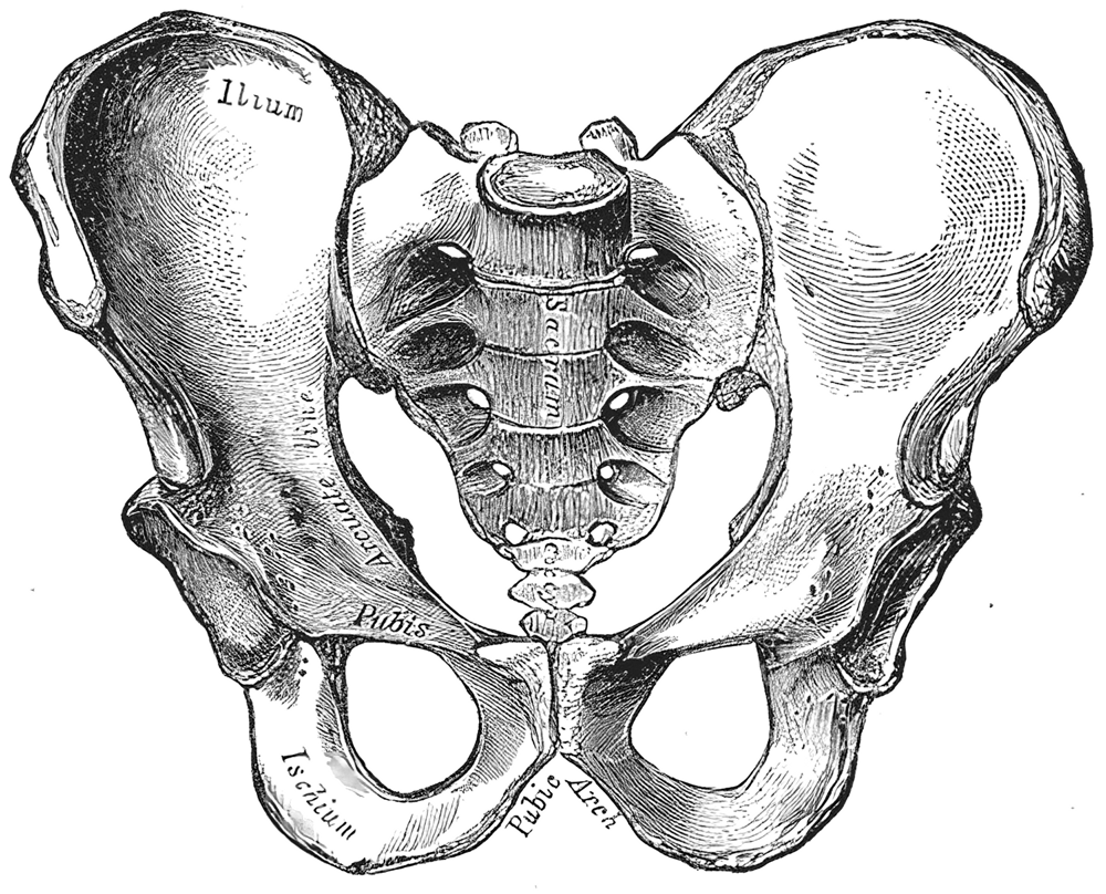
The pelvic region of the trunk is the lower part of the trunk, between the abdomen and the thighs.[1] It includes several structures: the bony pelvis, the pelvic cavity, the pelvic floor, and the perineum. The bony pelvis (pelvic skeleton) is the part of the skeleton embedded in the pelvic region of the trunk. It is subdivided into the pelvic girdle and the pelvic spine. The pelvic girdle is composed of the appendicular hip bones (ilium, ischium, and pubis) oriented in a ring, and connects the pelvic region of the spine to the lower limbs. The pelvic spine consists of the sacrum and coccyx.[1]

Pelvic bone

[edit]
The skeleton of the human pelvis:
2–4. Hip bone (os coxae)
1. Sacrum (os sacrum), 2. Ilium (os ilium), 3. Ischium (os ischii)
4. Pubic bone (os pubis) (4a. corpus, 4b. ramus superior, 4c. ramus inferior, 4d. tuberculum pubicum)
5. Pubic symphysis, 6. Acetabulum (of the hip joint), 7. Obturator foramen, 8. Coccyx/tailbone (os coccygis)
Dotted. Linea terminalis of the pelvic brim.

The pelvic skeleton is formed posteriorly (in the area of the back), by the sacrum and the coccyx and laterally and anteriorly (forward and to the sides), by a pair of hip bones. Each hip bone consists of three sections: ilium, ischium, and pubis. During childhood, these sections are separate bones, joined by the triradiate cartilage. During puberty, they fuse together to form a single bone.

Pelvic cavity

[edit]

The pelvic cavity is a body cavity that is bounded by the bones of the pelvis and which primarily contains reproductive organs and the rectum.

A distinction is made between the lesser or true pelvis inferior to the terminal line, and the greater or false pelvis above it. The pelvic inlet or superior pelvic aperture, which leads into the lesser pelvis, is bordered by the promontory, the arcuate line of ilium, the iliopubic eminence, the pecten of the pubis, and the upper part of the pubic symphysis. The pelvic outlet or inferior pelvic aperture is the region between the subpubic angle or pubic arch, the ischial tuberosities and the coccyx. [3]

Alternatively, the pelvis is divided into three planes: the inlet, midplane, and outlet.[4]

Pelvic floor

[edit]
Female perineum
Male perineum

The pelvic floor has two inherently conflicting functions: One is to close the pelvic and abdominal cavities and bear the load of the visceral organs; the other is to control the openings of the rectum and urogenital organs that pierce the pelvic floor and make it weaker. To achieve both these tasks, the pelvic floor is composed of several overlapping sheets of muscles and connective tissues.[5]

The pelvic diaphragm is composed of the levator ani and the coccygeus muscle. These arise between the symphysis and the ischial spine and converge on the coccyx and the anococcygeal ligament which spans between the tip of the coccyx and the anal hiatus. This leaves a slit for the anal and urogenital openings. Because of the width of the genital aperture, which is wider in females, a second closing mechanism is required. The urogenital diaphragm consists mainly of the deep transverse perineal which arises from the inferior ischial and pubic rami and extends to the urogenital hiatus. The urogenital diaphragm is reinforced posteriorly by the superficial transverse perineal.[6]

The external anal and urethral sphincters close the anus and the urethra. The former is surrounded by the bulbospongiosus which narrows the vaginal introitus in females and surrounds the corpus spongiosum in males. Ischiocavernosus squeezes blood into the corpora cavernosa penis and clitoridis.[7]

Variation

[edit]

Modern humans are to a large extent characterized by bipedal locomotion and large brains. Because the pelvis is vital to both locomotion and childbirth, natural selection has been confronted by two conflicting demands: a wide birth canal and locomotion efficiency, a conflict referred to as the "obstetrical dilemma". The female pelvis, or gynecoid pelvis,[8] has evolved to its maximum width for childbirth—a wider pelvis would make human females unable to walk. In contrast, human male pelvises are not constrained by the need to give birth and therefore are more optimized for bipedal locomotion.[9]

The principal differences between male and female true and false pelvis include:

  • The female pelvis is larger and broader than the male pelvis which is taller, narrower, and more compact.[10] The female pelvis is lighter and thinner than the male pelvis.[11]
  • The female inlet is larger and oval in shape, while the male sacral promontory projects further (i.e. the male inlet is more heart-shaped).[10]
  • The sides of the male pelvis converge from the inlet to the outlet, whereas the sides of the female pelvis are wider apart.[12]
  • The angle between the inferior pubic rami is acute (70 degrees) in males, but obtuse (90–100 degrees) in females. Accordingly, the angle is called subpubic angle in males and pubic arch in females.[10] Additionally, the bones forming the angle/arch are more concave in females but straight in males.[13]
  • The distance between the ischia bones is small in males, making the outlet narrow, but large in females, who have a relatively large outlet. The ischial spines and tuberosities are heavier and project farther into the pelvic cavity in males. The greater sciatic notch is wider in females.[13]
  • The iliac crests are higher and more pronounced in males, making the male false pelvis deeper and more narrow than in females.[13]
  • The male sacrum is long, narrow, more straight, and has a pronounced sacral promontory. The female sacrum is shorter, wider, more curved posteriorly, and has a less pronounced promontory.[13]
  • The acetabula are wider apart in females than in males.[13] In males, the acetabulum faces more laterally, while it faces more anteriorly in females. Consequently, when males walk the leg can move forwards and backwards in a single plane. In females, the leg must swing forward and inward, from where the pivoting head of the femur moves the leg back in another plane. This change in the angle of the femoral head gives the female gait its characteristic (i.e. swinging of hips).[14]

Development

[edit]

Each side of the pelvis is formed as cartilage, which ossifies as three main bones which stay separate through childhood: ilium, ischium, pubis. At birth the whole of the hip joint (the acetabulum area and the top of the femur) is still made of cartilage (but there may be a small piece of bone in the great trochanter of the femur); this makes it difficult to detect congenital hip dislocation by X-raying.

"In terms of comparative anatomy the human scapula represents two bones that have become fused together; the (dorsal) scapula proper and the (ventral) coracoid. The epiphyseal line across the glenoid cavity is the line of fusion. They are the counterparts of the ilium and ischium of the pelvic girdle."

— R. J. LastLast's Anatomy

There is preliminary evidence that the pelvis continues to widen over the course of a lifetime.[15][16]

Functions

[edit]

The skeleton of the pelvis is a basin-shaped ring of bones connecting the vertebral column to the femora. It is then connected to two hip bones.

Its primary functions are to bear the weight of the upper body when sitting and standing, transferring that weight from the axial skeleton to the lower appendicular skeleton when standing and walking, and providing attachments for and withstanding the forces of the powerful muscles of locomotion and posture. Compared to the shoulder girdle, the pelvic girdle is thus strong and rigid.[1]

Its secondary functions are to contain and protect the pelvic and abdominopelvic viscera (inferior parts of the urinary tracts, internal reproductive organs), providing attachment for external reproductive organs and associated muscles and membranes.[1]

As a mechanical structure

[edit]
Anatomical drawing of the female pelvis

The pelvic girdle consists of the two hip bones. The hip bones are connected to each other anteriorly at the pubic symphysis, and posteriorly to the sacrum at the sacroiliac joints to form the pelvic ring. The ring is very stable and allows very little mobility, a prerequisite for transmitting loads from the trunk to the lower limbs.[17]

As a mechanical structure the pelvis may be thought of as four roughly triangular and twisted rings. Each superior ring is formed by the iliac bone; the anterior side stretches from the acetabulum up to the anterior superior iliac spine; the posterior side reaches from the top of the acetabulum to the sacroiliac joint; and the third side is formed by the palpable iliac crest. The lower ring, formed by the rami of the pubic and ischial bones, supports the acetabulum and is twisted 80–90 degrees in relation to the superior ring.[18]

An alternative approach is to consider the pelvis part of an integrated mechanical system based on the tensegrity icosahedron as an infinite element. Such a system is able to withstand omnidirectional forces—ranging from weight-bearing to childbearing—and, as a low energy requiring system, is favoured by natural selection.[19]

The pelvic inclination angle is the single most important element of the human body posture and is adjusted at the hips. It is also one of the rare things that can be measured at the assessment of the posture. A simple method of measurement was described by the British orthopedist Philip Willes and is performed by using an inclinometer.

As an anchor for muscles

[edit]

The lumbosacral joint, between the sacrum and the last lumbar vertebra, has, like all vertebral joints, an intervertebral disc, anterior and posterior ligaments, ligamenta flava, interspinous and supraspinous ligaments, and synovial joints between the articular processes of the two bones. In addition to these ligaments the joint is strengthened by the iliolumbar and lateral lumbosacral ligaments. The iliolumbar ligament passes between the tip of the transverse process of the fifth lumbar vertebra and the posterior part of the iliac crest. The lateral lumbosacral ligament, partly continuous with the iliolumbar ligament, passes down from the lower border of the transverse process of the fifth vertebra to the ala of the sacrum. The movements possible in the lumbosacral joint are flexion and extension, a small amount of lateral flexion (from 7 degrees in childhood to 1 degree in adults), but no axial rotation. Between ages 2–13 the joint is responsible for as much as 75% (about 18 degrees) of flexion and extension in the lumbar spine. From age 35 the ligaments considerably limit the range of motions. [20]

The three extracapsular ligaments of the hip joint—the iliofemoral, ischiofemoral, and pubofemoral ligaments—form a twisting mechanism encircling the neck of the femur. When sitting, with the hip joint flexed, these ligaments become lax permitting a high degree of mobility in the joint. When standing, with the hip joint extended, the ligaments get twisted around the femoral neck, pushing the head of the femur firmly into the acetabulum, thus stabilizing the joint.[21] The zona orbicularis assists in maintaining the contact in the joint by acting like a buttonhole on the femoral head.[22] The intracapsular ligament, the ligamentum teres, transmits blood vessels that nourish the femoral head.[23]

Junctions

[edit]
Coronal section through pubic symphysis

The two hip bones are joined anteriorly at the pubic symphysis by a fibrous cartilage covered by a hyaline cartilage, the interpubic disk, within which a non-synovial cavity might be present. Two ligaments, the superior and inferior pubic ligaments, reinforce the symphysis.[3]

Both sacroiliac joints, formed between the auricular surfaces of the sacrum and the two hip bones. are amphiarthroses, almost immobile joints enclosed by very taut joint capsules. This capsule is strengthened by the ventral, interosseous, and dorsal sacroiliac ligaments.[3] The most important accessory ligaments of the sacroiliac joint are the sacrospinous and sacrotuberous ligaments which stabilize the hip bone on the sacrum and prevent the promonotory from tilting forward. Additionally, these two ligaments transform the greater and lesser sciatic notches into the greater and lesser foramina, a pair of important pelvic openings.[24] The iliolumbar ligament is a strong ligament which connects the tip of the transverse process of the fifth lumbar vertebra to the posterior part of the inner lip of the iliac crest. It can be thought of as the lower border of the thoracolumbar fascia and is occasionally accompanied by a smaller ligamentous band passing between the fourth lumbar vertebra and the iliac crest. The lateral lumbosacral ligament is partly continuous with the iliolumbar ligament. It passes between the transverse process of the fifth vertebra to the ala of the sacrum where it intermingle with the anterior sacroiliac ligament.[25]

The joint between the sacrum and the coccyx, the sacrococcygeal symphysis, is strengthened by a series of ligaments. The anterior sacrococcygeal ligament is an extension of the anterior longitudinal ligament (ALL) that run down the anterior side of the vertebral bodies. Its irregular fibers blend with the periosteum. The posterior sacrococcygeal ligament has a deep and a superficial part, the former is a flat band corresponding to the posterior longitudinal ligament (PLL) and the latter corresponds to the ligamenta flava. Several other ligaments complete the foramen of the last sacral nerve.[26]

Shoulder and intrinsic back

[edit]
Intrinsic back muscles

The inferior parts of latissimus dorsi, one of the muscles of the upper limb, arises from the posterior third of the iliac crest.[27] Its action on the shoulder joint are internal rotation, adduction, and retroversion. It also contributes to respiration (i.e. coughing).[28] When the arm is adducted, latissimus dorsi can pull it backward and medially until the back of the hand covers the buttocks.[27]

In a longitudinal osteofibrous canal on either side of the spine there is a group of muscles called the erector spinae which is subdivided into a lateral superficial and a medial deep tract. In the lateral tract, the iliocostalis lumborum and longissimus thoracis originates on the back of the sacrum and the posterior part of the iliac crest. Contracting these muscles bilaterally extends the spine and unilaterally contraction bends the spine to the same side. The medial tract has a "straight" (interspinales, intertransversarii, and spinalis) and an "oblique" (multifidus and semispinalis) component, both of which stretch between vertebral processes; the former acts similar to the muscles of the lateral tract, while the latter function unilaterally as spine extensors and bilaterally as spine rotators. In the medial tract, the multifidi originates on the sacrum.[29]

Abdomen

[edit]

The muscles of the abdominal wall are subdivided into a superficial and a deep group.

The superficial group is subdivided into a lateral and a medial group. In the medial superficial group, on both sides of the centre of the abdominal wall (the linea alba), the rectus abdominis stretches from the cartilages of ribs V-VII and the sternum down to the pubic crest. At the lower end of the rectus abdominis, the pyramidalis tenses the linea alba. The lateral superficial muscles, the transversus and external and internal oblique muscles, originate on the rib cage and on the pelvis (iliac crest and inguinal ligament) and are attached to the anterior and posterior layers of the sheath of the rectus.[30]

Flexing the trunk (bending forward) is essentially a movement of the rectus muscles, while lateral flexion (bending sideways) is achieved by contracting the obliques together with the quadratus lumborum and intrinsic back muscles. Lateral rotation (rotating either the trunk or the pelvis sideways) is achieved by contracting the internal oblique on one side and the external oblique on the other. The transversus' main function is to produce abdominal pressure in order to constrict the abdominal cavity and pull the diaphragm upward.[30]

There are two muscles in the deep or posterior group. Quadratus lumborum arises from the posterior part of the iliac crest and extends to the rib XII and lumbar vertebrae I–IV. It unilaterally bends the trunk to the side and bilaterally pulls the 12th rib down and assists in expiration. The iliopsoas consists of psoas major (and occasionally psoas minor) and iliacus, muscles with separate origins but a common insertion on the lesser trochanter of the femur. Of these, only iliacus is attached to the pelvis (the iliac fossa). However, psoas passes through the pelvis and because it acts on two joints, it is topographically classified as a posterior abdominal muscle but functionally as a hip muscle. Iliopsoas flexes and externally rotates the hip joints, while unilateral contraction bends the trunk laterally and bilateral contraction raises the trunk from the supine position.[31]

Hip and thigh

[edit]
Posterior hip muscles Anterior hip muscles
Posterior hip muscles
Muscles of the hip.
Anterior view for the top-left and right diagrams.
Posterior view for the bottom-left diagram

The muscles of the hip are divided into a dorsal and a ventral group.

The dorsal hip muscles are either inserted into the region of the lesser trochanter (anterior or inner group) or the greater trochanter (posterior or outer group). Anteriorly, the psoas major (and occasionally psoas minor) originates along the spine between the rib cage and pelvis. The iliacus originates on the iliac fossa to join psoas at the iliopubic eminence to form the iliopsoas which is inserted into the lesser trochanter.[32] The iliopsoas is the most powerful hip flexor.[33]

The posterior group includes the gluteus maximus, gluteus medius, and gluteus minimus. Maximus has a wide origin stretching from the posterior part of the iliac crest and along the sacrum and coccyx, and has two separate insertions: a proximal which radiates into the iliotibial tract and a distal which inserts into the gluteal tuberosity on the posterior side of the femoral shaft. It is primarily an extensor and lateral rotator of the hip joint, but, because of its bipartite insertion, it can both adduct and abduct the hip. Medius and minimus arise on the external surface of the ilium and are both inserted into the greater trochanter. Their anterior fibers are medial rotators and flexors while the posterior fibers are lateral rotators and extensors. The piriformis has its origin on the ventral side of the sacrum and is inserted on the greater trochanter. It abducts and laterally rotates the hip in the upright posture and assists in extension of the thigh.[32] The tensor fasciae latae arises on the anterior superior iliac spine and inserts into the iliotibial tract.[34] It presses the head of the femur into the acetabulum and flexes, medially rotates, and abducts the hip.[32]

The ventral hip muscles are important in the control of the body's balance. The internal and external obturator muscles together with the quadratus femoris are lateral rotators of the hip. Together they are stronger than the medial rotators and therefore the feet point outward in the normal position to achieve a better support. The obturators have their origins on either sides of the obturator foramen and are inserted into the trochanteric fossa on the femur. Quadratus arises on the ischial tuberosity and is inserted into the intertrochanteric crest. The superior and inferior gemelli, arising from the ischial spine and ischial tuberosity respectively, can be thought of as marginal heads of the obturator internus, and their main function is to assist this muscle.[32]

Anterior and posterior thigh muscles

The muscles of the thigh can be subdivided into adductors (medial group), extensors (anterior group), and flexors (posterior group). The extensors and flexors act on the knee joint, while the adductors mainly act on the hip joint.

The thigh adductors have their origins on the inferior ramus of the pubic bone and are, with the exception of gracilis, inserted along the femoral shaft. Together with sartorius and semitendinosus, gracilis reaches beyond the knee to their common insertion on the tibia.[35]

The anterior thigh muscles form the quadriceps which is inserted on the patella with a common tendon. Three of the four muscles have their origins on the femur, while rectus femoris arises from the anterior inferior iliac spine and is thus the only of the four acting on two joints.[36]

The posterior thigh muscles have their origins on the inferior ischial ramus, with the exception of the short head of the biceps femoris. The semitendinosus and semimembranosus are inserted on the tibia on the medial side of the knee, while biceps femoris is inserted on the fibula, on the knee's lateral side.[37]

In pregnancy and childbirth

[edit]

In later stages of pregnancy the fetus's head aligns inside the pelvis.[38] Also joints of bones soften due to the effect of pregnancy hormones.[39] These factors may cause pelvic joint pain (symphysis pubis dysfunction or SPD).[40][41] As the end of pregnancy approaches, the ligaments of the sacroiliac joint loosen, letting the pelvis outlet widen somewhat; this is easily noticeable in the cow.

During childbirth (unless by Cesarean section) the fetus passes through the maternal pelvic opening.[42]

Clinical significance

[edit]

Hip fractures often affect the elderly and occur more often in females; this is frequently due to osteoporosis. There are also different types of pelvic fracture, often resulting from traffic accidents.

Pelvic pain can affect anybody and has a variety of causes, including bowel adhesions, irritable bowel syndrome, interstitial cystitis, and endometriosis in women.

There are many anatomical variations of the pelvis. In the female the pelvis can be of a much larger size than normal, known as a giant pelvis or pelvis justo major, or it can be much smaller, known as a reduced pelvis or pelvis justo minor.[43] Other variations include an android pelvis, a pelvis of the normal male shape in a female, which can prove problematic in childbirth.

History

[edit]

Caldwell–Moloy classification

[edit]

Throughout the 20th century pelvimetric measurements were made on pregnant women to determine whether a natural birth would be possible, a practice today limited to cases where a specific problem is suspected or following a caesarean delivery. William Edgar Caldwell and Howard Carmen Moloy studied collections of skeletal pelves and thousands of stereoscopic radiograms and finally recognized three types of female pelves plus the masculine type. In 1933 and 1934 they published their typology, including the Greek names since then frequently quoted in various handbooks: Gynaecoid (gyne, woman), anthropoid (anthropos, human being), platypelloid (platys, flat), and android (aner, man).[44][45]

  • The gynaecoid pelvis is the so-called normal female pelvis. Its inlet is either slightly oval, with a greater transverse diameter, or round. The interior walls are straight, the subpubic arch wide, the sacrum shows an average to backward inclination, and the greater sciatic notch is well rounded. Because this type is spacious and well proportioned there is little or no difficulty in the birth process. Caldwell and his co-workers found gynaecoid pelves in about 50 per cent of specimens. This gives a round shape to the gluteus region, circle shape to hip region, and circle shape side way profile https://www.ncbi.nlm.nih.gov/books/NBK519068/#
  • The platypelloid pelvis has a transversally wide, flattened shape, is wide anteriorly, greater sciatic notches of male type, and has a short sacrum that curves inwards reducing the diameters of the lower pelvis. This is similar to the rachitic pelvis where the softened bones widen laterally because of the weight from the upper body resulting in a reduced anteroposterior diameter. Giving birth with this type of pelvis is associated with problems, such as transverse arrest. Less than 3 per cent of women have this pelvis type. This gives an inverted triangle shape to the gluteus region, hip region and straight sideway profile https://www.ncbi.nlm.nih.gov/books/NBK519068/#
  • The android pelvis is a female pelvis with masculine features, including a wedge or heart shaped inlet caused by a prominent sacrum and a triangular anterior segment. The reduced pelvis outlet often causes problems during child birth. In 1939 Caldwell found this type in one-third of white women and in one-sixth of non-white women. The android pelvis is found in the vast majority of subsaharan african women . The android pelvis gives the human skeleton a triangle shape looking at it from the front and a android shape looking at it from the back these features result in giving steatopygia the basis of its unique specific shape with added relation to the femur.[46] also gives a trapezoidal shape to the gluteus region, hip region, and side way profile https://www.ncbi.nlm.nih.gov/books/NBK519068/# This trapezoidal shape is what gives the genetic characteristic steatopygia its specific shape.
  • The anthropoid pelvis is characterized by an oval shape with a greater anteroposterior diameter. It has straight walls, a small subpubic arch, and large sacrosciatic notches. The sciatic spines are placed widely apart and the sacrum is usually straight resulting in deep non-obstructed pelvis. Caldwell found this type in one-quarter of white women and almost half of non-white women.[47] This gives a square shape to the gluteus region, also hip region and side way profile https://www.ncbi.nlm.nih.gov/books/NBK519068/#

However, Caldwell and Moloy then complicated this simple fourfold scheme by dividing the pelvic inlet into posterior and anterior segments. They named a pelvis according to the anterior segment and affixed another type according to the character of the posterior segment (i.e. anthropoid-android) and ended up with no less than 14 morphologies. Notwithstanding the popularity of this simple classification, the pelvis is much more complicated than this as the pelvis can have different dimensions at various levels of the birth canal.[48]

Caldwell and Moloy also classified the physique of women according to their types of pelves: the gynaecoid type has small shoulders, a small waist and wide hips; the android type looks square-shaped from behind; and the anthropoid type has wide shoulders and narrow hips.[49] Lastly, in their article they described all non-gynaecoid or "mixed" types of pelves as "abnormal", a word which has stuck in the medical world even though at least 50 per cent of women have these "abnormal" pelves.[50]

The classification of Caldwell and Moloy was influenced by earlier classifications attempting to define the ideal female pelvis, treating any deviations from this ideal as dysfunctions and the cause of obstructed labour. In the 19th century anthropologists and others saw an evolutionary scheme in these pelvic typologies, a scheme since then refuted by archaeology. Since the 1950s malnutrition is thought to be one of the chief factors affecting pelvic shape in the Third World even though there are at least some genetic component to variation in pelvic morphology.[51]

Nowadays obstetric suitability of the female pelvis is assessed by ultrasound. The dimensions of the head of the fetus and of the birth canal are accurately measured and compared, and the feasibility of labor can be predicted.

Other animals

[edit]
The pelvic girdle of the dinosaur Falcarius utahensis

The pelvic girdle was present in early vertebrates, and can be tracked back to the paired fins of fish that were some of the earliest chordates.[52]

The shape of the pelvis, most notably the orientation of the iliac crests and shape and depth of the acetabula, reflects the style of locomotion and body mass of an animal. In bipedal mammals, the iliac crests are parallel to the vertically oriented sacroiliac joints, where in quadrupedal mammals they are parallel to the horizontally oriented sacroiliac joints. In heavy mammals, especially in quadrupeds, the pelvis tend to be more vertically oriented because this allows the pelvis to support greater weight without dislocating the sacroiliac joints or adding torsion to the vertebral column.

In ambulatory mammals, the acetabula are shallow and open to allow a wider range of hip movements, including significant abduction, than in cursorial mammals. The lengths of the ilium and ischium and their angles relative to the acetabulum are functionally important as they determine the moment arms for the hip extensor muscles that provide momentum during locomotion.[53]

In addition to this, the relatively wide shape (front to back) of the pelvis provides greater leverage for the gluteus medius and minimus. These muscles are responsible for hip abduction which plays an integral role in upright balance.

Primates

[edit]

In primates, the pelvis consists of four parts - the left and the right hip bones which meet in the mid-line ventrally and are fixed to the sacrum dorsally and the coccyx. Each hip bone consists of three components, the ilium, the ischium, and the pubis, and at the time of sexual maturity these bones become fused together, though there is never any movement between them. In humans, the ventral joint of the pubic bones is closed.

Larger apes, such as Pongo (orangutans), Gorilla (gorillas), Australopithecus afarensis, and Pan troglodytes (chimpanzees), have longer three-pelvic planes with a maximum diameter in the sagittal plane.[54]

Evolution

[edit]

The present-day morphology of the pelvis is inherited from the pelvis of our quadrupedal ancestors. The most striking feature of evolution of the pelvis in primates is the widening and the shortening of the blade called the ilium. Because of the stresses involved in bipedal locomotion, the muscles of the thigh move the thigh forward and backward, providing the power for bi-pedal and quadrupedal locomotion.[55]

The drying of the environment of East Africa in the period since the creation of the Red Sea and the African Rift Valley saw open woodlands replace the previous closed canopy forest. The apes in this environment were compelled to travel from one clump of trees to another across open country. This led to a number of complementary changes to the human pelvis. It is suggested that bipedalism was the result.

Additional images

[edit]

See also

[edit]

Notes

[edit]

References

[edit]
[edit]
Revisions and contributorsEdit on WikipediaRead on Wikipedia
from Grokipedia
The pelvis is the bony basin located at the base of the spine in the , situated between the superiorly and the thighs inferiorly, serving as the structural link between the trunk and lower limbs. It comprises the bony pelvis, , , and , with the bony component forming a ring-like structure from paired hip bones (each fusing the ilium, , and pubis), the , and the . This framework protects the pelvic and abdominopelvic organs, including the , reproductive organs, and , while providing attachment sites for numerous muscles of the trunk and lower limbs. The primary functions of the pelvis include supporting the weight of the upper body and transferring it to the lower extremities during standing, sitting, and walking, thereby facilitating locomotion and maintaining posture. It also encloses and safeguards vital viscera, such as the urinary bladder, pelvic colon, (in females), and associated neurovascular structures, while the muscles—primarily the and coccygeus—support these organs and regulate bodily openings like the , , and . In females, the pelvis is adapted for , featuring a wider, shallower basin with a round or oval inlet to accommodate passage of the . Structurally, the pelvis is divided into the greater (false) pelvis above the , which extends into the , and the lesser (true) pelvis below, which houses the pelvic organs and is bounded inferiorly by the . The bones articulate posteriorly with the at the sacroiliac joints and anteriorly at the , stabilized by strong ligaments to distribute mechanical loads effectively. Notable exists: the male pelvis is typically taller, narrower, and heart-shaped with a subpubic less than 70 degrees, optimized for leverage in bipedal , whereas the female pelvis is broader, with a subpubic greater than 80 degrees and a wider outlet, prioritizing obstetric capacity. Physiologic variants in pelvic shape, such as android (narrow, heart-shaped) or gynecoid (round, wide), further influence and clinical considerations like delivery.

Anatomy

Bones

The pelvis is composed of four primary bones: two hip bones (also known as innominate or coxal bones), the sacrum, and the coccyx. Each hip bone is formed by the fusion of three separate bones—the ilium, ischium, and pubis—which ossify together at the acetabulum during development, creating a single robust structure that contributes to the pelvic girdle. The ilium constitutes the superior and largest portion of the hip bone, featuring a broad, wing-like flare with the prominent iliac crest along its superior border, which extends between the anterior superior iliac spine and posterior superior iliac spine. The ischium forms the posterior-inferior part, including the robust ischial tuberosity and the lesser sciatic notch superior to it. The pubis makes up the anterior-inferior component, consisting of a body and superior and inferior rami that project medially to meet the contralateral pubis at the pubic symphysis, a fibrocartilaginous joint. A key feature shared among these elements is the acetabulum, a deep, cup-shaped cavity on the lateral aspect of each hip bone, formed by contributions from the ilium, ischium, and pubis, which serves as the socket for the femoral head. Additionally, the pubis and ischium enclose the large, oval obturator foramen in the medial aspect of the hip bone, providing passage for neurovascular structures. The is a triangular located posteriorly, formed by the fusion of five sacral vertebrae into a single unit, with a concave anterior surface (pelvic surface) and convex posterior surface; it features the sacral at its superior border, four pairs of sacral foramina, and lateral alae extending from the first sacral segment. The , or tailbone, articulates inferiorly with the via the sacrococcygeal and consists of three to five fused coccygeal segments that decrease in size inferiorly, forming a small triangular structure. The , the superior opening of the true pelvis, is bounded by the sacral promontory posteriorly, the arcuate line of the ilium laterally, and the pubic crest anteriorly; its key dimensions include an anteroposterior of approximately 11 cm (anatomical conjugate), a transverse of 13 cm, and an oblique of 12 cm. The , the inferior opening, is delineated by the anteriorly, ischial tuberosities laterally, and the tip of the posteriorly, with dimensions averaging 11 cm in the anteroposterior direction and 10 cm transversely. These bones collectively articulate to form the stable pelvic girdle, as detailed further in the joints and ligaments section.

Joints and Ligaments

The pelvis features three primary joints that connect its bony components: the sacroiliac joints, the , and the sacrococcygeal joint. These articulations, supported by a network of ligaments, provide stability while allowing limited motion essential for load transfer and pelvic mechanics. The sacroiliac joints link the to the ilia bilaterally, the unites the pubic bones anteriorly, and the sacrococcygeal joint joins the to the posteriorly. The is a diarthrodial characterized by a , , and articular surfaces lined with on the sacral side and on the iliac side. Its auricular surfaces, formed by the irregular, L-shaped facets of the and ilium, interlock to enhance stability, with motion restricted to approximately 2-4 mm of and 2-3 degrees of rotation. The anterior sacroiliac , a thickening of the , spans the anterior aspect of the , while the posterior sacroiliac , comprising long and short components, reinforces the posterior surface from the and to the at levels S3-S4. The interosseous sacroiliac , the strongest of these, fills the irregular space deep to the posterior and primarily prevents anterior and inferior displacement of the . The pubic symphysis is a secondary cartilaginous joint, or symphysis, consisting of a thick fibrocartilaginous interpubic disc that occupies the gap between the medial surfaces of the pubic bones, which are covered by hyaline cartilage. This midline joint allows slight anteroposterior and vertical mobility, particularly during pregnancy, but is reinforced to maintain overall pelvic ring integrity. Supporting ligaments include the superior pubic ligament, which extends along the pubic crests superior to the joint; the anterior pubic ligament, covering the anterior surface; the posterior pubic ligament, strengthening the posterior aspect; and the inferior (arcuate) pubic ligament, which arches inferiorly across the joint to provide the primary stability. The sacrococcygeal joint is also a secondary cartilaginous , formed by the articulation between the inferior apex of the (S5) and the base of the first coccygeal , separated by a fibrocartilaginous disc and surrounded by on the articular surfaces. This joint permits minimal flexion-extension motion, contributing to subtle adjustments in pelvic posture. It is stabilized by the anterior sacrococcygeal , a continuation of the that connects the anterior to the anterior ; the posterior sacrococcygeal , which mirrors the and spans the dorsal surfaces; and paired lateral sacrococcygeal ligaments that bridge the lateral aspects. Key ligaments beyond those directly associated with the joints further stabilize the pelvic girdle. The sacrospinous ligament is a thin, triangular band arising from the lateral margins of the sacrum and coccyx, inserting onto the ischial spine, where it divides the greater sciatic notch into the greater and lesser sciatic foramina. The sacrotuberous ligament, a broad, fan-shaped structure, originates from the posterolateral sacrum, lateral sacral crest, and posterior coccyx, blending with the posterior sacroiliac ligament before inserting onto the ischial tuberosity. The iliolumbar ligament, thick and V-shaped, extends from the transverse process of the fifth lumbar vertebra to the iliac crest and adjacent anterior sacroiliac ligament, restricting lumbosacral rotation and vertebral slippage. These ligaments collectively enhance joint stability by resisting excessive rotation and shear forces. Biomechanically, the exhibits complex, coupled motions, with — the anterior-inferior tilting of the relative to the ilia, accompanied by posterior movement of the —primarily resisted by the sacrotuberous and sacrospinous ligaments. Counternutation, the reverse motion involving posterior-superior sacral tilting and anterior coccygeal movement, is countered by the long posterior sacroiliac and iliolumbar ligaments. These motions occur along a transverse axis in the anterior-posterior plane, with total range limited to about 3 degrees of flexion-extension, 1.5 degrees of , and 0.8 degrees of lateral bending due to the joint's interlocking geometry and ligamentous constraints. The and sacrococcygeal joint contribute minimally to motion but help distribute forces across the pelvic ring. Overall, these joints and ligaments facilitate force transmission from the lumbar spine to the lower extremities, attenuating compressive loads and converting vertical forces into horizontal stability, thereby acting as a for the .

Pelvic Cavity

The is the basin-shaped space within the pelvis that lies inferior to the and serves as a compartment for several visceral organs. It is divided by the , a curved line marking the superior boundary of the true pelvis, into the false pelvis (greater pelvis) superiorly and the true pelvis (lesser pelvis) inferiorly. The false pelvis is a shallow, expansive region that primarily accommodates abdominal contents like portions of the small and large intestines, while the true pelvis forms a narrower, more cylindrical space that houses the pelvic viscera. The boundaries of the true pelvic cavity define its enclosure: anteriorly by the and pubic bones, posteriorly by the and , laterally by the innominate bones (including the iliac fossae, arcuate lines of the ilia, and obturator foramina), and inferiorly by the . The , or inlet, forms the superior boundary, extending from the sacral promontory posteriorly to the anteriorly, with lateral extensions along the pectineal and arcuate lines. These boundaries, formed by the pelvic bones (see Bones), create a protective enclosure for the contained structures. The dimensions of the pelvic cavity vary across its superior, middle, and inferior planes, influencing its capacity and shape. The , at the level of the brim, has an average transverse of 13 cm (between the widest points of the innominate lines), an anteroposterior of approximately 11 cm (from sacral promontory to ), and oblique diameters of about 12 cm (from to ). The midpelvis, at the level of the ischial spines, features a narrower transverse of roughly 10-12 cm and anteroposterior of 11-12 cm, representing the plane of least dimensions. The , bounded inferiorly, measures an average transverse of 11 cm (between ischial tuberosities), anteroposterior of 9.5-12.5 cm (from to ), and includes a posterior sagittal component of about 7-9 cm. These measurements provide a for the cavity's , with actual values varying by individual morphology. The contents of the pelvic cavity include the urinary positioned anteriorly, the situated posteriorly, and the internal reproductive organs centrally, such as the , ovaries, and fallopian tubes in females or the gland in males, along with segments of the and ureters traversing the space. This arrangement positions the against the anterior wall, the along the posterior , and reproductive structures within the central pelvic basin, separated by fascial layers and spaces.

Pelvic Floor

The pelvic floor, also known as the pelvic diaphragm, consists primarily of the levator ani and coccygeus muscles, along with associated fascial layers that form a supportive hammock-like structure spanning the pelvic outlet. The levator ani muscle is the largest component and is subdivided into three main parts: the pubococcygeus, which originates from the posterior aspect of the pubic body and superior fascia of the levator ani, inserting into the anococcygeal raphe and coccyx; the iliococcygeus, arising from the tendinous arch of the levator ani and obturator internus fascia, also inserting into the coccyx and anococcygeal ligament; and the puborectalis, which originates from the inferior pubic ramus and forms a sling around the anorectal junction. The coccygeus muscle, sometimes termed ischiococcygeus, originates from the ischial spine and sacrospinous ligament, inserting into the lateral margin of the coccyx and lowest part of the sacrum. These muscles attach to the pelvic bones, including the pubis, ischium, and sacrum, providing anchorage for the pelvic floor. The pelvic floor is enveloped by multiple fascial layers that contribute to its integrity. The superior layer, known as the endopelvic fascia, is a condensation of covering the superior surfaces of the and coccygeus muscles, extending to visceral supports like the pubocervical and rectovaginal fasciae. The inferior fascia lines the undersurface of the pelvic diaphragm, blending with the perineal body and coccygeal attachments. Beneath this lies the , a dense fibromuscular sheet stretching between the anteriorly and the ischial tuberosities posteriorly, reinforced by the deep . The pelvic floor features specific openings that accommodate visceral structures. The urogenital hiatus, located anteriorly, is a gap in the through which the and, in females, the pass. Posteriorly, the pelvic floor surrounds the , which traverses the via the levator hiatus, guarded by the ( continuous with the rectal circular layer) and the ( with subcutaneous, superficial, and deep components). Innervation of the pelvic floor arises mainly from the , with contributions from direct branches of S2-S4 spinal nerves to the iliococcygeus and pubococcygeus, while the puborectalis and receive supply from the (S2-S4). Blood supply is provided by the , a branch of the , which gives off inferior rectal and perineal arteries to the muscles and fascia; venous drainage parallels this via the internal pudendal veins to the .

Sexual Dimorphism and Variations

The human pelvis exhibits pronounced , primarily adapted to differing reproductive roles, with the pelvis generally wider and more spacious to facilitate , while the male pelvis is narrower and more robust for mechanical support. In males, the is typically heart-shaped and narrower, with a more angular structure that emphasizes transverse posterior positioning, whereas in females, it is oval-shaped and broader, allowing for greater transverse and anteroposterior diameters. This dimorphism arises largely during , as prepubertal pelves in both sexes show only moderate differences and follow similar developmental paths, but females experience greater widening of key dimensions post-puberty to accommodate obstetric demands. Key morphological metrics highlight these differences: the subpubic angle is wider in , averaging approximately 90 degrees compared to about 60 degrees in males, contributing to a more open ; the is broader and shallower in (often exceeding 80 degrees in angle) versus narrower and deeper in males; and the pubic bones, particularly the inferior rami, are longer in to expand the outlet dimensions, while males tend to have relatively longer pubic symphyses scaled to overall stature. These features result in a female pelvis that is broader and flatter overall, with a wider and shallower , in contrast to the taller, narrower, and deeper configuration in males. The in measures roughly 11-13 cm transversely and 11 cm anteroposteriorly on average, compared to 10-12 cm transverse and 11 cm anteroposterior in males, underscoring the obstetric adaptations. Individual and ethnic variations further diversify pelvic morphology, often classified into four primary types based on inlet and overall shape: gynecoid (oval inlet, most common in females and associated with European populations, comprising about 41% of cases); android (heart-shaped inlet, more male-like and narrower, seen in some males and atypical females); anthropoid (elongated anteroposteriorly, oval inlet, prevalent in about 40% of sub-Saharan African females); and platypelloid (flat and transversely wide, less common but noted in various groups). These types reflect a continuum of variation influenced by genetics, stature, and population-specific traits, with no single "ideal" form, though gynecoid is traditionally viewed as optimal for vaginal delivery. Beyond sex, age-related changes include a broader, more circular pelvis in children that narrows and differentiates sexually after puberty, with ossification beginning around the seventh fetal week already showing early dimorphic cues in inlet breadth and notch width.

Development

Embryonic Formation

The embryonic formation of the pelvis begins with the differentiation of mesodermal tissues during early . The pelvic girdle primarily originates from the (LPM), specifically the somatopleure, which undergoes an epithelial-to-mesenchymal transition (EMT) to form the mesenchymal core of the developing structure. This LPM contribution is evident at somite levels 26 to 35 in models, with no direct skeletal elements derived from s, though paraxial mesoderm provides essential signaling cues for development. By the fourth week of , lower limb buds emerge as protrusions from the lateral body wall, marking the initiation of pelvic fin precursors. These buds consist of loosely packed covered by , with the hindlimb bud appearing around day 28 post-fertilization, slightly later than the forelimb bud. The positioning of the hindlimb bud at the lumbar-sacral transition is crucial for pelvic girdle formation, driven by interactions between (RA) gradients and 8 (Fgf8) expression. Within the hindlimb bud , the prospective pelvic girdle condenses around the future . Chondrification centers for the ilium, , and pubis arise simultaneously at Carnegie stage 18 (approximately 44-48 days), located peripherally around the , and expand radially to form a Y-shaped cartilaginous template by stage 23. Genetic regulation plays a pivotal role in patterning the pelvic girdle during these stages. Hox genes, particularly those in the Hox9-11 paralogous groups, establish axial identity and limb positioning by regulating T-box transcription factors like Tbx4 in the hindlimb LPM, ensuring the pelvic girdle forms at the appropriate somitic level. Sonic hedgehog (Shh) signaling, expressed in the zone of polarizing activity (ZPA) of the limb bud, is essential for anteroposterior patterning, including the proximal-distal specification of girdle elements; its activation in the mesoderm is required for proper mesenchymal proliferation and differentiation. These pathways interact dynamically, with Hox genes influencing Shh onset and both contributing to the segregation of ilium, ischium, and pubis precursors from shared chondrogenic condensations.

Ossification and Growth

The ossification of the pelvic bones commences during fetal development, with the primary of the ilium appearing at approximately 8 weeks of , followed by those of the and pubis at around 4 to 5 months. The forms through the triradiate , a Y-shaped structure that connects the ilium, , and pubis and serves as a growth plate until . These primary centers arise from the mesenchymal precursors established earlier in embryonic development, as detailed in the section on embryonic formation. Fusion of the pelvic components occurs progressively postnatally. The and pubis unite at the ischiopubic in childhood, typically between 4 and 9 years in females and 7 and 13 years in males, though this process can extend to 15 years with variability. Subsequently, the ilium fuses with the ischiopubis at the triradiate around 11-15 years in females and 14-17 years in males, completing the formation of the os coxae by 15-17 years overall. For the , which integrates with the pelvis via the sacroiliac joints, begins from five primary centers, with neural arch fusions starting in childhood and full vertebral body fusion achieving a unified adult by approximately 25 years of age. Hormonal factors significantly influence pelvic bone growth and maturation. Estrogen accelerates epiphyseal closure and bone elongation during puberty, contributing to earlier fusion in females, while testosterone drives periosteal expansion and cortical bone growth, particularly in males. Mechanical stress from weight-bearing activities further modulates ossification by promoting bone remodeling and density through mechanotransduction pathways in osteoblasts and osteoclasts. Variations in ossification timing exist across sexes and populations, though patterns remain broadly consistent. Females generally exhibit earlier onset and completion of fusions—such as iliac crest epiphysis by 15-18 years compared to 17-20 years in males—due to higher estrogen levels. Population differences, including slight delays in some ethnic groups, arise from genetic and environmental factors but do not substantially alter the overall timeline.

Functions

Structural Support and Mechanics

The pelvis functions as a key intermediary in the body's kinetic chain, primarily responsible for transferring compressive and shear loads from the lumbar spine to the lower limbs. This load transfer occurs predominantly through the (SIJ), where forces from the upper body are transmitted to the ilium and then distributed to the femurs via the hip joints and . During activities such as standing or walking, the SIJ experiences compression forces up to several times body weight, ensuring efficient weight-bearing while minimizing stress concentrations in the spinal column. The pelvic ring's architecture, including the interlocking surfaces of the and ilia, provides inherent stability through form closure, which resists under vertical loads. Pelvic tilt and inclination play crucial roles in modulating spinal alignment and overall mechanics. Anterior pelvic tilt, often involving muscle imbalances such as tight hip flexors, hip adductors (particularly their anterior fibers), and lower back muscles paired with weak glutes, hamstrings, and core muscles, characterized by forward rotation of the pelvis around the joints, increases the lordosis angle, potentially exacerbating lower back strain in prolonged postures. Conversely, posterior pelvic tilt flattens the lumbar curve by posteriorly rotating the pelvis, which can alleviate excessive but may alter load distribution to the lower extremities if extreme. The pelvic inclination, measured as the angle between the anterior pelvic plane and the horizontal, influences acetabular orientation and coverage, thereby affecting force vectors during weight transfer. These adjustments are vital for maintaining balance and optimizing energy efficiency in upright positions. Stress distribution within the pelvis is facilitated by its bony morphology, which dissipates forces to prevent localized failure. The flared iliac wings act as broad levers that spread compressive loads from the SIJ across a larger surface area, with high cortical stresses concentrated near muscle attachment sites and trabecular in the central regions absorbing lower-intensity forces. This configuration enhances shock absorption during , such as heel strike in . The , formed by the inferior rami of the pubic bones, contributes to anterior ring integrity by channeling shear stresses through the , where helps buffer vertical impacts and maintain pelvic symmetry under asymmetric loads. Biomechanical models, particularly finite element analysis (FEA), provide foundational insights into pelvic stress without relying on cadaveric testing. These models simulate the three-dimensional pelvic geometry using computed tomography data to predict strain patterns under various loading conditions, such as one-legged stance or simulated walking cycles. For instance, FEA reveals that primary stresses (up to 20 MPa in cortical bone) occur along the superior acetabular rim and pubic regions during load transfer, highlighting vulnerabilities to fractures. Such analyses underscore the pelvis's sandwich-like construction, where a dense cortical shell carries most loads while trabecular cores provide lightweight support.

Muscle Attachments

The pelvis provides attachment points for a variety of muscles essential to core stability, lower limb movement, and pelvic support, with origins and insertions primarily on the ilium, , pubis, and . These attachments are grouped by anatomical region for clarity. Abdominal muscles. The originates from the and adjacent regions of the ilium, blending with the psoas major to form the complex, which inserts on the lesser trochanter of the . The psoas major, while primarily originating from the , contributes to the attachment via its fusion with the iliacus on the ilium. The rectus abdominis originates from the and superior ramus of the pubis, extending upward to insert on the costal cartilages and . Back muscles. The erector spinae group originates in part from the posterior surface of the , the of the ilium, and spinous processes of the , with insertions varying along the spine and to support extension. The arises from the , , and , inserting on the spinous processes of higher vertebrae for segmental stabilization. Hip and thigh muscles. The originates from the ilium (between the posterior and anterior gluteal lines), posterior ilium, , , and , inserting primarily on the of the and . The originates from the external surface of the ilium between the anterior and posterior gluteal lines, inserting on the of the . The arises from the ilium between the anterior and inferior gluteal lines and the margin of the , also inserting on the . The piriformis originates from the anterior surface of the and , passing through the greater sciatic to insert on the . The obturator internus originates from the pelvic surface of the obturator membrane and surrounding bones (including the and pubis), inserting via its tendon on the after passing through the lesser sciatic . Perineal muscles. The originates from the perineal body and median raphe, with attachments to the inferior fascia of the and, in females, the (involving the and pubis); it inserts on structures of the external genitalia. The ischiocavernosus originates from the inner surface of the and (), inserting on the crus of the or . These perineal muscles contribute to the broader , as detailed in the section.

Role in Locomotion and Posture

The pelvis plays a crucial role in the gait cycle by undergoing coordinated movements in multiple planes to ensure efficient locomotion. During walking, the pelvis rotates in the , completing one full cycle per stride with an average of approximately 9.5° (range 3–14°), which helps minimize vertical displacement of the center of mass and promotes energy-efficient progression. In the frontal plane, lateral , or obliquity, exhibits a similar cyclic pattern with a mean of 7.4° (range 6–11°), where the pelvis drops slightly on the swing limb side to maintain trunk stability and reduce the energetic cost of limb swing. These motions are amplified during running, with greater overall pelvic compared to walking, facilitating higher stride lengths and shock absorption. At initial foot contact, the ipsilateral pelvis internally rotates due to forward foot placement, followed by external rotation through the stance phase, reaching less than 5° by contralateral foot contact. In maintaining balance, particularly during single-leg stance, the pelvis compensates through obliquity adjustments to prevent excessive trunk lean and preserve upright posture. The hip abductors on the stance side actively control pelvic drop, limiting lateral tilt to counteract gravitational forces and keep the center of mass over the base of support. Even minor asymmetries, such as induced leg length discrepancies as small as 5 mm, can increase frontal plane pelvic rotation and alter tilt, highlighting the pelvis's sensitivity in dynamic balance tasks. This compensation is essential for weight transfer during gait, where pelvic displacement directly influences stability indices and gait speed; greater anterior-posterior or lateral shifts correlate with reduced balance ability (e.g., higher stability index with eyes closed, r=0.32, p<0.05). The pelvis integrates with the spine and lower limbs to sustain upright posture by aligning the axes for optimal load distribution. In individuals, and incidence regulate spinal curvatures, with the pelvis acting as a hinge to maintain the cranial sagittal vertical axis over the femoral heads despite variations in thoracic or flexion. This alignment ensures ergonomic standing, where pelvic retroversion compensates for forward trunk tilt, coordinating with lumbar lordosis and hip extension to position the gravity line within the base of support. Disruptions in this integration, such as limited pelvic compensation capacity, lead to adaptive changes in spinal and lower limb angles to preserve global balance. Pathomechanically, anterior pelvic tilt contributes to exaggerated lumbar lordosis, a posture associated with chronic low back pain by increasing segmental stress on the lumbar spine. This tilt enhances lordotic curvature through heightened activity in lumbar stabilizers like the multifidus and erector spinae (e.g., 23.9% MVC for multifidus), potentially exacerbating pain during prolonged standing or gait. In clinical observations, maneuvers inducing anterior tilt can provoke pain responses in up to 25% of low back pain patients, indicating poorer baseline status and altered biomechanics.

Adaptation in Pregnancy and Childbirth

During pregnancy, the hormone relaxin, primarily secreted by the and later by the , plays a key role in preparing the pelvis for by increasing , particularly in the sacroiliac joints and . This relaxation is further supported by elevated levels of progesterone and , which soften the fibers in these ligaments, allowing for greater mobility and joint separation to accommodate the growing and facilitate delivery. These hormonal changes lead to structural adaptations in the pelvis, including an overall increase in pelvic capacity at the and outlet from approximately gestational week 20 to week 32, with dimensions expanding by up to 0.9 cm in the midplane and outlet when transitioning to upright positions like . To compensate for the forward shift in the center of gravity caused by the enlarging , the angle increases, enhancing and maintaining postural balance while distributing the added weight more effectively across the lower spine and pelvis. In childbirth, these adaptations enable the fetal head to descend through the sequential planes of the pelvis—the inlet, midpelvis, and outlet—via the cardinal movements of labor. The process begins with engagement, where the fetal head enters the in an occiput-transverse position, followed by descent driven by and maternal pushing; flexion aligns the head's suboccipital region with the for smoother passage; internal rotation orients the occiput anteriorly to match the pelvic curve; extension allows the head to deliver under the ; external rotation repositions the shoulders; and finally, expulsion completes the delivery. Postpartum, pelvic ligaments gradually tighten as relaxin levels decline, typically returning toward baseline within 4 to 12 weeks, though elevated concentrations may persist longer during , supporting tissue remodeling and joint stabilization. This recovery process can also involve resolution of abdominal wall separations like , where the rectus abdominis muscles reconnect over 8 to 24 weeks through natural healing and targeted core strengthening, indirectly aiding integrity.

Clinical Significance

Common Disorders and Injuries

Pelvic fractures represent a significant category of injuries to the pelvic girdle, often resulting from high-energy trauma such as motor vehicle accidents or falls from height, which can disrupt the pelvic ring or acetabulum. Acetabular fractures specifically involve the socket of the hip joint and are classified using systems like the Letournel classification, leading to symptoms including severe pain in the hip or groin, inability to bear weight, and potential limb length discrepancies. Pelvic ring fractures, which affect the stability of the sacroiliac joints and pubic symphysis, commonly present with hemodynamic instability, lower abdominal pain, and associated visceral injuries due to the high-energy mechanisms involved. Stress fractures of the pelvis, prevalent among athletes particularly in high-impact sports like running, arise from repetitive microtrauma and overuse, manifesting as insidious groin or buttock pain that worsens with activity and may include local tenderness or a positive FABER test. Risk factors for these stress fractures include sudden increases in training intensity, low bone mineral density, and biomechanical issues such as leg length discrepancies, with women facing 1.5 to 3.5 times higher incidence than men. Inflammatory conditions of the pelvis include , an inflammation of the often linked to spondyloarthropathies like , presenting with lower back or buttock that radiates to the or legs and exacerbates with prolonged sitting, standing, or rotational movements. Causes encompass traumatic injury, pregnancy-related joint laxity from relaxin hormone, and infectious or autoimmune processes, with risk factors including inflammatory bowel diseases such as and . , also known as osteitis pubis, involves chronic inflammation of the pubic symphysis due to repetitive stress or muscle imbalances between the rectus abdominis and adductors, commonly affecting athletes in like soccer where incidence reaches 10-18%. Symptoms typically include anterior aggravated by hip flexion, kicking, or transitioning from sitting to standing, alongside adductor tightness and a waddling , with men affected 2-5 times more frequently than women. Pelvic floor disorders encompass conditions like urinary incontinence and pelvic organ prolapse, stemming from weakening of the pelvic floor muscles and connective tissues. Urinary incontinence, particularly stress type, occurs when pelvic floor laxity allows urine leakage during activities like coughing or sneezing, often caused by vaginal childbirth that damages nerves and supportive structures. Pelvic organ prolapse involves descent of organs such as the bladder or uterus into the vaginal canal due to similar weakening, leading to sensations of pelvic pressure, heaviness, or a vaginal bulge, with urinary urgency or incomplete emptying as common symptoms. Key risk factors include vaginal delivery—especially with high birth weight infants or prolonged labor—aging-related muscle atrophy, obesity, and chronic straining from constipation, with multiparous women over age 30 at elevated risk. Congenital anomalies affecting the pelvis include , a condition where the fails to fully form around the , leading to instability and potential long-term issues like early . Symptoms in infancy may include uneven leg folds or limited abduction, while untreated cases cause limping or pain in ; risk factors encompass breech presentation, female sex (4-8 times higher incidence), and family history, with low also contributing. occulta, a mild form of involving incomplete sacral vertebral closure, often remains asymptomatic but can subtly impact pelvic function through tethered cord effects, which may result in and , particularly in cases with neurological involvement. This may manifest as symptoms or recurrent infections, with risk factors including and maternal , though most individuals experience no overt pelvic skeletal changes.

Diagnostic and Surgical Considerations

Diagnosis of pelvic conditions often begins with imaging modalities tailored to the clinical context. X-ray imaging, particularly pelvimetry, measures pelvic dimensions to assess adequacy for childbirth or detect structural abnormalities, though its use has declined due to radiation exposure concerns. Computed tomography (CT) provides detailed visualization of bony structures, such as fractures, and enables three-dimensional pelvimetry for precise measurements of the pelvic inlet, midpelvis, and outlet. Magnetic resonance imaging (MRI) excels in evaluating soft tissues, ligaments, and organs without ionizing radiation, making it valuable for assessing pelvic floor disorders, tumors, or pregnancy-related issues. Ultrasound serves as a safe, non-invasive option during pregnancy to monitor fetal position relative to the pelvis and evaluate soft tissue integrity. Clinical examinations complement imaging by providing functional insights. Pelvic tilt assessment, typically performed using an aligned with the anterior superior iliac spines and posterior superior iliac spines in a standing or , evaluates sagittal alignment and its contribution to postural or pain issues. Digital rectal examination assesses muscle strength, tone, and tenderness by palpating the and other muscles, aiding in the diagnosis of dysfunction or weakness. Surgical interventions address structural pelvic issues when conservative measures fail. Periacetabular corrects acetabular by reorienting the hip socket to improve coverage, preserving function in young adults. Sacroiliac fusion stabilizes the joint to alleviate from or instability, often via minimally invasive techniques using image guidance and implants. , while primarily for uterine conditions, can impact support by altering ligamentous integrity and nerve supply, potentially increasing risks of or incontinence over time. Advancements in modern techniques enhance precision and recovery. Three-dimensional (3D) modeling from CT or MRI data facilitates custom prosthetic design for pelvic reconstruction post-tumor resection or trauma, improving fit and stability. Minimally invasive repairs, including robotic-assisted sacrocolpopexy or ventral mesh rectopexy, reduce operative trauma, shorten hospital stays, and lower complication rates for prolapse or incontinence as of 2025 standards.

History

Anatomical Descriptions

The term "pelvis" originates from the Latin pelvis, meaning "basin," a descriptor that aptly captures the basin-shaped cavity formed by the pelvic girdle bones in humans. This nomenclature, first adopted in anatomical texts around the , highlighted the structure's resemblance to a shallow vessel, though early misconceptions persisted, such as equating the entire pelvis with the singular "os coxae" (), which actually refers only to each innominate bone comprising the girdle. In , (c. 460–370 BCE) offered foundational insights into fractures and dislocations in his broader orthopedic treatises, emphasizing conservative management through reduction and immobilization. Building on this, the Roman physician (c. 129–c. 216 CE) advanced pelvic anatomy by detailing the ligaments in works like On the Usefulness of the Parts of the Body, where he identified key stabilizing structures such as those supporting the , based on his dissections of animal and human cadavers. The marked a pivotal shift with ' De Humani Corporis Fabrica (1543), which included precise illustrations of the pelvic bones, showcasing their articulated form and correcting prior inaccuracies through direct human dissection. These depictions, rendered with artistic realism, portrayed the pelvis as a dynamic skeletal framework integrating the ilium, , and pubis. By the , comparative anatomists like expanded understandings through systematic analyses of the pelvis across vertebrates, as outlined in his Hunterian lectures, where he compared its homologous structures in mammals to elucidate evolutionary homologies and functional adaptations.

Classification Systems

The Caldwell-Moloy classification, introduced in 1933, categorizes female pelvic morphology into four primary types based on the shape of the pelvic inlet: gynecoid (rounded, oval inlet, considered ideal for childbirth, occurring in approximately 50% of cases), android (heart-shaped inlet resembling the male pelvis), anthropoid (oval inlet elongated anteroposteriorly, similar to that in great apes), and platypelloid (wide transversely but flat anteroposteriorly). This system was developed through radiographic analysis of over 500 women to correlate pelvic shape with obstetric outcomes, emphasizing the gynecoid type's facilitation of labor. Critiques of the Caldwell-Moloy system highlight its methodological flaws, including a biased sample primarily from urban white populations and overstated predictive value for childbirth complications, as pelvic shape alone does not reliably determine delivery success due to soft tissue adaptability and fetal positioning. The classification perpetuates outdated racial stereotypes by associating anthropoid and platypelloid types with non-European ancestries, implying inherent obstetric risks, which lacks empirical support and raises ethical concerns regarding racialized medical biases. Modern studies using computed tomography (CT) scans demonstrate pelvic shapes form a continuous spectrum rather than discrete categories, undermining the system's validity. Updates to pelvic assessment have shifted toward quantitative 3D biometric methods, such as MRI and CT-based pelvimetry, which measure dimensions like the obstetric conjugate (the anteroposterior distance from the sacral promontory to the posterior symphysis pubis, ideally over 11 cm) more precisely than categorical typing, allowing for personalized obstetric planning without typological assumptions. These approaches, including Bauer's emphasis on accurate obstetric conjugate measurements via clinical examination, provide alternatives to shape-based systems by focusing on functional dimensions critical for labor. Other historical systems, such as those incorporating racial morphology (e.g., linking pelvic types to ancestry groups), have been largely abandoned due to their pseudoscientific basis and contribution to discriminatory practices in , with contemporary ethics prioritizing individual variation over group generalizations. In current applications, elements of these classifications persist in forensics for skeletal identification, where pelvic dimorphism aids estimation through features like the sciatic notch and subpubic angle, achieving over 95% accuracy in adults, and in orthopedics for preoperative assessment of pelvic alignment in procedures like total hip arthroplasty.

Comparative Anatomy

In Primates

In non-human , the pelvis generally features a narrower ilium compared to s, with iliac blades that are tall and oriented parallel to the vertebral column, facilitating quadrupedal locomotion and arboreal activities. For instance, in monkeys (Cercopithecidae), the is often elongated to provide attachment points for muscles, supporting balance and prehensile functions during arboreal quadrupedalism. This contrasts with the broader, shorter human ilium, which is adapted for bipedal stability and . Among apes, the pelvis exhibits further specializations tied to locomotor modes. Chimpanzees (Pan troglodytes) possess a long, narrow pelvis with an elongated and tall, flat iliac blades oriented coronally, enhancing leverage for brachiation and vertical climbing in arboreal environments. Gorillas (Gorilla gorilla), adapted for terrestrial , have a relatively broader pelvis than chimpanzees but still retain elongated ischia and mediolaterally compressed iliac blades to support and occasional suspension. These features underscore the human pelvis's uniqueness in its short, wide ilium and shortened ischium, which prioritize bipedal posture over the suspensory and quadrupedal demands seen in apes. Sexual dimorphism in the pelvis is generally less pronounced than in humans, where female pelves are markedly wider to accommodate . In most non-human , differences are subtler, primarily involving overall size with males exhibiting slightly larger pelves for muscular attachments, as documented in comparative analyses across . For example, in prosimians like lemurs, dimorphism is minimal, reflecting lower obstetric constraints. Functional correlates of the pelvis in prosimians highlight adaptations for , such as vertical clinging and leaping. Strepsirrhine , including lorises and galagos, feature robust pelves with wider ilia in larger vertical clingers and leapers to resist torsional stresses during leaps, while the maintains moderate length for climbing leverage. These structures support agile, orthograde postures in forested canopies, differing from the more planar quadrupedal orientations in monkeys and the suspensory emphases in apes.

Evolutionary Development

The pelvis in tetrapods originated from the pelvic fins of ancestral sarcopterygian , which served primarily for stabilization and maneuvering in aquatic environments. Fossil evidence from transitional forms like roseae, a 375-million-year-old species, reveals an expanded pelvic girdle with a robust ilium that foreshadowed the weight-bearing structures of land-dwelling vertebrates, marking the initial shift toward dominance before full . This evolutionary precursor allowed for enhanced muscular attachments and joint mobility, facilitating the eventual transition from fin-based propulsion to limb-supported movement on land. The move to terrestrial in early hominins drove significant pelvic remodeling, as seen in specimens like the 3.2-million-year-old "" (AL 288-1). This species exhibited a broadened, bowl-shaped pelvis with laterally flared ilia, an adaptation that supported upright walking by distributing body weight more effectively over the hips and improving stability during bipedal strides. Compared to earlier hominoids, the A. afarensis pelvis showed increased mediolateral width, which enhanced leverage for the lower limbs while retaining some arboreal features, reflecting a transitional phase in locomotor around 3-4 million years ago. Key adaptations in the hominin pelvis further optimized bipedalism, including iliac flaring that repositioned the gluteus medius and minimus muscles for better lateral balance and hip abduction during walking. This lateral expansion of the iliac blades, evident from Australopithecus onward, provided greater mechanical advantage to the gluteal musculature, countering pelvic tilt and enabling efficient weight transfer between legs without excessive energy expenditure. Concurrently, the development of sacral curvature—characterized by increased kyphosis and anterior tilt—integrated the sacrum more firmly with the lumbar spine, promoting forward projection of the center of gravity and maintaining postural equilibrium in upright postures. These modifications collectively transformed the pelvis into a stable platform for bipedal locomotion, distinguishing hominins from quadrupedal primates. In modern humans, these evolutionary changes embody the obstetrical dilemma, a functional trade-off where pelvic widening for obstetric passage of large-brained neonates compromises locomotor efficiency by increasing rotational inertia during gait—a hypothesis that remains debated among researchers. The broader birth canal, necessary for encephalized infants, has been proposed to elevate the energetic cost of bipedalism compared to narrower-hipped ancestors; however, biomechanical studies indicate that human pelvic morphology permits effective walking without increased metabolic costs. This enduring tension underscores how selection pressures for both mobility and reproduction shaped the human pelvis over millions of years.

References

Add your contribution
Related Hubs
User Avatar
No comments yet.