Recent from talks
Synovial membrane
Knowledge base stats:
Talk channels stats:
Members stats:
Synovial membrane
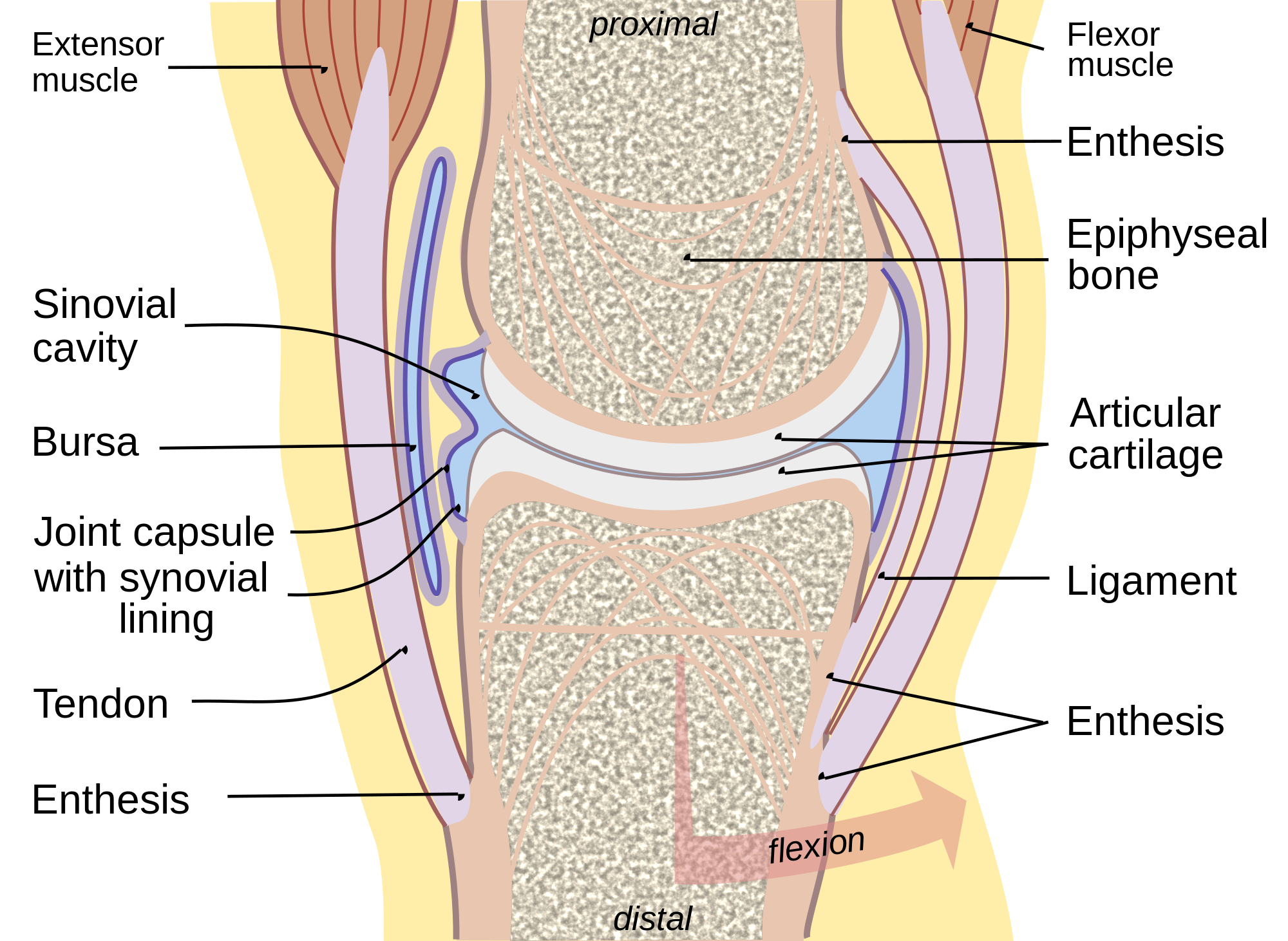
The synovial membrane (also known as the synovial stratum, synovium or stratum synoviale) is a specialized connective tissue that lines the inner surface of capsules of synovial joints, tendon sheaths, and synovial bursas. It makes direct contact with the fibrous membrane on the outside surface and with the synovial fluid lubricant on the inside surface.
In contact with the synovial fluid at the tissue surface are many rounded macrophage-like synovial cells (type A) and also type B cells, which are also known as fibroblast-like synoviocytes (FLS). Type A cells maintain the synovial fluid by removing wear-and-tear debris. The FLS (type B cells) produce hyaluronan, as well as other extracellular components in the synovial fluid.
The synovial membrane is variable but often has two layers:
Where the underlying subintima is loose, the intima sits on a pliable membrane, giving rise to the term synovial membrane.
This membrane, together with the cells of the intima, provides something like an inner tube, sealing the synovial fluid from the surrounding tissue (effectively stopping the joints from being squeezed dry when subject to impact, such as running).
Just outside the intima, most synovium has a dense net of fenestrated small blood vessels that provide nutrients not only for synovium but also for the avascular cartilage.
In any one position, much of the cartilage is close enough to get nutrition directly from the synovium.[citation needed]
Some areas of cartilage have to obtain nutrients indirectly and may do so either from diffusion through cartilage or possibly by 'stirring' of synovial fluid.
Hub AI
Synovial membrane AI simulator
(@Synovial membrane_simulator)
Synovial membrane
The synovial membrane (also known as the synovial stratum, synovium or stratum synoviale) is a specialized connective tissue that lines the inner surface of capsules of synovial joints, tendon sheaths, and synovial bursas. It makes direct contact with the fibrous membrane on the outside surface and with the synovial fluid lubricant on the inside surface.
In contact with the synovial fluid at the tissue surface are many rounded macrophage-like synovial cells (type A) and also type B cells, which are also known as fibroblast-like synoviocytes (FLS). Type A cells maintain the synovial fluid by removing wear-and-tear debris. The FLS (type B cells) produce hyaluronan, as well as other extracellular components in the synovial fluid.
The synovial membrane is variable but often has two layers:
Where the underlying subintima is loose, the intima sits on a pliable membrane, giving rise to the term synovial membrane.
This membrane, together with the cells of the intima, provides something like an inner tube, sealing the synovial fluid from the surrounding tissue (effectively stopping the joints from being squeezed dry when subject to impact, such as running).
Just outside the intima, most synovium has a dense net of fenestrated small blood vessels that provide nutrients not only for synovium but also for the avascular cartilage.
In any one position, much of the cartilage is close enough to get nutrition directly from the synovium.[citation needed]
Some areas of cartilage have to obtain nutrients indirectly and may do so either from diffusion through cartilage or possibly by 'stirring' of synovial fluid.
Recent media