Hubbry Logo
Anatomical terms of motionAnatomical terms of motionMain
Open search
Anatomical terms of motion
Community hub
Anatomical terms of motion
logo
8 pages, 0 posts
0 subscribers
Be the first to start a discussion here.
Contribute something
Anatomical terms of motion
from Wikipedia

Motion, the process of movement, is described using specific terms. Motion includes movement of organs, joints, limbs, and specific sections of the body. The terminology used describes this motion according to its direction relative to the anatomical position of the body parts involved. Anatomists and others use a unified set of terms to describe most of the movements, although other, more specialized terms are necessary for describing unique movements such as those of the hands, feet, and eyes.

In general, motion is classified according to the anatomical plane it occurs in. Flexion and extension are examples of angular motions, in which two axes of a joint are brought closer together or moved further apart. Rotational motion may occur at other joints, for example the shoulder, and are described as internal or external. Other terms, such as elevation and depression, describe movement above or below the horizontal plane. Many anatomical terms derive from Latin terms with the same meaning.

Classification

[edit]

Motions are classified after the anatomical planes they occur in,[1] although movement is more often than not a combination of different motions occurring simultaneously in several planes.[2] Motions can be split into categories relating to the nature of the joints involved:

Apart from this, motions can also be divided into:

  • Linear motions (or translatory motions), which move in a line between two points. Rectilinear motion is motion in a straight line between two points, whereas curvilinear motion is motion following a curved path.[2]
  • Angular motions (or rotary motions), which occur when an object is around another object increasing or decreasing the angle.[clarification needed] The different parts of the object do not move the same distance. Examples include a movement of the knee, where the lower leg changes angle compared to the femur, or movements of the ankle.[2]

The study of movement in the human body is known as kinesiology.[4] A categoric list of movements and the muscles involved can be found at list of movements of the human body.

Abnormal motion

[edit]
Hyperextended finger in hypermobility spectrum disorder
Hyperextended thumb in hypermobility spectrum disorder

The prefix hyper- is sometimes added to describe movement beyond the normal limits, such as in hypermobility, hyperflexion or hyperextension. The range of motion describes the total range of motion that a joint is able to do.[5] For example, if a part of the body such as a joint is overstretched or "bent backwards" because of exaggerated extension motion, then it can be described as hyperextended. Hyperextension increases the stress on the ligaments of a joint, and is not always because of a voluntary movement. It may be a result of accidents, falls, or other causes of trauma. It may also be used in surgery, such as in temporarily dislocating joints for surgical procedures.[6] Or it may be used as a pain compliance method to force a person to take a certain action, such as allowing a police officer to take him into custody.

General motion

[edit]

These are general terms that can be used to describe most movements the body makes. Most terms have a clear opposite, and so are treated in pairs.[7]

Flexion and extension

[edit]
Flexion and extension

Flexion and extension are movements that affect the angle between two parts of the body. These terms come from the Latin words with the same meaning.[a]

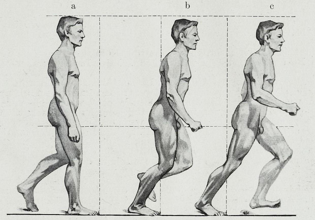
Flexion is a bending movement that decreases the angle between a segment and its proximal segment.[9] For example, bending the elbow, or clenching a hand into a fist, are examples of flexion. When a person is sitting down, the knees are flexed. When a joint can move forward and backward, such as the neck and trunk, flexion is movement in the anterior direction.[10] When the chin is against the chest, the neck is flexed, and the trunk is flexed when a person leans forward.[10] Flexion of the shoulder or hip is movement of the arm or leg forward.[11]

Extension is the opposite of flexion, a straightening movement that increases the angle between body parts.[12] For example, when standing up, the knees are extended. When a joint can move forward and backward, such as the neck and trunk, extension is movement in the posterior direction.[10] Extension of the hip or shoulder moves the arm or leg backward.[11] Even for other upper extremity joints – elbow and wrist, backward movement results in extension. The knee, ankle, and wrist are exceptions, where the distal end has to move in the anterior direction for it to be called extension.[13][page needed]

For the toes, flexion is curling them downward whereas extension is uncurling them or raising them.[14]

Abduction and adduction

[edit]
Abduction and adduction

Abduction is the motion of a structure away from the midline while adduction is motion towards the center of the body.[15] The center of the body is defined as the midsagittal or longitudinal plane.[3] These terms come from Latin words with similar meanings, ab- being the Latin prefix indicating 'away', ad- indicating 'toward', and ducere meaning 'to draw or pull'.[b]

Abduction is a motion that pulls a structure or part away from the midline of the body, carried out by one or more abductor muscles. In the case of fingers and toes, it is spreading the digits apart, away from the centerline of the hand or foot.[15] For example, raising the arms up, such as when tightrope-walking, is an example of abduction at the shoulder.[11] When the legs are splayed at the hip, such as when doing a star jump or doing a split, the legs are abducted at the hip.[3]

Adduction is a motion that pulls a structure or part towards the midline of the body, or towards the midline of a limb, carried out by one or more adductor muscles. In the case of fingers and toes, it is bringing the digits together, towards the centerline of the hand or foot. Dropping the arms to the sides, and bringing the knees together, are examples of adduction.[15]

Adduction of the wrist is also known as ulnar deviation which moves the hand towards the ulnar styloid (or, towards the little finger). Abduction of the wrist is also called radial deviation which moves the hand moving towards the radial styloid (or, towards the thumb).[15][17]

Elevation and depression

[edit]

Elevation and depression are movements above and below the horizontal. The words derive from the Latin terms with similar meanings.[c]

Elevation is movement in a superior direction.[19] For example, shrugging is an example of elevation of the scapula.[20]

Depression is movement in an inferior direction, the opposite of elevation.[21]

Rotation

[edit]
Rotation

Rotation of body parts may be internal or external, that is, towards or away from the center of the body.[22]

Internal rotation (medial rotation or intorsion) is rotation towards the axis of the body,[22] carried out by internal rotators.

External rotation (lateral rotation or extorsion) is rotation away from the center of the body,[22] carried out by external rotators.

Internal and external rotators make up the rotator cuff, a group of muscles that help to stabilize the shoulder joint.
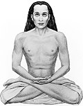
Other

[edit]
  • Anterograde and retrograde flow refer to movement of blood or other fluids in a normal (anterograde) or abnormal (retrograde) direction.[23]
  • Circumduction is a conical movement of a body part, such as a ball and socket joint or the eye. Circumduction is a combination of flexion, extension, adduction and abduction. Circumduction can be best performed at ball and socket joints, such as the hip and shoulder, but may also be performed by other parts of the body such as fingers, hands, feet, and head.[24] For example, circumduction occurs when spinning the arm when performing a serve in tennis or bowling a cricket ball.[25]
  • Reduction is a motion returning a bone to its original state,[26] such as a shoulder reduction following shoulder dislocation, or reduction of a hernia.

Special motion

[edit]

Hands and feet

[edit]

Flexion and extension of the foot

[edit]

Dorsiflexion and plantar flexion refer to extension or flexion of the foot at the ankle. These terms refer to flexion in direction of the "back" of the foot, which is the upper surface of the foot when standing, and flexion in direction of the sole of the foot. These terms are used to resolve confusion, as technically extension of the joint is dorsiflexion, which could be considered counter-intuitive as the motion reduces the angle between the foot and the leg.[27]

Dorsiflexion is where the toes are brought closer to the shin. This decreases the angle between the dorsum of the foot and the leg.[28] Dorsiflexion helps in assuming a deep squat position, i.e. squatting without the heels lifting off the ground.[29]

Plantar flexion or plantarflexion is the movement which decreases the angle between the sole of the foot and the back of the leg; for example, the movement when depressing a car pedal or standing on tiptoes.[27]

Flexion and extension of the hand

[edit]

Palmarflexion and dorsiflexion refer to movement of the flexion (palmarflexion) or extension (dorsiflexion) of the hand at the wrist.[30] These terms refer to flexion between the hand and the body's dorsal surface, which in anatomical position is considered the back of the arm; and flexion between the hand and the body's palmar surface, which in anatomical position is considered the anterior side of the arm.[31] The direction of terms are opposite to those in the foot because of embryological rotation of the limbs in opposite directions.[10]

Palmarflexion is flexion of the wrist towards the palm and ventral side of forearm.[30]

Dorsiflexion is hyperextension of the wrist joint, towards the dorsal side of forearm.[30]

Pronation and supination

[edit]

Pronation and supination refer generally to the prone (facing down) or supine (facing up) positions. In the extremities, they are the rotation of the forearm or foot so that in the standard anatomical position the palm or sole is facing anteriorly when in supination and posteriorly when in pronation.[32] As an example, when a person is typing on a computer keyboard, their hands are pronated; when washing their face, they are supinated.

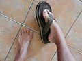
Pronation at the forearm is a rotational movement where the hand and upper arm are turned so the thumbs point towards the body. When the forearm and hand are supinated, the thumbs point away from the body. Pronation of the foot is turning of the sole outwards, so that weight is borne on the medial part of the foot.[33]

Supination of the forearm occurs when the forearm or palm are rotated outwards. Supination of the foot is turning of the sole of the foot inwards, shifting weight to the lateral edge.[34]
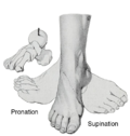
Inversion and eversion

[edit]

Inversion and eversion are movements that tilt the sole of the foot away from (eversion) or towards (inversion) the midline of the body.[35]

Eversion is the movement of the sole of the foot away from the median plane.[36] Inversion is the movement of the sole towards the median plane. For example, inversion describes the motion when an ankle is twisted.[28]

Eyes

[edit]

Unique terminology is also used to describe the eye. For example:

  • A version is an eye movement involving both eyes moving synchronously and symmetrically in the same direction.[37]
  • Torsion is eye movement that affects the vertical axis of the eye,[38] such as the movement made when looking in to the nose.

Jaw and teeth

[edit]

Other

[edit]

Other terms include:

  • Nutation and counternutation[d] are movement of the sacrum defined by the rotation of the promontory downwards and anteriorly, as with lumbar extension (nutation); or upwards and posteriorly, as with lumbar flexion (counternutation).[42]
  • Opposition is the movement that involves grasping of the thumb and fingers.[43]
  • Protraction and retraction is an anterior (protraction) or posterior (retraction) movement,[44] such as of the arm at the shoulders, although these terms have been criticised as non-specific.[45]
  • Reciprocal motion is alternating motions in opposing directions.[46]
  • Reposition is restoring an object to its natural condition.[47]

See also

[edit]

Notes

[edit]

References

[edit]

Sources

[edit]
[edit]
Revisions and contributorsEdit on WikipediaRead on Wikipedia
from Grokipedia
Anatomical terms of motion constitute a standardized in , , and for describing the directional and qualitative aspects of joint movements in the relative to the anatomical position. These terms enable precise communication among healthcare professionals, researchers, and educators by specifying how body segments move around axes in defined planes, such as the sagittal, frontal, and transverse planes. The anatomical position serves as the reference point for all such descriptions: the body is upright, facing forward, with the feet parallel and arms hanging at the sides, palms facing anteriorly. Movements are categorized primarily as angular (linear changes in angle) or rotary (circular paths around an axis), with angular motions further divided by plane—for instance, flexion and extension occur primarily in the , abduction and adduction in the frontal plane, and medial and lateral rotation in the . Key angular movements include flexion, which decreases the angle between two body parts (e.g., bending the ), and extension, which increases it (e.g., straightening the ). Abduction moves a limb away from the body's midline or median plane (e.g., raising the arm laterally), while adduction brings it closer (e.g., lowering the arm). Circumduction combines flexion, extension, abduction, and adduction to produce a conical or circular path at the joint, as seen in arm circling. Rotational movements encompass medial rotation (internal rotation, turning toward the body's center) and lateral rotation (external rotation, turning away from the center), often occurring at ball-and-socket joints like the or . Specialized terms address unique motions: protraction moves a body part anteriorly (e.g., thrusting the forward), retraction posteriorly (e.g., pulling the back), elevation upward (e.g., shrugging the s), and depression downward. For the foot, inversion turns the sole medially, and eversion laterally; in the , supination rotates the palm superiorly, and pronation inferiorly. These terms are foundational in clinical practice for assessing , diagnosing musculoskeletal disorders, and developing therapeutic interventions, as they provide a universal framework independent of individual anatomical variations.

Fundamentals

Anatomical position

The anatomical position is the standard reference posture used in to describe the location of body structures and the direction of movements. In this position, the body stands upright with the feet parallel and together, facing directly forward, the upper limbs positioned at the sides with the palms facing anteriorly (forward), and the lower limbs extended and close together. This erect stance ensures a consistent baseline for anatomical descriptions across medical and scientific contexts. Key features of the anatomical position include the head and eyes oriented forward, the mouth closed, and the upper limbs hanging relaxed with the pointing laterally away from the body (indicating supination of the forearms). The lower limbs are aligned with the feet pointing forward, and the long bones of the body are in their straight, neutral alignment. These elements collectively define a neutral, reproducible orientation that avoids ambiguity in spatial referencing. The importance of the anatomical position lies in its role as the neutral starting point from which all body motions and positional relationships are described; for instance, movements are termed relative to this posture, and deviations from it necessitate adjusted to maintain precision. This facilitates clear communication among anatomists, clinicians, and researchers, reducing errors in , , and . Without this reference, descriptions of body orientation would vary widely, complicating interdisciplinary work. Historically, the anatomical position emerged as part of broader 19th-century efforts to standardize amid growing chaos in . Anatomists like contributed through influential texts such as his 1858 Anatomy: Descriptive and Surgical, which popularized consistent descriptive practices, while formal codification occurred with the Basle Nomina Anatomica (BNA) in 1895, selecting and defining key terms including this reference posture. This late-19th-century standardization marked a pivotal shift toward international uniformity in anatomical .

Planes and axes of motion

In human anatomy, planes and axes of motion provide a standardized framework for describing the direction and orientation of body movements, relative to the anatomical position where the body stands upright with feet together, arms at the sides, and palms facing forward. These imaginary reference lines and surfaces intersect at the body's , typically near the second sacral , allowing for precise analysis of actions in three dimensions. The three cardinal planes—sagittal, frontal (coronal), and transverse (horizontal)—are mutually perpendicular and bisect the body, while the corresponding axes are lines perpendicular to each plane around which rotation occurs during movement. The , also known as the median or paramedian plane, divides the body into left and right halves, with the midsagittal plane creating equal portions. Movements in this plane, such as forward and backward bending, occur around a mediolateral (frontal) axis that runs horizontally from side to side through the body. This axis is perpendicular to the , enabling rotations that primarily affect anterior-posterior directions. The frontal plane, alternatively called the , divides the body into anterior (front) and posterior (back) sections. Motions within this plane, involving side-to-side deviations, take place around an anteroposterior (sagittal) axis that extends horizontally from front to back. This axis is oriented to the frontal plane, facilitating lateral rotations relative to the body's midline. The , also referred to as the horizontal plane, separates the body into superior (upper) and inferior (lower) parts. Rotational movements in this plane occur around a vertical (longitudinal) axis that passes through the body from top to bottom, to the transverse plane itself. This setup supports twisting actions that maintain the body's height while altering orientation. In addition to the cardinal planes, oblique planes exist at angles not aligned with the primary three, accommodating more complex, multi-directional movements that do not fit neatly into sagittal, frontal, or transverse categories. These non-cardinal planes are less commonly used in basic descriptions but are relevant for analyzing diagonal actions in biomechanics. Visual representations, such as diagrams illustrating the planes intersecting at the center of mass, are essential for understanding these concepts, often depicting the body in anatomical position with colored overlays for each plane and axis.

General motions

Flexion and extension

Flexion and extension are fundamental movements in the of the body, occurring around a transverse or frontal axis and involving changes in the angle between two adjacent body segments or parts. These motions are defined relative to the anatomical position, where the body stands upright with arms at the sides, palms facing forward, and feet parallel, serving as the neutral reference for describing joint actions. In contrast to frontal plane movements like abduction and adduction, flexion and extension primarily describe anterior-posterior bending and straightening. Flexion refers to the bending movement that decreases the angle between two body parts at a joint, in the sagittal plane. For example, flexion at the elbow joint brings the forearm closer to the upper arm, reducing the angle from approximately 180 degrees in the anatomical position. This motion is facilitated by muscles such as the biceps brachii and is essential for activities like reaching or grasping. Extension is the opposing motion to flexion, increasing the angle between body parts and generally directed posteriorly to return the joint toward or beyond its anatomical position. A common example is extension at the knee joint, where the lower leg straightens away from the thigh, restoring the leg to a fully extended posture during walking or standing. Hyperextension occurs when extension exceeds the normal anatomical range, potentially leading to joint strain; for instance, hyperextension of the spine during back arching can occur in yoga poses but risks injury if excessive. These movements vary by joint type: hinge joints, such as the and , primarily permit flexion and extension in a single plane with limited deviation. Ball-and-socket joints, like those at the and , allow flexion and extension alongside other motions, enabling greater versatility in compound actions such as swinging. Examples include trunk flexion during forward bending at the waist, which flexes the vertebral column, and extension at the ankle, such as pointing the toes downward.

Abduction and adduction

Abduction refers to the movement of a limb or body part away from the midline of the body, also known as the , typically occurring in the frontal or around a sagittal axis. Adduction is the opposing motion, bringing the limb or body part toward the midline in the same plane and around the same axis. These terms are defined relative to the anatomical position, where the body stands upright with arms at the sides and palms facing forward, establishing the midline as the central reference. At major joints such as the and , abduction involves lateral deviation of the appendage, for instance, raising the arm laterally from the side at the or spreading the legs apart at the joint. Adduction reverses these actions, such as lowering the arm back to the side or bringing the legs together at the . Unlike flexion and extension, which occur in the as anterior-posterior bending motions, abduction and adduction describe medial-lateral shifts in the frontal plane. For the digits, abduction and adduction apply to spreading or approximating the fingers and toes relative to a central axis, typically the second digit; for example, spreading the fingers apart constitutes abduction, while bringing them together is adduction. Hyperabduction extends abduction beyond the typical range of 180 degrees, such as positioning the hand over the opposite or behind the head, which can contribute to instability or injury like inferior .

Protraction and retraction

Protraction and retraction describe anterior-posterior gliding movements of body parts relative to the midline, typically occurring in the . Protraction involves forward (anterior) movement away from the midline, while retraction entails backward (posterior) movement toward the midline. These motions differ from , which produces around an axis, whereas protraction and retraction represent linear translations without significant . Common sites for these movements include the , , and . For the , protraction occurs during forward movements, such as in a motion, where the glides anterolaterally along the . Retraction of the , conversely, pulls the blades posteriorly and medially, as seen in posture correction exercises where the shoulders are drawn back. protraction is exemplified by pushing the forward, protruding the lower jaw anteriorly. Retraction returns the to its neutral position by drawing it posteriorly. For the , protraction advances it forward, as in protrusion during speech or , while retraction pulls it backward. These gliding actions primarily facilitate functional adjustments, enabling efficient positioning for activities like reaching or mastication without altering angles substantially. Unlike abduction and adduction, which involve mediolateral separation, or and depression, which address vertical shifts, protraction and retraction emphasize horizontal anterior-posterior dynamics.

Elevation and depression

Elevation and depression refer to the superior (upward) and inferior (downward) movements of body structures relative to the anatomical position, typically occurring within the frontal (coronal) plane and involving displacement along the vertical axis. These motions enable essential functions such as respiration, , and , distinguishing them from horizontal shifts like protraction and retraction, which occur in the . Elevation involves lifting a structure toward the head or superior aspect of the body. A primary example is the elevation of the during shoulder shrugging, where the moves upward to protect the or express tension. Similarly, the facilitates elevation of the eyebrows, raising the superior portion of the to convey surprise or alertness. In the neck region, , such as the mylohyoid, elevate the during , stabilizing the floor of the mouth and aiding in tongue propulsion. For respiration, elevate the during inspiration, expanding the to increase volume. Depression, conversely, lowers a structure away from the head or toward the inferior aspect. Lowering the shoulders after shrugging depresses the , relaxing the and returning the to neutral. Mandibular depression occurs when the drops to open the , primarily driven by muscles like the digastric during actions such as yawning or speaking, allowing for increased oral cavity volume. The can also undergo depression via like the sternohyoid, which lowers it post-swallowing to facilitate laryngeal descent. In breathing, internal and depress the ribs during expiration, reducing thoracic volume to expel air.

Rotation

Rotation is an angular movement in which a or body segment turns around its longitudinal (long axis), occurring primarily in the about a vertical axis. This motion allows for twisting actions that are essential for orientation and locomotion, distinguishing it from linear or bending movements like flexion and extension. Medial rotation, also termed internal rotation, describes the turning of a limb or body part toward the midline of the body. For instance, at the , internal rotation directs the anterior surface of the medially, such as when bringing the hand across the body with the elbow flexed at 90 degrees. Lateral rotation, or external rotation, involves the opposite action, moving the limb away from the midline; an example is rotating the so the thumb points laterally. These movements occur at several synovial joints, including the (glenohumeral), (acetabulofemoral), (cervical vertebrae), and (radioulnar, though specific forearm twisting is addressed separately). At the shoulder, the typical range is 70–90 degrees for both internal and external when the arm is abducted. The hip permits approximately 30–40 degrees of internal and 40–60 degrees of external . Neck allows up to 80 degrees to each side. Practical examples include rotating the head to look over the , which utilizes cervical rotation for visual scanning. In gait, thigh rotation at the —external during the swing phase and internal during stance—contributes to foot placement and balance. Pure rotation contrasts with circumduction, which combines rotation with other motions to trace a conical path.

Circumduction

Circumduction is a complex movement in which a distal body part traces a conical or circular path around a central point while the proximal end remains relatively fixed, resulting from the sequential integration of other fundamental motions such as flexion, extension, abduction, and adduction. This motion is not a singular action but a coordinated pattern that requires freedom across multiple planes, allowing the limb or to describe a with its apex at the center. The direction of circumduction is typically described as clockwise or counterclockwise when viewed from the anatomical position, enabling a full or partial circular depending on the joint's range. It occurs primarily at multiaxial ball-and-socket joints, such as the glenohumeral () and coxal () joints, which permit extensive multiplanar motion; limited circumduction is also possible at biaxial condyloid joints like the or saddle joints such as the metacarpophalangeal joints of the thumb. Unlike isolated , which involves twisting around a single axis, circumduction combines movements across sagittal, frontal, and transverse planes, emphasizing its role as a multi-axis pattern rather than a pure . Representative examples include the shoulder's ability to perform full circumduction, as seen when drawing large circles with the extended, or the hip's partial circumduction during a limping to compensate for lower limb weakness by swinging the leg in a semicircular arc. At the , circumduction manifests as a smaller conical motion of the hand, combining flexion, extension, radial and ulnar deviation. These motions highlight circumduction's dependence on synovial joints with sufficient articular freedom to integrate all three anatomical planes and axes simultaneously.

Special motions

Pronation and supination

Pronation and supination are anatomical terms describing specific rotational movements of the that occur at the proximal and distal radioulnar joints, altering the orientation of the palm relative to the anatomical position. Pronation involves medial of the around the , positioning the palm to face posteriorly or downward, as in the action of turning a doorknob. In contrast, supination entails lateral of the , orienting the palm anteriorly or upward, such as when holding a of to prevent spilling. These movements are opposite and complementary, enabling functional hand positioning for daily activities like pouring water (pronation) or using a (supination). The mechanism of pronation and supination relies on the pivot-like articulation between the and , facilitated by the annular ligament at the proximal joint and the ulnar notch at the distal joint. During pronation, the distal end of the crosses over the , rotating medially while the remains relatively fixed; in supination, the uncrosses and rotates laterally around the . This rotation is supported by the , which binds the two bones and transmits forces between them throughout the motion. These actions occur in the , perpendicular to the long axis of the , distinguishing them as specialized forms of rotation. The typical for both pronation and supination is approximately 80-90 degrees from the neutral position, though individual variations exist, with supination often reaching up to 85 degrees and pronation around 70-80 degrees in healthy adults. This range allows for full mobility, essential for tasks requiring precise hand manipulation, and is measured using a aligned with the forearm's long axis.

Inversion and eversion

Inversion and eversion are special movements of the foot that occur primarily at the , involving tilting of the sole medially or laterally relative to the midline of the body. Inversion refers to the medial tilting of the foot, where the sole faces inward toward the midline, as in turning the foot inward during activities. Eversion is the opposite motion, characterized by lateral tilting of the foot so that the sole faces away from the midline, such as when turning the foot outward to adapt to terrain. These actions are analogous to adduction and abduction in other appendages but are specifically termed for the foot's transverse orientation. The mechanism of inversion and eversion centers on the , formed between the talus and , which permits rotational gliding of the relative to the talus along an oblique axis. During inversion, the rolls and glides medially under the talus, elevating the lateral border of the foot, while eversion involves lateral rolling and gliding of the , raising the medial border. These isolated motions contribute to the composite movements of foot pronation (which incorporates eversion) and supination (which incorporates inversion), enhancing overall foot adaptability. Both occur in the frontal plane of the body, perpendicular to the used for flexion and extension. The typical at the is approximately 30 degrees of inversion and 15-20 degrees of eversion, though individual variation exists based on and morphology. Inversion sprains are common injuries resulting from excessive inversion force, often stretching or tearing the lateral ankle ligaments like the during activities such as landing awkwardly or stepping on uneven surfaces. Eversion plays a key role in walking on uneven ground, allowing the foot to conform and absorb shock by laterally tilting to maintain balance and propulsion.

Dorsiflexion and plantarflexion

Dorsiflexion and plantarflexion are specialized terms describing hinge-like movements at the ankle joint, where the foot flexes or extends relative to the in the . Dorsiflexion refers to the flexion of the foot such that the toes point superiorly, effectively lifting the anterior aspect of the foot toward the shin. In contrast, plantarflexion involves the extension of the foot, directing the toes inferiorly and pointing the foot away from the shin. These motions parallel the general concepts of flexion and extension but are tailored to the ankle's functional . The primary mechanism for these movements occurs at the talocrural joint, a synovial formed by the articulation of the talus with the and . Dorsiflexion is primarily driven by the , with contributions from the extensor hallucis longus, extensor digitorum longus, and peroneus tertius muscles acting concentrically to elevate the foot. Plantarflexion, on the other hand, is powered mainly by the gastrocnemius (medial and lateral heads) and soleus muscles, which form the triceps surae and generate force through the to depress the foot. Both actions facilitate essential locomotor functions, such as propulsion and shock absorption during . The typical at the talocrural allows approximately 20 degrees of dorsiflexion and 50 degrees of plantarflexion from a neutral position. Practical examples include walking, which emphasizes sustained dorsiflexion to maintain the toes elevated off the ground, and the pointe position, which demands extreme plantarflexion to align the over the toes. These movements are integral to activities requiring precise foot control in the , underscoring their role in balance and mobility.

Radial and ulnar deviation

Radial deviation, also known as wrist abduction, involves the lateral movement of the hand toward the and , elevating the ulnar aspect of the wrist. This motion positions the hand away from the body's midline in the anatomical position. Conversely, ulnar deviation, or wrist adduction, directs the hand medially toward the and , raising the radial side of the wrist. Both movements occur within the frontal ( of the body, facilitating side-to-side bending at the wrist . The biomechanical mechanism of radial and ulnar deviation relies on the gliding of the proximal carpal row—primarily the scaphoid, lunate, and triquetrum—across the articular surfaces of the distal and ulnar head. During radial deviation, the carpals shift ulnarly relative to the , while ulnar deviation prompts a radial shift of the carpals. These motions are constrained by the radial and s of the wrist; the radial collateral ligament resists excessive ulnar deviation, and the limits excessive radial deviation. Additional stability is provided by the dorsal and palmar radiocarpal ligaments, which prevent overextension during deviation. Normal ranges of motion for these deviations vary slightly by individual factors such as age and occupation, but typically, radial deviation measures about 20 degrees from neutral, while ulnar deviation permits 30 to 40 degrees. These asymmetries reflect the anatomical structure of the wrist, where the ulnar side allows greater excursion due to the complex accommodating more translation. Practical examples illustrate these motions in daily activities: ulnar deviation occurs when striking a tennis ball during a forehand swing, as the wrist bends toward the pinky side to generate power and control. Radial deviation is evident in the prayer position, where the palms are pressed together in front of the chest, tilting the wrists toward the thumbs to maintain alignment. These wrist-specific deviations parallel the broader concepts of abduction and adduction but are adapted to the hand's fine motor demands.

Opposition and reposition

Opposition is a specialized movement of the thumb in which the pad of the thumb is brought into contact with the pads of the fingers of the same hand, facilitating a precision grip such as pinching or holding small objects. This motion is essential for fine motor tasks and is primarily achieved through a combination of flexion and abduction at the thumb's joints. Reposition, the reciprocal movement, involves returning the thumb to its anatomical position alongside the fingers, reversing the opposition to release the grip. The primary mechanism enabling opposition occurs at the carpometacarpal (CMC) joint of the thumb, a saddle-shaped synovial joint formed between the first metacarpal and the trapezium bone of the carpus. This unique saddle configuration allows for multiplanar motion, including approximately 50-60 degrees of flexion-extension, 40-50 degrees of abduction-adduction, and up to 70 degrees of internal rotation, collectively permitting the thumb to rotate and flex toward the fingertips. The CMC joint's structure, supported by ligaments such as the anterior oblique and dorsal radial, provides both mobility and stability during these movements. A related but less commonly used term is , which refers to a variant of opposition where the is positioned against the lateral side of the rather than the pad-to-pad contact with other fingers, often used in specific grasping patterns. This distinction highlights subtle differences in positioning for varied manipulative tasks, though opposition remains the dominant term for pad-to-pad interaction. The capacity for opposition is a key evolutionary in , particularly pronounced in humans, enabling advanced tool use and dexterity that distinguished early hominins from other around two million years ago. Biomechanical studies indicate that enhanced thumb opposition efficiency in facilitated precise manipulation, contributing to cultural and technological advancements by improving and object control. In practical examples, opposition is crucial during activities like writing, where opposes the pen held by the fingers, or pinching small items such as a . Reposition occurs when releasing the grip, such as opening the hand after holding an object. The full range of opposition typically allows for about 90 degrees of effective at the CMC joint, though individual variation exists based on factors like age and joint health.

Jaw movements

Jaw movements occur at the (TMJ), a specialized classified as ginglymoarthrodial, which combines hinge-like rotation and gliding translation to enable mastication and related functions. This joint's unique structure allows the to perform , depression, protrusion, retraction, and lateral , adapting general anatomical terms of motion to the demands of oral activities. Unlike simpler joints, the TMJ's dual articulations—between the mandibular condyle and the temporal bone's mandibular fossa, separated by an articular disc—facilitate these combined motions without dislocation under normal loads. Elevation of the closes the mouth and is driven primarily by the masseter, temporalis, and medial pterygoid muscles, which contract to approximate the mandibular teeth with the maxillary teeth. Depression opens the mouth and relies on gravity, along with such as the digastric, mylohyoid, and geniohyoid, plus the inferior head of the lateral pterygoid to initiate translation. The normal range of depression measures approximately 40-50 mm interincisally, while returns to a resting position of near occlusion. Common examples include clenching the teeth during stress, which emphasizes , and yawning, which maximizes depression for airway expansion. Protrusion shifts the forward along the , aiding in actions like into food, and is produced by bilateral contraction of the lateral pterygoid muscles, assisted by the anterior fibers of the masseter and medial pterygoid. Retraction pulls the backward to its resting position and is achieved mainly by the posterior fibers of the , with contributions from the posterior masseter and . The typical range for protrusion is 5-10 mm, measured from the midline, while retraction covers a similar but slightly lesser . For instance, protruding the facilitates into an apple by advancing the incisors. Lateral excursion involves side-to-side grinding motions essential for tougher foods, resulting from unilateral where the contralateral lateral pterygoid protrudes one side while the ipsilateral temporalis retracts the other, creating a Bennett movement or shift. This excursion typically ranges from 8-12 mm to each side, allowing the to deviate laterally relative to the without rotation. Such movements enable the grinding phase of mastication, as seen when shifting the sideward to process fibrous .

Eye movements

Eye movements refer to the coordinated rotations of the eyeball within the , enabling visual fixation, tracking, and scanning of the environment. These motions are distinct from general body rotations, as they involve precise control by specialized to maintain and stabilize gaze. Unlike appendicular rotations, eye movements occur in specific planes: horizontal displacements in the and vertical displacements in the frontal plane. Ductions describe monocular movements of a single eye, independent of the other. Key ductions include adduction, which moves the eye medially toward the ; abduction, which moves it laterally away from the ; supraduction (), which raises the eye upward; and infraduction (depression), which lowers it downward. These monocular rotations form the basis for more complex binocular coordination. Versions involve binocular movements where both eyes rotate in the same direction, ensuring conjugate . For example, dextroversion shifts both eyes to the right, while levoversion shifts them to the left; similar paired terms apply for upward (supraversion) and downward (infraversion) . Vergences, in contrast, are binocular dysconjugate movements where the eyes rotate in opposite directions, such as convergence (both eyes for near vision) or divergence (both abduct for distant vision). These mechanisms support and focus adjustment. The primary mechanism driving these movements is the six , innervated by III (oculomotor), IV (trochlear), and VI (abducens). The medial rectus (CN III) primarily adducts the eye, while the lateral rectus (CN VI) abducts it; the superior rectus (CN III) elevates (supraducts) primarily in abduction, and the inferior rectus (CN III) depresses (infraducts) primarily in abduction. The superior oblique (CN IV) depresses primarily in adduction and intorts the eye, whereas the inferior oblique (CN III) elevates primarily in adduction and extorts it. These muscles work in synergistic pairs for balanced motion. Specific examples of eye movements include saccades, which are rapid, ballistic shifts in to redirect fixation; , which tracks slowly moving objects with continuous velocity matching; and the vestibulo-ocular reflex, which stabilizes during head movements by counter-rotating the eyes. These patterns rely on ductions, , and vergences to function effectively in the transverse and frontal planes.

Abnormal motions

Definitions and types

Abnormal motions in anatomy refer to variations of standard joint movements that either exceed the normal physiological range or are restricted beyond typical limits, often due to injury, pathology, or structural changes. These differ from normal anatomical terms of motion, which describe controlled actions within expected ranges, such as flexion or extension. Examples include hyperextension, which is excessive extension of a joint beyond its normal range, potentially causing injury to ligaments or tissues, and hyperflexion, excessive flexion that can lead to similar damage. Pathological conditions can produce abnormal variants of these motions, such as spastic flexion, where increased from lesions causes involuntary contraction and resists extension, or , resulting in diminished or absent motion due to damage. Other types include , a fusion of the leading to complete loss of motion, and s, fixed joint positions from shortened muscles or tissues. Classification may consider (e.g., traumatic, degenerative) or appearance (e.g., excessive range, reduced amplitude), aiding while aligning with normative motion descriptions.

Pathological examples

In , spastic flexion due to results in persistent flexed postures at joints like the elbows and knees, limiting extension and contributing to abnormalities such as scissoring. This arises from lesions, causing velocity-dependent increases in that resist stretching. Joint , as in or post-traumatic cases, eliminates flexion and extension, fixing the joint in a neutral or deviated position and restricting functional range, often requiring surgical correction. Contractures lead to fixed abnormal positions, such as persistent flexion at the fingers or plantarflexion at the ankles, from prolonged immobility or shortening soft tissues like muscles and tendons. These limit and may cause secondary deformities, common in chronic neurological conditions. In hemiplegic gait after , compensatory circumduction of the paretic swings the limb in a lateral arc to avoid toe drag, despite weakness in dorsiflexion and knee control, increasing energy expenditure. Observing these abnormal motions aids , as patterns like or fixed contractures indicate underlying musculoskeletal or neurological issues, guiding interventions like or .

References

Add your contribution
Related Hubs
Contribute something
User Avatar
No comments yet.