Recent from talks
Brodmann area 45
Knowledge base stats:
Talk channels stats:
Members stats:
Brodmann area 45
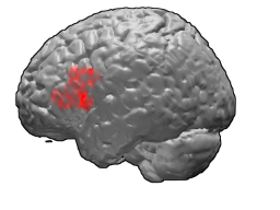
Brodmann area 45 (BA45), is part of the frontal cortex in the human brain. It is situated on the lateral surface, inferior to BA9 and adjacent to BA46.
This area in humans occupies the triangular part of inferior frontal gyrus (H) and, surrounding the anterior horizontal limb of the lateral sulcus (H), a portion of the orbital part of the inferior frontal gyrus (H). Bounded caudally by the anterior ascending limb of the lateral sulcus (H), it borders on the insula in the depth of the lateral sulcus.
In terms of cytoarchitecture, it is bounded caudally by the opercular part of inferior frontal gyrus (Brodmann area 44 (BA44)), rostrodorsally by the middle frontal area 46 (BA46), and ventrally by the orbital part of inferior frontal gyrus (Brodmann area 47 BA47).
The left-hemisphere Brodmann area 44 and Brodmann area 45 make up Broca's area, a region that is active in semantic tasks, such as semantic decision tasks (determining whether a word represents an abstract or a concrete entity) and generation tasks (generating a verb associated with a noun).
The precise role of BA45 in semantic tasks remains controversial. For some researchers, its role would be to subserve semantic retrieval or semantic working memory processes. Under this view, BA44 and BA45 would together guide recovery of semantic information and evaluate the recovered information with regard to the criterion appropriate to a given context. A slightly modified account of this view is that activation of BA45 is needed only under controlled semantic retrieval, when strong stimulus-stimulus associations are absent. For other researchers, BA45's role is not restricted to semantics per se, but to all activities that require task-relevant representations from among competing representations. Lesions of the BA45 lead to the characteristic findings of expressive aphasia in individuals who are left hemispheric dominant.
A strong correlation has been found between speech-language and the anatomically asymmetric pars triangularis. Foundas, et al. showed that language function can be localized to one region of the brain, as Paul Broca had done before them, but they also supported the idea that one side of the brain is more involved with language than the other. The human brain has two hemispheres, and each one looks similar to the other; that is, it looks like one hemisphere is a mirror image of the other. However, Foundas, et al. found that the pars triangularis in Broca's area is actually larger than the same region in the right side of the brain. This "leftward asymmetry" corresponded both in form and function, which means that the part of the brain that is active during language processing is larger. In almost all the test subjects, this was the left side. In fact, the only subject tested that had right-hemispheric language dominance was found to have a rightward asymmetry of the pars triangularis.
Certain other researchers, however, have found no volumetric asymmetries in the pars triangularis. They have challenged previous findings that pars triangularis asymmetry exists and have suggested that inconsistencies in previous findings may be due to great variability in inter-individual pars triangularis morphology. That is, these regions tend to vary in size and shape much more than other areas of the brain, such as deep cortical nuclei. Furthermore, while these researcher found statistically significant asymmetries in the pars opercularis and the planum temporale, they found no correlations between asymmetries of these brain regions with that of the pars triangularis.
At least one study demonstrated a high degree of connectivity between the three subregions of the inferior frontal gyrus (IFG). By stimulating one region of the IFG and measuring the response in distinct regions, these researchers were able to demonstrate the existence of numerous pathways between the pars triangularis and pars opercularis. Also, stimulation of one region of the pars triangularis elicited a response in distinct regions of the pars triangularis, illustrating the presence of networks within the subgyral region.
Hub AI
Brodmann area 45 AI simulator
(@Brodmann area 45_simulator)
Brodmann area 45
Brodmann area 45 (BA45), is part of the frontal cortex in the human brain. It is situated on the lateral surface, inferior to BA9 and adjacent to BA46.
This area in humans occupies the triangular part of inferior frontal gyrus (H) and, surrounding the anterior horizontal limb of the lateral sulcus (H), a portion of the orbital part of the inferior frontal gyrus (H). Bounded caudally by the anterior ascending limb of the lateral sulcus (H), it borders on the insula in the depth of the lateral sulcus.
In terms of cytoarchitecture, it is bounded caudally by the opercular part of inferior frontal gyrus (Brodmann area 44 (BA44)), rostrodorsally by the middle frontal area 46 (BA46), and ventrally by the orbital part of inferior frontal gyrus (Brodmann area 47 BA47).
The left-hemisphere Brodmann area 44 and Brodmann area 45 make up Broca's area, a region that is active in semantic tasks, such as semantic decision tasks (determining whether a word represents an abstract or a concrete entity) and generation tasks (generating a verb associated with a noun).
The precise role of BA45 in semantic tasks remains controversial. For some researchers, its role would be to subserve semantic retrieval or semantic working memory processes. Under this view, BA44 and BA45 would together guide recovery of semantic information and evaluate the recovered information with regard to the criterion appropriate to a given context. A slightly modified account of this view is that activation of BA45 is needed only under controlled semantic retrieval, when strong stimulus-stimulus associations are absent. For other researchers, BA45's role is not restricted to semantics per se, but to all activities that require task-relevant representations from among competing representations. Lesions of the BA45 lead to the characteristic findings of expressive aphasia in individuals who are left hemispheric dominant.
A strong correlation has been found between speech-language and the anatomically asymmetric pars triangularis. Foundas, et al. showed that language function can be localized to one region of the brain, as Paul Broca had done before them, but they also supported the idea that one side of the brain is more involved with language than the other. The human brain has two hemispheres, and each one looks similar to the other; that is, it looks like one hemisphere is a mirror image of the other. However, Foundas, et al. found that the pars triangularis in Broca's area is actually larger than the same region in the right side of the brain. This "leftward asymmetry" corresponded both in form and function, which means that the part of the brain that is active during language processing is larger. In almost all the test subjects, this was the left side. In fact, the only subject tested that had right-hemispheric language dominance was found to have a rightward asymmetry of the pars triangularis.
Certain other researchers, however, have found no volumetric asymmetries in the pars triangularis. They have challenged previous findings that pars triangularis asymmetry exists and have suggested that inconsistencies in previous findings may be due to great variability in inter-individual pars triangularis morphology. That is, these regions tend to vary in size and shape much more than other areas of the brain, such as deep cortical nuclei. Furthermore, while these researcher found statistically significant asymmetries in the pars opercularis and the planum temporale, they found no correlations between asymmetries of these brain regions with that of the pars triangularis.
At least one study demonstrated a high degree of connectivity between the three subregions of the inferior frontal gyrus (IFG). By stimulating one region of the IFG and measuring the response in distinct regions, these researchers were able to demonstrate the existence of numerous pathways between the pars triangularis and pars opercularis. Also, stimulation of one region of the pars triangularis elicited a response in distinct regions of the pars triangularis, illustrating the presence of networks within the subgyral region.
Recent media