Hubbry Logo
BronchophonyBronchophonyMain
Open search
Bronchophony
Community hub
Bronchophony
logo
8 pages, 0 posts
0 subscribers
Be the first to start a discussion here.
Bronchophony
Bronchophony
from Wikipedia
Bronchophony
Other namesBronchiloquy[1]
Front view of cartilages of larynx, trachea, and bronchi.
SpecialtyRespirology

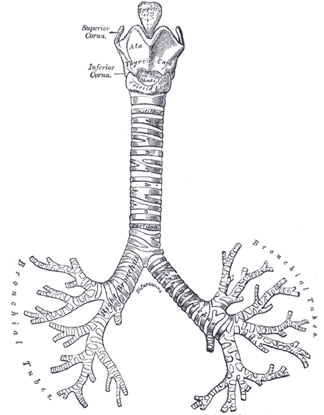
Bronchophony is the abnormal transmission of sounds from the lungs or bronchi. Bronchophony is a type of pectoriloquy.

It is a general medical sign detected by auscultation.

Technique

[edit]

The patient is requested to repeat a word several times while the provider auscultates symmetrical areas of each lung. The numbers "ninety-nine" or "sixty-six" are traditionally mentioned. The choice of "ninety-nine" is the result of a literal translation. The test was originally described by a German physician who used the phrase "neun und neunzig" (pronounced [ˈnɔʏnʊntˈnɔʏntsɪç]), which he found would cause maximum vibration of the chest. The translation, "ninety-nine", has fewer vowels and is less effective in evoking the phenomenon. Better phrases in English include "toy boat”, "Scooby Doo", and “blue balloons".[2]

Interpretation

[edit]

Normally, the sound of the patient's voice becomes less distinct as the auscultation moves peripherally; bronchophony is the phenomenon of the patient's voice remaining loud at the periphery of the lungs or sounding louder than usual over a distinct area of consolidation, such as in pneumonia. This is a valuable tool in physical diagnosis used by medical personnel when auscultating the chest.

Often, the patient does not have to speak for the provider to hear signs of bronchophony. Rather, the normal breath sounds are increased in loudness – referred to by doctors as "increased breath sounds" – over the affected area of the lungs is indicative.

Causes

[edit]

Bronchophony may be caused by a solidification of lung tissue around the bronchi – which may indicate lung cancer – or by fluid in the alveoli, which may indicate pneumonia. However, it may also have benign causes, such as wide bronchi. As such, it is usually an indication for further investigation rather than the main basis of a diagnosis.

Other observations

[edit]

Other tools used in auscultation include listening for egophony, whispered pectoriloquy, crackles, rhonchi or wheezing. Percussion is also often used to determine diseases of the chest.

References

[edit]
[edit]
Revisions and contributorsEdit on WikipediaRead on Wikipedia
from Grokipedia
Bronchophony is a clinical sign observed during the physical examination of the lungs, characterized by an abnormal increase in the intensity, clarity, and transmission of the patient's spoken voice sounds through the chest wall when auscultated with a stethoscope. This phenomenon occurs because pathological changes in the lung tissue, such as the replacement of air with fluid or solid material, enhance the conduction of sound vibrations from the bronchi to the surface of the chest. Unlike normal lung auscultation, where voice sounds are muffled and indistinct due to the air-filled alveoli acting as a barrier, bronchophony results in the voice being perceived as louder and more articulate, often resembling direct speech. To elicit bronchophony, the examiner places the diaphragm of the over symmetric areas of the chest while instructing the patient to repeat a phrase such as "ninety-nine" or to speak in a normal voice. The test is performed bilaterally to compare findings, with positive bronchophony noted when the sounds are distinctly louder and clearer over the affected field compared to the unaffected side or normal baseline. This assessment is part of a broader evaluation of vocal , which includes related signs like (a nasal or bleating quality to the voice) and (clear transmission of whispered words). Clinically, bronchophony is a key indicator of , most commonly associated with conditions such as , where alveolar spaces fill with inflammatory , or other processes like and that alter the air-fluid dynamics in the lungs. It is often accompanied by other abnormal auscultatory findings, including bronchial breath sounds and increased tactile fremitus, helping clinicians differentiate consolidated lung tissue from normal aerated lung or pleural effusions, where voice transmission may be diminished. Early detection through bronchophony can guide diagnostic imaging and treatment, underscoring its value in bedside assessment despite the rise of advanced imaging modalities.

Introduction

Definition

Bronchophony refers to the abnormal increase in the intensity and clarity of spoken voice sounds transmitted through the chest and heard via over the fields, resulting from altered tissue density that enhances conduction. In this phenomenon, normally muffled vocal sounds become louder and more distinct due to pathological changes, such as or consolidation replacing air-filled alveoli, which act as a in healthy . This increased transmission indicates underlying respiratory affecting propagation. Bronchophony is distinguished from the broader category of pectoriloquy by its focus on voiced, spoken sounds rather than whispered ones. Specifically, while general pectoriloquy encompasses enhanced voice transmission, bronchophony involves the patient's spoken words—such as repeating "ninety-nine"—being perceived as unusually clear and resonant, often as if the voice is directed straight into the stethoscope. In contrast, whispered pectoriloquy tests unvoiced whispers for similar clarity, providing a more specific indicator of severe consolidation. The term bronchophony derives from the Greek roots "broncho-" meaning airway or bronchus, and "-phony" meaning sound, reflecting the bronchial-like quality of the transmitted voice. It was first described in the early 19th century by French physician René Laënnec in his 1819 treatise A Treatise on the Diseases of the Chest and Mediate Auscultation, where he termed it "bronchophonism" as part of his pioneering work on stethoscopic auscultation of voice-generated sounds in lung diseases.

Clinical Relevance

Bronchophony serves as a valuable bedside test in physical examinations for detecting , such as consolidation, by assessing the transmission of spoken voice sounds through the chest without requiring modalities. This non-invasive technique is particularly useful in resource-limited settings where access to or advanced diagnostics may be restricted, allowing clinicians to identify potential abnormalities like those seen in through alone. The assessment of bronchophony was integrated into standard lung auscultation protocols in the early 19th century, following René Laennec's invention of the in 1816 and his detailed descriptions of abnormal voice transmission sounds, including bronchophony, in his 1819 treatise De l'Auscultation Médiate. These advancements enabled more precise correlation of auscultatory findings with underlying pulmonary conditions, establishing bronchophony as a foundational element of respiratory evaluation. Studies evaluating bronchophony's diagnostic performance for lung consolidation in report variable sensitivity ranging from 11% to 69% and specificity from 61% to 96%, indicating it is more reliable for confirming when present but less effective at ruling out . Its accuracy can diminish in obese patients due to reduced sound transmission through thicker chest walls and is further compromised by suboptimal technique. Despite its utility, bronchophony's interpretation remains subjective, prone to inter-observer variability, and heavily influenced by patient cooperation, examiner experience, and factors like ambient noise or quality. These limitations underscore the need for confirmatory tests in clinical decision-making.

Assessment Technique

Procedure

Bronchophony is assessed as part of the auscultatory component of the pulmonary , utilizing vocal resonance to evaluate lung transmission of spoken sounds. The test is typically performed after and percussion of the chest but prior to other specialized vocal assessments, such as or . A standard acoustic is preferred for this traditional evaluation, with the diaphragm side applied firmly to the skin to optimize detection of high-frequency components in voice transmission. To conduct the test, position the patient either seated upright or , ensuring the chest is exposed for direct contact. The systematically places the over symmetric areas, beginning with the posterior chest fields (from apex to base), followed by anterior and lateral regions, to allow for bilateral . At each site, instruct the patient to repeat a resonant , such as "ninety-nine" or "one-one-one," in a normal speaking voice rather than a whisper, as whispering is reserved for assessing . The patient should speak steadily while the listens attentively for the clarity and intensity of the transmitted sounds, moving methodically across comparable sites on both sides of the to identify any asymmetry. The phrase "ninety-nine" is commonly used because it contains voiced consonants that produce distinct vibrations, facilitating the detection of increased transmission over consolidated lung areas. Throughout the procedure, ensure the patient maintains consistent volume and avoids extraneous movements that could introduce artifacts. If clear transmission of words is noted at any point, it may suggest pathological enhancement, though detailed interpretation is addressed elsewhere.

Patient Preparation

The examiner should begin by explaining the bronchophony assessment to in simple, clear terms, emphasizing that it involves listening to their voice through a placed on the chest to evaluate sounds, thereby promoting cooperation and reducing anxiety. Patients are instructed to remove any upper clothing or ensure the chest is bare to allow direct contact of the with the skin, which enhances acoustic transmission and accuracy. Additionally, patients are directed to speak test phrases, such as "ninety-nine," at a normal conversational volume without straining or shouting, as excessive effort can distort the findings. Optimal patient positioning facilitates comprehensive access to lung fields while maintaining comfort. For posterior and lateral auscultation, the sitting upright or semi-recumbent position is preferred, allowing the patient to lean slightly forward if needed. Anterior auscultation may be performed with the patient , though the sitting position remains ideal overall to promote even breathing and minimize gravitational effects on lung expansion. Throughout the procedure, patients should breathe normally and remain relaxed to avoid influencing respiratory . The examiner must prepare the environment and equipment to ensure reliable results. A quiet room is essential to eliminate ambient noise that could obscure subtle voice transmissions. The stethoscope's diaphragm should be warmed by rubbing it between the palms before application to prevent discomfort from cold metal on the skin. follows a systematic , typically mapping 6 to 8 points per side across anterior, posterior, and lateral chest fields to systematically compare symmetry and cover all lobes. While bronchophony assessment is non-invasive and generally safe, it is not advisable in patients with acute respiratory distress, where positioning and speaking may exacerbate symptoms or prove impossible. Similarly, in cases of severe , such as advanced , the test may be infeasible due to inability to follow instructions, necessitating alternative diagnostic approaches.

Physiological Basis

Normal Voice Transmission

In healthy lungs, voice vibrations originate in the and propagate through the trachea and bronchi as sound waves, which are then significantly attenuated by the air-filled alveoli and surrounding before reaching the chest wall. This attenuation results in muffled, indistinct sounds that are typically unintelligible to the listener during . Acoustically, the air density in the alveoli acts as a , permitting low-frequency components of the voice (below 200 Hz) to pass with reduced intensity while dampening higher frequencies (above 400 Hz), producing a characteristic low-intensity, hollow quality to the transmitted sounds. The gas-filled structure of normal tissue scatters and absorbs , further contributing to this filtered transmission. Several factors can subtly influence the normalcy of voice transmission without altering its indistinct nature. For instance, advancing age is associated with systematic changes in respiratory sound characteristics, potentially leading to slight variations in transmission efficiency due to alterations in elasticity and chest wall compliance. Similarly, body habitus affects sound propagation, with thinner chest walls or reduced (as in lean individuals) allowing marginally better transmission compared to , which further diminishes intensity through increased tissue damping. Despite these influences, transmitted voice sounds remain muffled and bilateral in healthy individuals. This normal voice transmission differs from breath sounds, which are also filtered by the lungs but exhibit a vesicular pattern characterized by soft, low-pitched inspiration longer than expiration, reflecting turbulent in the smaller airways rather than vocal cord vibrations.

Abnormal Transmission

In pathological conditions such as lung consolidation, the normally air-filled alveolar spaces are replaced by solid or fluid-filled tissue, which alters the transmission of vocal vibrations from the to the chest wall. Unlike air, which poorly conducts sound waves due to its low density, consolidated tissue—exemplified by the in —serves as a more efficient medium for acoustic , thereby reducing and enhancing the clarity and intensity of transmitted voice sounds. This change in the pulmonary medium leads to distinct acoustic modifications, where spoken voice assumes a bronchial quality characterized by increased and audibility. Higher-frequency components of voiced sounds, typically filtered out in healthy lungs, are preserved and amplified in consolidated areas, resulting in a qualitative elevation in perceived without precise measurements dominating the description. The denser tissue minimizes the dispersive effects of air, allowing vibrations to reach the with greater fidelity and a tubular reminiscent of sounds heard directly over the larger airways. The bronchi play a pivotal role in this abnormal transmission, particularly when airways remain patent or become relatively dilated within the consolidated . These open bronchial passages act as resonant tubes that channel and amplify the vibrations originating from the , facilitating their direct conduction through the surrounding solidified rather than diffuse scattering. This tubular effect intensifies the bronchophonic quality, as the bronchi effectively bridge the path from the central airways to the peripheral site. Compared to normal voice transmission, which involves extensive damping and low-pass filtering by aerated lung tissue to produce muffled, indistinct sounds, abnormal transmission in consolidation shifts toward unhindered conduction akin to endobronchial . This pathological enhancement eliminates the healthy lung's protective , transforming remote vocal into proximal, articulate signals that signal underlying disease.

Interpretation of Findings

Normal Results

In healthy individuals, bronchophony testing reveals faint, muffled, and indistinct transmission of spoken words through the , resembling a distant whisper where no clear syllables or consonants are audible, as air-filled tissue acts as a that attenuates higher-frequency voice components. These findings are typically symmetrical bilaterally over corresponding fields, though slight variations may occur between upper and lower lobes due to anatomical differences in proximity to the trachea and bronchial structures. The intensity of these muffled sounds can be somewhat louder in children or thin adults owing to reduced subcutaneous tissue and chest wall thickness, but they remain indistinct and never achieve clarity. Such transmission is absent over non-pulmonary structures like the healthy heart or liver, which lack air-filled parenchyma. Normal bronchophony correlates with clear vesicular breath sounds—soft, low-pitched, and predominantly inspiratory—and resonant percussion over the lung fields, confirming intact air-filled alveoli. In contrast, pathological clarity of voice sounds indicates enhanced transmission due to underlying disease.

Pathological Findings

In pathological conditions, bronchophony manifests as an abnormal increase in the intensity and clarity of transmitted voice sounds during , where spoken words become louder and more articulate than the muffled quality heard over healthy tissue. This enhancement allows consonants and syllables, such as the "ty" in "ninety-nine," to be distinctly audible, often resembling the quality of sounds heard directly over the or mouth. The finding is typically localized to the specific area of lung pathology, such as over a consolidated lobe, and may be unilateral in cases of asymmetric disease when compared to the unaffected side. The degree of bronchophony can vary based on the extent of underlying lung density changes, ranging from mildly increased vocal intensity to clear word recognition in pronounced cases. Bronchophony frequently correlates with other physical exam abnormalities, including dullness to percussion over the affected region and the presence of bronchial breath sounds, reflecting consolidated lung tissue that facilitates sound conduction. This abnormal transmission arises from mechanisms where fluid or solid density replaces air-filled alveoli, reducing sound filtration (as detailed in Abnormal Transmission).

Etiology and Causes

Pulmonary Consolidation

Pulmonary consolidation represents the primary etiology associated with bronchophony, where the lung becomes solidified due to the accumulation of fluid, cells, or other material in the alveoli and small airways, thereby improving the conduction of high-frequency voice sounds to the chest wall. This phenomenon is most commonly observed in , including bacterial forms such as those caused by , viral infections like , and resulting from inhaled gastric contents or oral secretions, all of which lead to alveolar filling with or inflammatory debris. Other forms of consolidation that can produce bronchophony include atelectasis, characterized by lung collapse due to airway obstruction or compression, which densifies the affected lung tissue and facilitates sound transmission similar to that in pneumonia. Pulmonary infarction, often secondary to pulmonary embolism, results in ischemic necrosis and hemorrhagic consolidation of lung segments, altering acoustic properties to enhance vocal resonance over the infarcted area. Additionally, mass lesions such as lung tumors can compress adjacent air spaces, creating localized consolidation that transmits voice sounds with increased clarity. Clinically, bronchophony in the setting of typically presents with an acute onset of symptoms, including high fever, productive with purulent , and pleuritic , often in the context of community-acquired infection. Confirmation of consolidation is achieved through chest , which reveals homogeneous opacities corresponding to the affected lobe or segment, indicating air space filling. Bronchophony is a frequent auscultatory finding in , appearing in the majority of cases as a classic sign of parenchymal involvement.

Other Conditions

Bronchophony can occur in when the accumulated fluid compresses the underlying tissue against the chest wall, particularly in shallow or small effusions, thereby increasing sound transmission similar to consolidation; however, large effusions typically dampen sounds and reduce vocal due to the insulating effect of the fluid layer. In chronic interstitial diseases such as , bronchophony arises from the replacement of air-filled alveoli with dense scar tissue, which enhances the conduction of voice sounds through the stiffened , mimicking the effects of acute consolidation. Neoplastic conditions, including primary tumors or metastatic lesions, may produce bronchophony by altering local tissue architecture through direct , compression, or secondary consolidation, leading to improved transmission of bronchial sounds to the chest .

Associated Clinical Signs

refers to the alteration in voice transmission where spoken vowels, particularly the letter "E," are perceived as having a nasal or bleating quality resembling an "A" sound during over areas of consolidation. This finding arises due to the increased of tissue, such as in , which distorts sound waves and enhances their transmission to the chest . Unlike bronchophony, which emphasizes the clarity and intensity of spoken words, specifically highlights the pitch and tonal shift, making it a complementary test for confirming pathological changes in the same regions. Whispered pectoriloquy involves the auscultation of whispered speech, such as numbers like "ninety-nine," which becomes unusually clear and intelligible over consolidated lung areas rather than muffled as in healthy tissue. This phenomenon occurs because fluid or solid replacement of air in the alveoli improves sound conduction, providing a more sensitive indicator of consolidation than standard voiced sounds. It is particularly useful in distinguishing dense lesions, as whispers normally attenuate significantly through aerated lung but transmit readily through denser media. Tactile is the palpable vibration of the chest wall elicited by a patient's vocalization, which correlates closely with bronchophony as both reflect enhanced transmission of low-frequency voice vibrations through consolidated . In areas of increased lung density, such as during , intensity rises because vibrations propagate more efficiently via solid or fluid-filled tissue compared to air-filled spaces. This palpatory finding often localizes to the same sites as positive bronchophony, aiding in initial bedside localization of abnormalities before . These auscultatory findings—, , and —are interrelated as manifestations of heightened vocal resonance due to increased pulmonary density, commonly co-occurring in conditions like consolidation where sound transmission is amplified across multiple modalities. Bronchophony typically aligns with these signs in the same pathological zones, collectively supporting the of underlying tissue alterations by demonstrating consistent patterns of enhanced voice conduction.

Differential Considerations

Bronchophony must be differentiated from conditions that alter vocal resonance differently, such as pleural effusion, which typically decreases tactile fremitus and vocal transmission due to fluid accumulation separating the lung from the chest wall, in contrast to the increased transmission seen in pulmonary consolidation. It also needs to be distinguished from normal variants, particularly in thin patients where a thinner chest wall may enhance sound transmission, mimicking pathological findings without underlying disease. When bronchophony is detected, it should be correlated with clinical symptoms such as dyspnea or fever to suggest acute processes like , prompting further evaluation with chest X-ray to confirm consolidation and to identify pathogens. Related auscultatory signs, such as , may accompany bronchophony and support the need for imaging. False positives for increased bronchophony can arise from poor acoustics in noisy environments that impair accurate . A positive finding of bronchophony often indicates treatable , such as , with resolution expected following appropriate therapy, whereas persistence may signal more chronic or serious conditions like or , warranting additional diagnostic pursuit.
Add your contribution
Related Hubs
User Avatar
No comments yet.