
Community hub
Recent from talks
Contribute something to knowledge base
Content stats: 0 posts, 0 articles, 1 media, 0 notes
Members stats: 0 subscribers, 0 contributors, 0 moderators, 0 supporters
Subscribers
Supporters
Contributors
Moderators
Hub AI
Cardiac catheterization AI simulator
(@Cardiac catheterization_simulator)
Hub AI
Cardiac catheterization AI simulator
(@Cardiac catheterization_simulator)
Cardiac catheterization
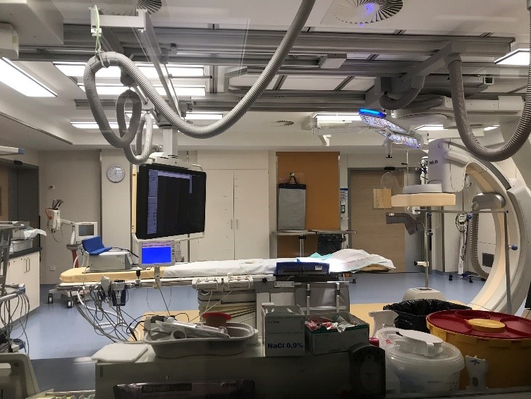
Cardiac catheterization (heart cath) is the insertion of a catheter into a chamber or vessel of the heart. This is done both for diagnostic and interventional purposes.
A common example of cardiac catheterization is coronary catheterization that involves catheterization of the coronary arteries for coronary artery disease and myocardial infarctions ("heart attacks"). Catheterization is most often performed in special laboratories with fluoroscopy and highly maneuverable tables. These "cath labs" are often equipped with cabinets of catheters, stents, balloons, etc. of various sizes to increase efficiency. Monitors show the fluoroscopy imaging, electrocardiogram (ECG), pressure waves, and more.
Coronary angiography is a diagnostic procedure that allows visualization of the coronary vessels. Fluoroscopy is used to visualize the lumens of the arteries as a 2-D projection. Should these arteries show narrowing or blockage, then techniques exist to open these arteries. Percutaneous coronary intervention is a blanket term that involves the use of mechanical stents, balloons, etc. to increase blood flow to previously blocked (or occluded) vessels.[citation needed]
Measuring pressures in the heart is also an important aspect of catheterization. The catheters are fluid filled conduits that can transmit pressures to outside the body to pressure transducers. This allows measuring pressure in any part of the heart that a catheter can be maneuvered into.[citation needed]
Measuring blood flow is also possible through several methods. Most commonly, flows are estimated using the Fick principle and thermodilution. These methods have drawbacks, but give invasive estimations of the cardiac output, which can be used to make clinical decisions (e.g., cardiogenic shock, heart failure) to improve the person's condition.[citation needed]
Cardiac catheterization can be used as part of a therapeutic regimen to improve outcomes for survivors of out-of-hospital cardiac arrest.
Cardiac catheterization often requires the use of fluoroscopy to visualize the path of the catheter as it enters the heart or as it enters the coronary arteries. The coronary arteries are known as "epicardial vessels" as they are located in the epicardium, the outermost layer of the heart. The use of fluoroscopy requires radiopaque contrast, which in rare cases can lead to contrast-induced kidney injury (see Contrast-induced nephropathy). People are constantly exposed to low doses of ionizing radiation during procedures. Ideal table positioning between the x-ray source and receiver, and radiation monitoring via thermoluminescent dosimetry, are two main ways of reducing a person's exposure to radiation. People with certain comorbidities (people who have more than one condition at the same time) have a higher risk of adverse events during the cardiac catheterization procedure. These comorbidity conditions include aortic aneurysm, aortic stenosis, extensive three-vessel coronary artery disease, diabetes, uncontrolled hypertension, obesity, chronic kidney disease, and unstable angina.
Left heart catheterization (LHC) is an ambiguous term and sometime clarification is required:[citation needed]
Cardiac catheterization
Cardiac catheterization (heart cath) is the insertion of a catheter into a chamber or vessel of the heart. This is done both for diagnostic and interventional purposes.
A common example of cardiac catheterization is coronary catheterization that involves catheterization of the coronary arteries for coronary artery disease and myocardial infarctions ("heart attacks"). Catheterization is most often performed in special laboratories with fluoroscopy and highly maneuverable tables. These "cath labs" are often equipped with cabinets of catheters, stents, balloons, etc. of various sizes to increase efficiency. Monitors show the fluoroscopy imaging, electrocardiogram (ECG), pressure waves, and more.
Coronary angiography is a diagnostic procedure that allows visualization of the coronary vessels. Fluoroscopy is used to visualize the lumens of the arteries as a 2-D projection. Should these arteries show narrowing or blockage, then techniques exist to open these arteries. Percutaneous coronary intervention is a blanket term that involves the use of mechanical stents, balloons, etc. to increase blood flow to previously blocked (or occluded) vessels.[citation needed]
Measuring pressures in the heart is also an important aspect of catheterization. The catheters are fluid filled conduits that can transmit pressures to outside the body to pressure transducers. This allows measuring pressure in any part of the heart that a catheter can be maneuvered into.[citation needed]
Measuring blood flow is also possible through several methods. Most commonly, flows are estimated using the Fick principle and thermodilution. These methods have drawbacks, but give invasive estimations of the cardiac output, which can be used to make clinical decisions (e.g., cardiogenic shock, heart failure) to improve the person's condition.[citation needed]
Cardiac catheterization can be used as part of a therapeutic regimen to improve outcomes for survivors of out-of-hospital cardiac arrest.
Cardiac catheterization often requires the use of fluoroscopy to visualize the path of the catheter as it enters the heart or as it enters the coronary arteries. The coronary arteries are known as "epicardial vessels" as they are located in the epicardium, the outermost layer of the heart. The use of fluoroscopy requires radiopaque contrast, which in rare cases can lead to contrast-induced kidney injury (see Contrast-induced nephropathy). People are constantly exposed to low doses of ionizing radiation during procedures. Ideal table positioning between the x-ray source and receiver, and radiation monitoring via thermoluminescent dosimetry, are two main ways of reducing a person's exposure to radiation. People with certain comorbidities (people who have more than one condition at the same time) have a higher risk of adverse events during the cardiac catheterization procedure. These comorbidity conditions include aortic aneurysm, aortic stenosis, extensive three-vessel coronary artery disease, diabetes, uncontrolled hypertension, obesity, chronic kidney disease, and unstable angina.
Left heart catheterization (LHC) is an ambiguous term and sometime clarification is required:[citation needed]
Recent media
Recent media