
Community hub
Recent from talks
Contribute something to knowledge base
Content stats: 0 posts, 0 articles, 1 media, 0 notes
Members stats: 0 subscribers, 0 contributors, 0 moderators, 0 supporters
Subscribers
Supporters
Contributors
Moderators
Hub AI
Cardiac muscle AI simulator
(@Cardiac muscle_simulator)
Hub AI
Cardiac muscle AI simulator
(@Cardiac muscle_simulator)
Cardiac muscle
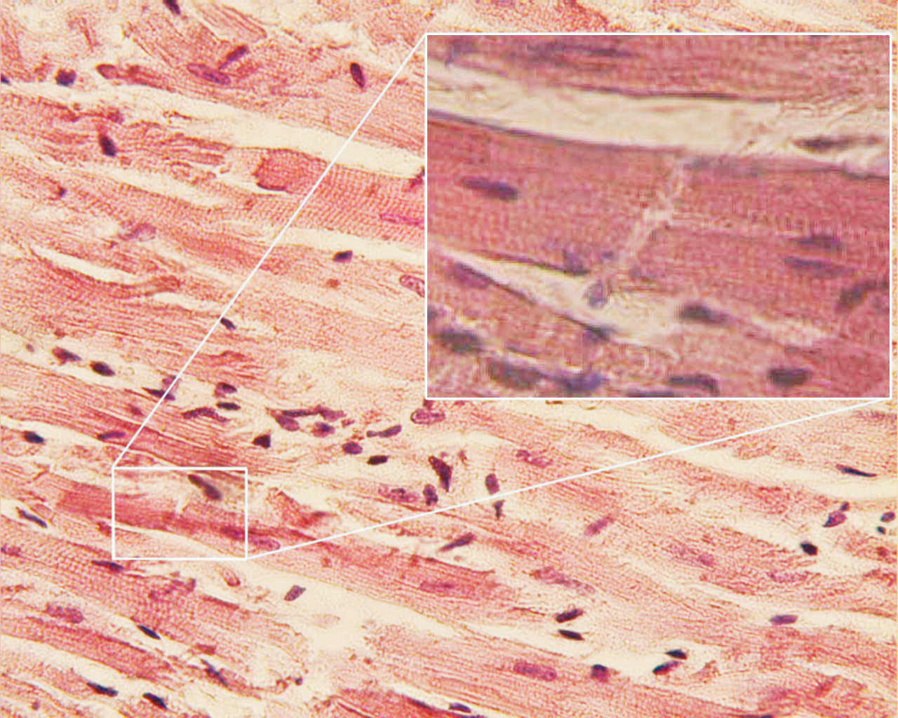
Cardiac muscle (also called heart muscle or myocardium) is one of three types of vertebrate muscle tissues, the others being skeletal muscle and smooth muscle. It is an involuntary, striated muscle that constitutes the main tissue of the wall of the heart. The cardiac muscle (myocardium) forms a thick middle layer between the outer layer of the heart wall (the pericardium) and the inner layer (the endocardium), with blood supplied via the coronary circulation. It is composed of individual cardiac muscle cells joined by intercalated discs, and encased by collagen fibers and other substances that form the extracellular matrix.
Cardiac muscle contracts in a similar manner to skeletal muscle, although with some important differences. Electrical stimulation in the form of a cardiac action potential triggers the release of calcium from the cell's internal calcium store, the sarcoplasmic reticulum. The rise in calcium causes the cell's myofilaments to slide past each other in a process called excitation-contraction coupling. Diseases of the heart muscle known as cardiomyopathies are of major importance. These include ischemic conditions caused by a restricted blood supply to the muscle such as angina, and myocardial infarction.
Cardiac muscle tissue or myocardium forms the bulk of the heart. The heart wall is a three-layered structure with a thick layer of myocardium sandwiched between the inner endocardium and the outer epicardium (also known as the visceral pericardium). The inner endocardium lines the cardiac chambers, covers the cardiac valves, and joins with the endothelium that lines the blood vessels that connect to the heart. On the outer aspect of the myocardium is the epicardium which forms part of the pericardial sac that surrounds, protects, and lubricates the heart.
Within the myocardium, there are several sheets of cardiac muscle cells or cardiomyocytes. The sheets of muscle that wrap around the left ventricle closest to the endocardium are oriented perpendicularly to those closest to the epicardium. When these sheets contract in a coordinated manner they allow the ventricle to squeeze in several directions simultaneously – longitudinally (becoming shorter from apex to base), radially (becoming narrower from side to side), and with a twisting motion (similar to wringing out a damp cloth) to squeeze the maximum possible amount of blood out of the heart with each heartbeat.
Contracting heart muscle uses a lot of energy, and therefore requires a constant flow of blood to provide oxygen and nutrients. Blood is brought to the myocardium by the coronary arteries. These originate from the aortic root and lie on the outer or epicardial surface of the heart. Blood is then drained away by the coronary veins into the right atrium.
Cardiac muscle cells (also called cardiomyocytes) are the contractile myocytes of the cardiac muscle. The cells are surrounded by an extracellular matrix produced by supporting fibroblast cells. Specialised modified cardiomyocytes known as pacemaker cells, set the rhythm of the heart contractions. The pacemaker cells are only weakly contractile without sarcomeres, and are connected to neighboring contractile cells via gap junctions. They are located in the sinoatrial node (the primary pacemaker) positioned on the wall of the right atrium, near the entrance of the superior vena cava. Other pacemaker cells are found in the atrioventricular node (secondary pacemaker).
Pacemaker cells carry the impulses that are responsible for the beating of the heart. They are distributed throughout the heart and are responsible for several functions. First, they are responsible for being able to spontaneously generate and send out electrical impulses. They also must be able to receive and respond to electrical impulses from the brain. Lastly, they must be able to transfer electrical impulses from cell to cell. Pacemaker cells in the sinoatrial node, and atrioventricular node are smaller and conduct at a relatively slow rate between the cells. Specialized conductive cells in the bundle of His, and the Purkinje fibers are larger in diameter and conduct signals at a fast rate.
The Purkinje fibers rapidly conduct electrical signals; coronary arteries to bring nutrients to the muscle cells, and veins and a capillary network to take away waste products.
Cardiac muscle
Cardiac muscle (also called heart muscle or myocardium) is one of three types of vertebrate muscle tissues, the others being skeletal muscle and smooth muscle. It is an involuntary, striated muscle that constitutes the main tissue of the wall of the heart. The cardiac muscle (myocardium) forms a thick middle layer between the outer layer of the heart wall (the pericardium) and the inner layer (the endocardium), with blood supplied via the coronary circulation. It is composed of individual cardiac muscle cells joined by intercalated discs, and encased by collagen fibers and other substances that form the extracellular matrix.
Cardiac muscle contracts in a similar manner to skeletal muscle, although with some important differences. Electrical stimulation in the form of a cardiac action potential triggers the release of calcium from the cell's internal calcium store, the sarcoplasmic reticulum. The rise in calcium causes the cell's myofilaments to slide past each other in a process called excitation-contraction coupling. Diseases of the heart muscle known as cardiomyopathies are of major importance. These include ischemic conditions caused by a restricted blood supply to the muscle such as angina, and myocardial infarction.
Cardiac muscle tissue or myocardium forms the bulk of the heart. The heart wall is a three-layered structure with a thick layer of myocardium sandwiched between the inner endocardium and the outer epicardium (also known as the visceral pericardium). The inner endocardium lines the cardiac chambers, covers the cardiac valves, and joins with the endothelium that lines the blood vessels that connect to the heart. On the outer aspect of the myocardium is the epicardium which forms part of the pericardial sac that surrounds, protects, and lubricates the heart.
Within the myocardium, there are several sheets of cardiac muscle cells or cardiomyocytes. The sheets of muscle that wrap around the left ventricle closest to the endocardium are oriented perpendicularly to those closest to the epicardium. When these sheets contract in a coordinated manner they allow the ventricle to squeeze in several directions simultaneously – longitudinally (becoming shorter from apex to base), radially (becoming narrower from side to side), and with a twisting motion (similar to wringing out a damp cloth) to squeeze the maximum possible amount of blood out of the heart with each heartbeat.
Contracting heart muscle uses a lot of energy, and therefore requires a constant flow of blood to provide oxygen and nutrients. Blood is brought to the myocardium by the coronary arteries. These originate from the aortic root and lie on the outer or epicardial surface of the heart. Blood is then drained away by the coronary veins into the right atrium.
Cardiac muscle cells (also called cardiomyocytes) are the contractile myocytes of the cardiac muscle. The cells are surrounded by an extracellular matrix produced by supporting fibroblast cells. Specialised modified cardiomyocytes known as pacemaker cells, set the rhythm of the heart contractions. The pacemaker cells are only weakly contractile without sarcomeres, and are connected to neighboring contractile cells via gap junctions. They are located in the sinoatrial node (the primary pacemaker) positioned on the wall of the right atrium, near the entrance of the superior vena cava. Other pacemaker cells are found in the atrioventricular node (secondary pacemaker).
Pacemaker cells carry the impulses that are responsible for the beating of the heart. They are distributed throughout the heart and are responsible for several functions. First, they are responsible for being able to spontaneously generate and send out electrical impulses. They also must be able to receive and respond to electrical impulses from the brain. Lastly, they must be able to transfer electrical impulses from cell to cell. Pacemaker cells in the sinoatrial node, and atrioventricular node are smaller and conduct at a relatively slow rate between the cells. Specialized conductive cells in the bundle of His, and the Purkinje fibers are larger in diameter and conduct signals at a fast rate.
The Purkinje fibers rapidly conduct electrical signals; coronary arteries to bring nutrients to the muscle cells, and veins and a capillary network to take away waste products.
Recent media
Recent media