Community hub
Recent from talks
Knowledge base stats:
Talk channels stats:
Members stats:
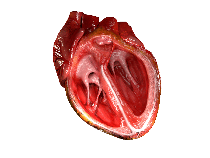
Heart valve
A heart valve (cardiac valve) is a biological one-way valve that allows blood to flow in one direction through the chambers of the heart. A mammalian heart usually has four valves. Together, the valves determine the direction of blood flow through the heart. Heart valves are opened or closed by a difference in blood pressure on each side.
The mammalian heart has two atrioventricular valves separating the upper atria from the lower ventricles: the mitral valve in the left heart, and the tricuspid valve in the right heart. The two semilunar valves are at the entrance of the arteries leaving the heart. These are the aortic valve at the aorta, and the pulmonary valve at the pulmonary artery.
The heart also has a coronary sinus valve and an inferior vena cava valve, not discussed here.
The heart valves and the chambers are lined with endocardium. Heart valves separate the atria from the ventricles, or the ventricles from a blood vessel. Heart valves are situated around the fibrous rings of the cardiac skeleton. The valves incorporate flaps called leaflets or cusps, similar to a duckbill valve or flutter valve, which are pushed open to allow blood flow and which then close together to seal and prevent backflow. The mitral valve has two cusps, whereas the others have three. There are nodules at the tips of the cusps that make the seal tighter.
The pulmonary valve has left, right, and anterior cusps. The aortic valve has left, right, and posterior cusps. The tricuspid valve has anterior, posterior, and septal cusps; and the mitral valve has just anterior and posterior cusps.
The valves of the human heart can be grouped in two sets:
The atrioventricular valves are the mitral valve, and the tricuspid valve, which are situated between the atria and the ventricles, and prevent backflow from the ventricles into the atria during systole. They are anchored to the walls of the ventricles by chordae tendineae, which prevent them from inverting.
The chordae tendineae are attached to papillary muscles that cause tension to better hold the valve. Together, the papillary muscles and the chordae tendineae are known as the subvalvular apparatus. The function of the subvalvular apparatus is to keep the valves from prolapsing into the atria when they close. The subvalvular apparatus has no effect on the opening and closure of the valves, however, which is caused entirely by the pressure gradient across the valve. The peculiar insertion of chords on the leaflet free margin, however, provides systolic stress sharing between chords according to their different thickness.
Hub AI
Heart valve AI simulator
(@Heart valve_simulator)
Heart valve
A heart valve (cardiac valve) is a biological one-way valve that allows blood to flow in one direction through the chambers of the heart. A mammalian heart usually has four valves. Together, the valves determine the direction of blood flow through the heart. Heart valves are opened or closed by a difference in blood pressure on each side.
The mammalian heart has two atrioventricular valves separating the upper atria from the lower ventricles: the mitral valve in the left heart, and the tricuspid valve in the right heart. The two semilunar valves are at the entrance of the arteries leaving the heart. These are the aortic valve at the aorta, and the pulmonary valve at the pulmonary artery.
The heart also has a coronary sinus valve and an inferior vena cava valve, not discussed here.
The heart valves and the chambers are lined with endocardium. Heart valves separate the atria from the ventricles, or the ventricles from a blood vessel. Heart valves are situated around the fibrous rings of the cardiac skeleton. The valves incorporate flaps called leaflets or cusps, similar to a duckbill valve or flutter valve, which are pushed open to allow blood flow and which then close together to seal and prevent backflow. The mitral valve has two cusps, whereas the others have three. There are nodules at the tips of the cusps that make the seal tighter.
The pulmonary valve has left, right, and anterior cusps. The aortic valve has left, right, and posterior cusps. The tricuspid valve has anterior, posterior, and septal cusps; and the mitral valve has just anterior and posterior cusps.
The valves of the human heart can be grouped in two sets:
The atrioventricular valves are the mitral valve, and the tricuspid valve, which are situated between the atria and the ventricles, and prevent backflow from the ventricles into the atria during systole. They are anchored to the walls of the ventricles by chordae tendineae, which prevent them from inverting.
The chordae tendineae are attached to papillary muscles that cause tension to better hold the valve. Together, the papillary muscles and the chordae tendineae are known as the subvalvular apparatus. The function of the subvalvular apparatus is to keep the valves from prolapsing into the atria when they close. The subvalvular apparatus has no effect on the opening and closure of the valves, however, which is caused entirely by the pressure gradient across the valve. The peculiar insertion of chords on the leaflet free margin, however, provides systolic stress sharing between chords according to their different thickness.