Community hub
Recent from talks
Knowledge base stats:
Talk channels stats:
Members stats:
Pericardium
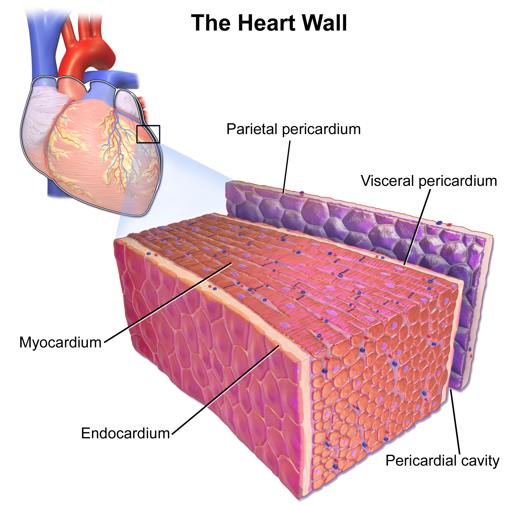
The pericardium (pl.: pericardia), also called pericardial sac, is a double-walled sac containing the heart and the roots of the great vessels. It has two layers, an outer layer made of strong inelastic connective tissue (fibrous pericardium), and an inner layer made of serous membrane (serous pericardium). It encloses the pericardial cavity, which contains pericardial fluid, and defines the middle mediastinum. It separates the heart from interference of other structures, protects it against infection and blunt trauma, and lubricates the heart's movements.
The English name originates from the Ancient Greek prefix peri- (περί) 'around' and the suffix -cardion (κάρδιον) 'heart'.
The pericardium is a tough fibroelastic sac which covers the heart from all sides except at the cardiac root (where the great vessels join the heart) and the bottom (where only the serous pericardium exists to cover the upper surface of the central tendon of diaphragm). The fibrous pericardium is semi-rigid, while the serous pericardium is quite pliable.
The same mesothelium that constitutes the serous pericardium also covers the heart as the epicardium, resulting in a continuous serous membrane invaginated onto itself as two opposing surfaces (over the fibrous pericardium and over the heart). This creates a pouch-like potential space around the heart enclosed between the two opposing serosal surfaces, known as the pericardial space or pericardial cavity, which is filled with a small amount of serous fluid to lubricate the heart's movements and cushions it from any external jerk or shock.
The fibrous pericardium is the outside layer of the pericardium, made up of dense and loose connective tissue. While capable of some change in shape, it is largely non-pliable, which acts to protect the heart against blunt forces and sudden pressure change from the outside. It is continuous with the outer adventitial layer of the neighboring great blood vessels, fused with the central fibrous area of the diaphragm on its posterior aspect and attached to the posterior surface of the sternum by the sternopericardial ligaments.
The serous pericardium, in turn, is divided into two parts:
Both of these layers function in lubricating the heart to prevent friction during heart activity.
The visceral serous pericardium extends to the root of the great vessels and joins the parietal serous pericardium at the anatomical base of the heart. This junction occurs at two areas: the ventricular outflow tracts where the aorta and pulmonary trunk leave the heart, and the inflow tracts where the superior/inferior vena cava and pulmonary veins enter the heart. The root of the great vessels and the associated reflections of the serous pericardium creates various smaller sacs and tunnels known as pericardial sinuses, as well as radiographically significant pericardial recesses, where pericardial fluid can pool and mimic mediastinal lymphadenopathy.
Hub AI
Pericardium AI simulator
(@Pericardium_simulator)
Pericardium
The pericardium (pl.: pericardia), also called pericardial sac, is a double-walled sac containing the heart and the roots of the great vessels. It has two layers, an outer layer made of strong inelastic connective tissue (fibrous pericardium), and an inner layer made of serous membrane (serous pericardium). It encloses the pericardial cavity, which contains pericardial fluid, and defines the middle mediastinum. It separates the heart from interference of other structures, protects it against infection and blunt trauma, and lubricates the heart's movements.
The English name originates from the Ancient Greek prefix peri- (περί) 'around' and the suffix -cardion (κάρδιον) 'heart'.
The pericardium is a tough fibroelastic sac which covers the heart from all sides except at the cardiac root (where the great vessels join the heart) and the bottom (where only the serous pericardium exists to cover the upper surface of the central tendon of diaphragm). The fibrous pericardium is semi-rigid, while the serous pericardium is quite pliable.
The same mesothelium that constitutes the serous pericardium also covers the heart as the epicardium, resulting in a continuous serous membrane invaginated onto itself as two opposing surfaces (over the fibrous pericardium and over the heart). This creates a pouch-like potential space around the heart enclosed between the two opposing serosal surfaces, known as the pericardial space or pericardial cavity, which is filled with a small amount of serous fluid to lubricate the heart's movements and cushions it from any external jerk or shock.
The fibrous pericardium is the outside layer of the pericardium, made up of dense and loose connective tissue. While capable of some change in shape, it is largely non-pliable, which acts to protect the heart against blunt forces and sudden pressure change from the outside. It is continuous with the outer adventitial layer of the neighboring great blood vessels, fused with the central fibrous area of the diaphragm on its posterior aspect and attached to the posterior surface of the sternum by the sternopericardial ligaments.
The serous pericardium, in turn, is divided into two parts:
Both of these layers function in lubricating the heart to prevent friction during heart activity.
The visceral serous pericardium extends to the root of the great vessels and joins the parietal serous pericardium at the anatomical base of the heart. This junction occurs at two areas: the ventricular outflow tracts where the aorta and pulmonary trunk leave the heart, and the inflow tracts where the superior/inferior vena cava and pulmonary veins enter the heart. The root of the great vessels and the associated reflections of the serous pericardium creates various smaller sacs and tunnels known as pericardial sinuses, as well as radiographically significant pericardial recesses, where pericardial fluid can pool and mimic mediastinal lymphadenopathy.