Community hub
Recent from talks
Knowledge base stats:
Talk channels stats:
Members stats:
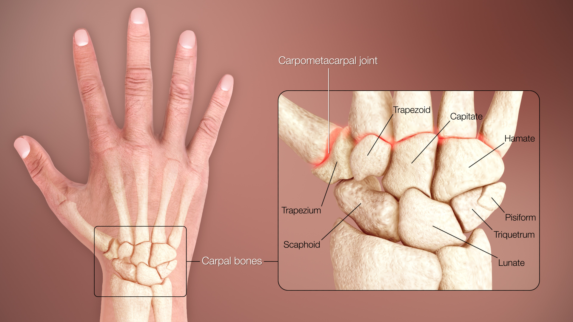
Carpal bones
The carpal bones are the eight small bones that make up the wrist (carpus) that connects the hand to the forearm. The terms "carpus" and "carpal" are derived from the Latin carpus and the Greek καρπός (karpós), meaning "wrist". In human anatomy, the main role of the carpal bones is to articulate with the radial and ulnar heads to form a highly mobile condyloid joint (i.e. wrist joint), to provide attachments for thenar and hypothenar muscles, and to form part of the rigid carpal tunnel which allows the median nerve and tendons of the anterior forearm muscles to be transmitted to the hand and fingers.
In tetrapods, the carpus is the sole cluster of bones in the wrist between the radius and ulna and the metacarpus. The bones of the carpus do not belong to individual fingers (or toes in quadrupeds), whereas those of the metacarpus do. The corresponding part of the foot is the tarsus. The carpal bones allow the wrist to move and rotate vertically.
The eight carpal bones may be conceptually organized as either two transverse rows, or three longitudinal columns.
When considered as paired rows, each row forms an arch which is convex proximally and concave distally. On the palmar side, the carpus is concave and forms the carpal tunnel, which is covered by the flexor retinaculum. The proximal row comprises the scaphoid, lunate, triquetral, and pisiform bones which articulate with the surfaces of the radius and distal carpal row, and thus constantly adapts to these mobile surfaces. Within the proximal row, each carpal bone has slight independent mobility. For example, the scaphoid contributes to midcarpal stability by articulating distally with the trapezium and the trapezoid. In contrast, the distal row is more rigid as its transverse arch moves with the metacarpals.
Biomechanically and clinically, the carpal bones are better conceptualized as three longitudinal columns:
In this context the pisiform is regarded as a sesamoid bone embedded in the tendon of the flexor carpi ulnaris. The ulnar column leaves a gap between the ulna and the triquetrum, and therefore, only the radial or scaphoid and central or capitate columns articulate with the radius. The wrist is more stable in flexion than in extension, mainly because of the strength of various capsules and ligaments than the interlocking parts of the skeleton.
Almost all carpals (except the pisiform) have six surfaces. Of these the palmar or anterior and the dorsal or posterior surfaces are rough, for ligamentous attachment; the dorsal surfaces being the broader, except in the lunate.
The superior or proximal, and inferior or distal surfaces are articular, the superior generally convex, the inferior concave; the medial and lateral surfaces are also articular where they are in contact with contiguous bones, otherwise they are rough and tuberculated.
Hub AI
Carpal bones AI simulator
(@Carpal bones_simulator)
Carpal bones
The carpal bones are the eight small bones that make up the wrist (carpus) that connects the hand to the forearm. The terms "carpus" and "carpal" are derived from the Latin carpus and the Greek καρπός (karpós), meaning "wrist". In human anatomy, the main role of the carpal bones is to articulate with the radial and ulnar heads to form a highly mobile condyloid joint (i.e. wrist joint), to provide attachments for thenar and hypothenar muscles, and to form part of the rigid carpal tunnel which allows the median nerve and tendons of the anterior forearm muscles to be transmitted to the hand and fingers.
In tetrapods, the carpus is the sole cluster of bones in the wrist between the radius and ulna and the metacarpus. The bones of the carpus do not belong to individual fingers (or toes in quadrupeds), whereas those of the metacarpus do. The corresponding part of the foot is the tarsus. The carpal bones allow the wrist to move and rotate vertically.
The eight carpal bones may be conceptually organized as either two transverse rows, or three longitudinal columns.
When considered as paired rows, each row forms an arch which is convex proximally and concave distally. On the palmar side, the carpus is concave and forms the carpal tunnel, which is covered by the flexor retinaculum. The proximal row comprises the scaphoid, lunate, triquetral, and pisiform bones which articulate with the surfaces of the radius and distal carpal row, and thus constantly adapts to these mobile surfaces. Within the proximal row, each carpal bone has slight independent mobility. For example, the scaphoid contributes to midcarpal stability by articulating distally with the trapezium and the trapezoid. In contrast, the distal row is more rigid as its transverse arch moves with the metacarpals.
Biomechanically and clinically, the carpal bones are better conceptualized as three longitudinal columns:
In this context the pisiform is regarded as a sesamoid bone embedded in the tendon of the flexor carpi ulnaris. The ulnar column leaves a gap between the ulna and the triquetrum, and therefore, only the radial or scaphoid and central or capitate columns articulate with the radius. The wrist is more stable in flexion than in extension, mainly because of the strength of various capsules and ligaments than the interlocking parts of the skeleton.
Almost all carpals (except the pisiform) have six surfaces. Of these the palmar or anterior and the dorsal or posterior surfaces are rough, for ligamentous attachment; the dorsal surfaces being the broader, except in the lunate.
The superior or proximal, and inferior or distal surfaces are articular, the superior generally convex, the inferior concave; the medial and lateral surfaces are also articular where they are in contact with contiguous bones, otherwise they are rough and tuberculated.