
Recent from talks
Cerebral angiography
Knowledge base stats:
Talk channels stats:
Members stats:
Cerebral angiography
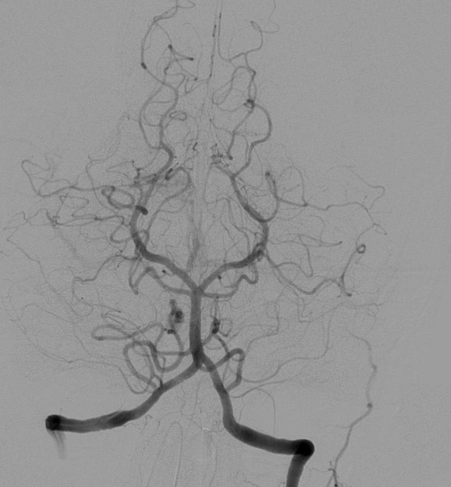
Cerebral angiography is a form of angiography which provides images of blood vessels in and around the brain, thereby allowing detection of abnormalities such as arteriovenous malformations and aneurysms. It was pioneered in 1927 by the Portuguese neurologist Egas Moniz at the University of Lisbon, who also helped develop thorotrast for use in the procedure.
Typically a catheter is inserted into a large artery (such as the femoral artery) and threaded through the circulatory system to the carotid artery, where a contrast agent is injected. A series of radiographs are taken as the contrast agent spreads through the brain's arterial system, then a second series as it reaches the venous system.
For some applications,[citation needed] cerebral angiography may yield better images than less invasive methods such as computed tomography angiography and magnetic resonance angiography. In addition, cerebral angiography allows certain treatments to be performed immediately, based on its findings. In recent decades, cerebral angiography has so assumed a therapeutic connotation thanks to the elaboration of endovascular therapeutic techniques. Embolization (a minimally invasive surgical technique) over time has played an increasingly significant role in the multimodal treatment of cerebral MAVs, facilitating subsequent microsurgical or radiosurgical treatment. Another type of treatment possible by angiography (if the images reveal an aneurysm) is the introduction of metal coils through the catheter already in place and maneuvered to the site of aneurysm; over time these coils encourage formation of connective tissue at the site, strengthening the vessel walls.
Prior to the advent of modern neuroimaging techniques such as MRI and CT in the mid-1970s, cerebral angiographies were frequently employed as a tool to infer the existence and location of certain kinds of lesions and hematomas by looking for secondary vascular displacement caused by the mass effect related to these medical conditions. This use of angiography as an indirect assessment tool is nowadays obsolete as modern non-invasive diagnostic methods are available to image many kinds of primary intracranial abnormalities directly. It is still widely used however for evaluating various types of vascular pathologies within the skull.
Cerebral angiography is used for diagnosis but may be followed by treatment procedures in the same setting. Cerebral angiography is used to image various intracranial (within the head) or extracranial (outside the head) diseases.
Intracranial diseases are: non-traumatic subarachnoid haemorrhage, non-traumatic intracerebral haemorrhage, intracranial aneurysm, stroke, cerebral vasospasm, cerebral arteriovenous malformation (for Spetzler-Martin grading and plan for intervention), dural arteriovenous fistula, embolisation of brain tumours such as meningioma, cavernous sinus haemangioma, for Wada test, and to obtain haemodynamics of cerebral blood flow such as cross flow, circulation time, and collateral flow.
Extracranial diseases are: Subclavian steal syndrome, rupture of the carotid artery, carotid artery stenosis, cervical spine trauma, epistaxis (nose bleeding) and plan for embolisation of juvenile nasopharyngeal angiofibroma before operation.
Although computed tomography angiography (CTA) and Magnetic resonance angiography (MRA) has been used widely in evaluation of intracranial disease, cerebral angiography provides higher resolution on the conditions of blood vessel lumens and vasculature. Cerebral angiography is also the standard of detecting intracranial aneurysm and evaluating the feasibility of endovascular coiling. Performing a cerebral angiogram by gaining access through the femoral artery or radial artery is feasible in order to treat cerebral aneurysms with a number of devices
Hub AI
Cerebral angiography AI simulator
(@Cerebral angiography_simulator)
Cerebral angiography
Cerebral angiography is a form of angiography which provides images of blood vessels in and around the brain, thereby allowing detection of abnormalities such as arteriovenous malformations and aneurysms. It was pioneered in 1927 by the Portuguese neurologist Egas Moniz at the University of Lisbon, who also helped develop thorotrast for use in the procedure.
Typically a catheter is inserted into a large artery (such as the femoral artery) and threaded through the circulatory system to the carotid artery, where a contrast agent is injected. A series of radiographs are taken as the contrast agent spreads through the brain's arterial system, then a second series as it reaches the venous system.
For some applications,[citation needed] cerebral angiography may yield better images than less invasive methods such as computed tomography angiography and magnetic resonance angiography. In addition, cerebral angiography allows certain treatments to be performed immediately, based on its findings. In recent decades, cerebral angiography has so assumed a therapeutic connotation thanks to the elaboration of endovascular therapeutic techniques. Embolization (a minimally invasive surgical technique) over time has played an increasingly significant role in the multimodal treatment of cerebral MAVs, facilitating subsequent microsurgical or radiosurgical treatment. Another type of treatment possible by angiography (if the images reveal an aneurysm) is the introduction of metal coils through the catheter already in place and maneuvered to the site of aneurysm; over time these coils encourage formation of connective tissue at the site, strengthening the vessel walls.
Prior to the advent of modern neuroimaging techniques such as MRI and CT in the mid-1970s, cerebral angiographies were frequently employed as a tool to infer the existence and location of certain kinds of lesions and hematomas by looking for secondary vascular displacement caused by the mass effect related to these medical conditions. This use of angiography as an indirect assessment tool is nowadays obsolete as modern non-invasive diagnostic methods are available to image many kinds of primary intracranial abnormalities directly. It is still widely used however for evaluating various types of vascular pathologies within the skull.
Cerebral angiography is used for diagnosis but may be followed by treatment procedures in the same setting. Cerebral angiography is used to image various intracranial (within the head) or extracranial (outside the head) diseases.
Intracranial diseases are: non-traumatic subarachnoid haemorrhage, non-traumatic intracerebral haemorrhage, intracranial aneurysm, stroke, cerebral vasospasm, cerebral arteriovenous malformation (for Spetzler-Martin grading and plan for intervention), dural arteriovenous fistula, embolisation of brain tumours such as meningioma, cavernous sinus haemangioma, for Wada test, and to obtain haemodynamics of cerebral blood flow such as cross flow, circulation time, and collateral flow.
Extracranial diseases are: Subclavian steal syndrome, rupture of the carotid artery, carotid artery stenosis, cervical spine trauma, epistaxis (nose bleeding) and plan for embolisation of juvenile nasopharyngeal angiofibroma before operation.
Although computed tomography angiography (CTA) and Magnetic resonance angiography (MRA) has been used widely in evaluation of intracranial disease, cerebral angiography provides higher resolution on the conditions of blood vessel lumens and vasculature. Cerebral angiography is also the standard of detecting intracranial aneurysm and evaluating the feasibility of endovascular coiling. Performing a cerebral angiogram by gaining access through the femoral artery or radial artery is feasible in order to treat cerebral aneurysms with a number of devices
Recent media