
Recent from talks
Dual-energy X-ray absorptiometry
Knowledge base stats:
Talk channels stats:
Members stats:
Dual-energy X-ray absorptiometry
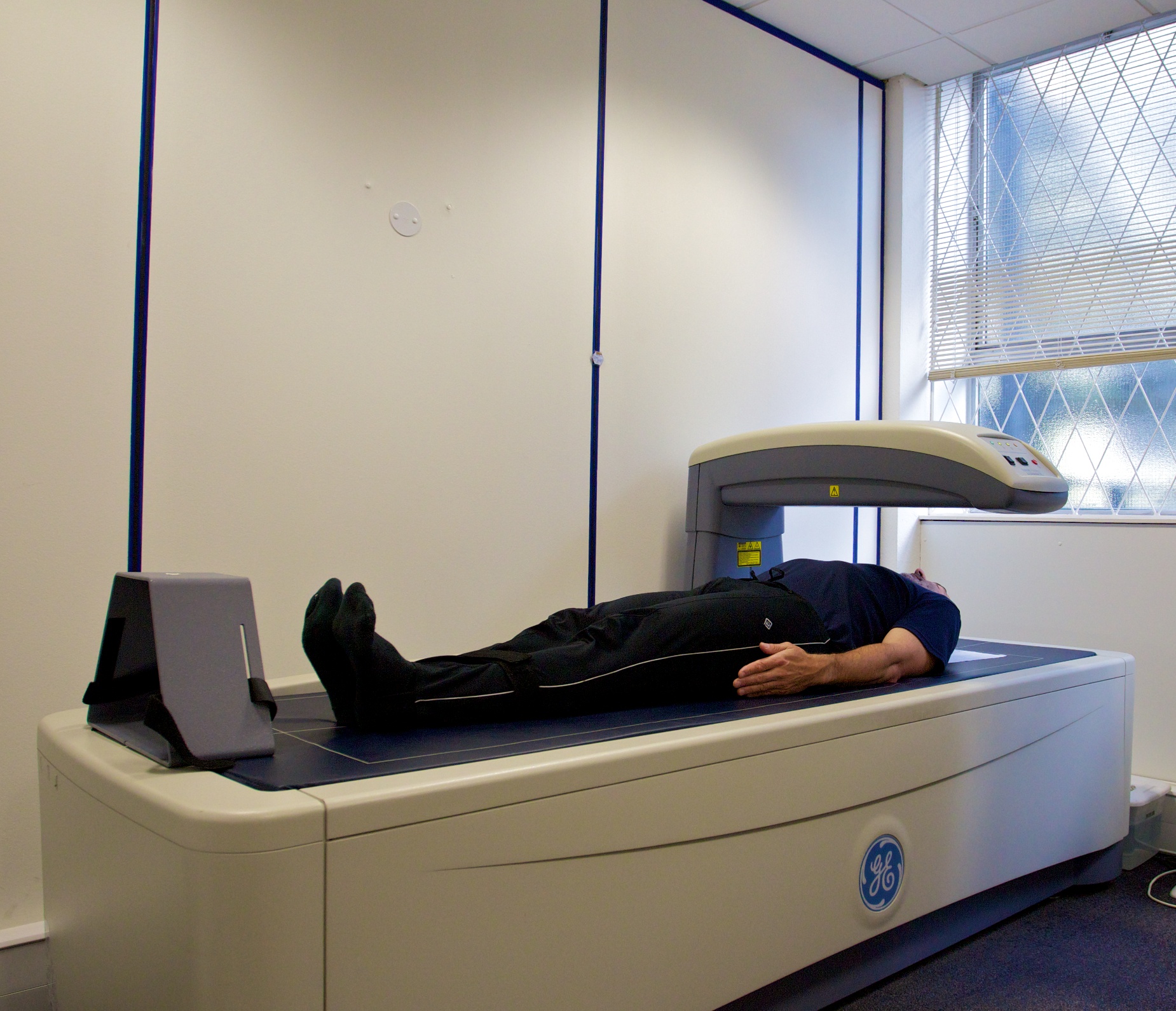
Dual-energy X-ray absorptiometry (DXA or DEXA; also BMD test, bone density test, bone densitometry, p-DEXA) is a means of measuring bone mineral density (BMD) with spectral imaging. Two X-ray beams, with different energy levels, are aimed at the patient's bones. When soft tissue absorption is subtracted, the bone mineral density (BMD) can be determined from the absorption of each beam by bone. Dual-energy X-ray absorptiometry is the most widely used and most thoroughly studied bone density measurement technology.
The DXA scan is typically used to diagnose and follow osteoporosis, as contrasted to the nuclear bone scan, which is sensitive to certain metabolic diseases of bones in which bones are trying to heal from infections, fractures, or tumors. It is also sometimes used to assess body composition.
Soft tissue and bone have different attenuation coefficients to X-rays. A single X-ray beam passing through the body is attenuated by both soft tissue and bone, and it is not possible to determine from a single beam how much attenuation is attributable to the bone. However, attenuation coefficients vary with the energy of the X-rays, and, crucially, the ratio of the attenuation coefficients also varies. DXA uses two energies of X-ray. The difference in total absorption between the two can be used, by suitable weighting, to subtract out the absorption by soft tissue, leaving just the absorption by bone, which is related to bone density.
One type of DXA scanner uses a cerium filter with a tube voltage of 80 kV, resulting in effective photon energies of about 40 and 70 keV. Another type of DXA scanner uses a samarium filter with a tube voltage of 100 kV, which produces effective energies of 47 and 80 keV. Also, the tube voltage can be continuously switched between a low (for example 70 kV) and high (for example 140 kV) value in synchronism with the frequency of the electrical mains, resulting in effective energies alternating between 45 and 100 keV.
The combination of dual X-ray absorptiometry and laser uses the laser to measure the thickness of the region scanned, allowing for varying proportions of lean soft tissue and adipose tissue within the soft tissue to be controlled for and improving the accuracy.
The U.S. Preventive Services Task Force recommends that women over the age of 65 should get a DXA scan. The age when men should be tested is uncertain, but some sources recommend age 70. At-risk women should consider getting a scan when their risk is equal to that of a normal 65-year-old woman.
A person's risk can be estimated with the University of Sheffield's FRAX calculator, which takes into account prior fragility fracture, use of glucocorticoids, heavy smoking, excess alcohol intake, rheumatoid arthritis, history of parental hip fracture, chronic renal and liver disease, chronic respiratory disease, long-term use of phenobarbital or phenytoin, celiac disease, inflammatory bowel disease, and other clinical risk factors.
The World Health Organization has defined the following categories based on bone density in white women:
Hub AI
Dual-energy X-ray absorptiometry AI simulator
(@Dual-energy X-ray absorptiometry_simulator)
Dual-energy X-ray absorptiometry
Dual-energy X-ray absorptiometry (DXA or DEXA; also BMD test, bone density test, bone densitometry, p-DEXA) is a means of measuring bone mineral density (BMD) with spectral imaging. Two X-ray beams, with different energy levels, are aimed at the patient's bones. When soft tissue absorption is subtracted, the bone mineral density (BMD) can be determined from the absorption of each beam by bone. Dual-energy X-ray absorptiometry is the most widely used and most thoroughly studied bone density measurement technology.
The DXA scan is typically used to diagnose and follow osteoporosis, as contrasted to the nuclear bone scan, which is sensitive to certain metabolic diseases of bones in which bones are trying to heal from infections, fractures, or tumors. It is also sometimes used to assess body composition.
Soft tissue and bone have different attenuation coefficients to X-rays. A single X-ray beam passing through the body is attenuated by both soft tissue and bone, and it is not possible to determine from a single beam how much attenuation is attributable to the bone. However, attenuation coefficients vary with the energy of the X-rays, and, crucially, the ratio of the attenuation coefficients also varies. DXA uses two energies of X-ray. The difference in total absorption between the two can be used, by suitable weighting, to subtract out the absorption by soft tissue, leaving just the absorption by bone, which is related to bone density.
One type of DXA scanner uses a cerium filter with a tube voltage of 80 kV, resulting in effective photon energies of about 40 and 70 keV. Another type of DXA scanner uses a samarium filter with a tube voltage of 100 kV, which produces effective energies of 47 and 80 keV. Also, the tube voltage can be continuously switched between a low (for example 70 kV) and high (for example 140 kV) value in synchronism with the frequency of the electrical mains, resulting in effective energies alternating between 45 and 100 keV.
The combination of dual X-ray absorptiometry and laser uses the laser to measure the thickness of the region scanned, allowing for varying proportions of lean soft tissue and adipose tissue within the soft tissue to be controlled for and improving the accuracy.
The U.S. Preventive Services Task Force recommends that women over the age of 65 should get a DXA scan. The age when men should be tested is uncertain, but some sources recommend age 70. At-risk women should consider getting a scan when their risk is equal to that of a normal 65-year-old woman.
A person's risk can be estimated with the University of Sheffield's FRAX calculator, which takes into account prior fragility fracture, use of glucocorticoids, heavy smoking, excess alcohol intake, rheumatoid arthritis, history of parental hip fracture, chronic renal and liver disease, chronic respiratory disease, long-term use of phenobarbital or phenytoin, celiac disease, inflammatory bowel disease, and other clinical risk factors.
The World Health Organization has defined the following categories based on bone density in white women:
Recent media