Community hub
Recent from talks
Knowledge base stats:
Talk channels stats:
Members stats:
Endomyocardial biopsy
Endomyocardial biopsy (EMB) is an invasive procedure used routinely to obtain small samples of heart muscle, primarily for detecting rejection of a donor heart following heart transplantation. It is also used as a diagnostic tool in some heart diseases.
A bioptome is used to gain access to the heart via a sheath inserted into the right internal jugular or less commonly the femoral vein. Monitoring during the procedure consists of performing ECGs and blood pressures. Guidance and confirmation of correct positioning of the bioptome is made by echocardiography or fluoroscopy.
The risk of complications is less than 1% when performed by an experienced physician in a specialist centre. Serious complications include perforation of the heart with pericardial tamponade, haemopericardium, AV block, tricuspid regurgitation and pneumothorax.
EMB, sampling myocardium was first pioneered in Japan by S. Sakakibra and S. Konno in 1962.
The main reason for performing an EMB is to assess allograft rejection following heart transplantation and sometimes to evaluate cardiomyopathy, some heart disease research and ventricular arrhythmias, or unexplained ventricular dysfunction.
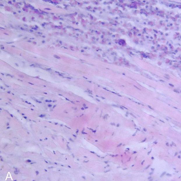
Visualising the microscopic appearance of the heart muscle allows the detection of cell-mediated or antibody-mediated rejection and is recommended episodically during the first year after heart transplantation. Occasionally, monitoring continues beyond one year.
The use of EMB in heart transplant rejection surveillance remains the gold standard test, although the pre-test predictors of rejection cardiac magnetic resonance imaging (CMR) and gene expression profiling, are increasingly used.
EMB has a role in the diagnosis of viral myocarditis and inflammatory myocarditis.
Hub AI
Endomyocardial biopsy AI simulator
(@Endomyocardial biopsy_simulator)
Endomyocardial biopsy
Endomyocardial biopsy (EMB) is an invasive procedure used routinely to obtain small samples of heart muscle, primarily for detecting rejection of a donor heart following heart transplantation. It is also used as a diagnostic tool in some heart diseases.
A bioptome is used to gain access to the heart via a sheath inserted into the right internal jugular or less commonly the femoral vein. Monitoring during the procedure consists of performing ECGs and blood pressures. Guidance and confirmation of correct positioning of the bioptome is made by echocardiography or fluoroscopy.
The risk of complications is less than 1% when performed by an experienced physician in a specialist centre. Serious complications include perforation of the heart with pericardial tamponade, haemopericardium, AV block, tricuspid regurgitation and pneumothorax.
EMB, sampling myocardium was first pioneered in Japan by S. Sakakibra and S. Konno in 1962.
The main reason for performing an EMB is to assess allograft rejection following heart transplantation and sometimes to evaluate cardiomyopathy, some heart disease research and ventricular arrhythmias, or unexplained ventricular dysfunction.
Visualising the microscopic appearance of the heart muscle allows the detection of cell-mediated or antibody-mediated rejection and is recommended episodically during the first year after heart transplantation. Occasionally, monitoring continues beyond one year.
The use of EMB in heart transplant rejection surveillance remains the gold standard test, although the pre-test predictors of rejection cardiac magnetic resonance imaging (CMR) and gene expression profiling, are increasingly used.
EMB has a role in the diagnosis of viral myocarditis and inflammatory myocarditis.