Recent from talks
Fetal membranes
Knowledge base stats:
Talk channels stats:
Members stats:
Fetal membranes
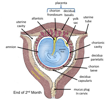
The fetal membranes are the four extraembryonic membranes, associated with the developing embryo, and fetus in humans and other mammals. They are the amnion, chorion, allantois, and yolk sac. The amnion and the chorion are the chorioamniotic membranes that make up the amniotic sac which surrounds and protects the embryo. The fetal membranes are four of six accessory organs developed by the conceptus that are not part of the embryo itself, the other two are the placenta, and the umbilical cord.
The fetal membranes surround the developing embryo and form the fetal-maternal interface. The fetal membranes are derived from the trophoblast layer (outer layer of cells) of the implanting blastocyst. The trophoblast layer differentiates into amnion and the chorion, which then comprise the fetal membranes. The amnion is the innermost layer and, therefore, contacts the amniotic fluid, the fetus and the umbilical cord. The internal pressure of the amniotic fluid causes the amnion to be passively attached to the chorion. The chorion functions to separate the amnion from the maternal decidua and uterus. The placenta develops from the chorion of the embryo and the uterine tissue of the mother.
Initially, the amnion is separated from the chorion by chorionic fluid. The fusion of the amnion and chorion is completed in the human at the 12th week of development.
From inside to outside, the fetal membranes consist of amnion and chorion. In addition, parts of decidua are often attached to the outside of the chorion.
The amnion is avascular, meaning it does not contain its own blood vessels. Therefore, it must obtain necessary nutrients and oxygen from nearby chorionic and amniotic fluid, and fetal surface vessels. The amnion is characterised by cuboidal and columnar epithelial layers. The columnar cells are located in the vicinity of the placenta, whereas the cuboidal cells are found in the periphery. During early pregnancy, the amnionic epithelium is sparsely covered in microvilli, which increase in number throughout pregnancy. The function of this microvillous surface is associated with a densely-packed glycocalix with anionic binding sites; these are thought to be involved with intra-amnionic lipid synthesis. This amnionic epithelium is connected to a basement membrane, which is then attached by filaments to a connective tissue layer.
The chorionic membrane is a fibrous tissue layer containing the fetal blood vessels. Chorionic villi form on the outer surface of the chorion, which maximise surface area for contact with maternal blood. The chorionic villi are involved in fetal-maternal exchange.
The yolk sac is a membranous sac attached to an embryo, formed by cells of the hypoblast layer of the bilaminar embryonic disc. This is alternatively called the umbilical vesicle. In humans, the yolk sac is important in early embryonic blood supply.
The human allantois is a caudal out-pouching of the yolk sac, which becomes surrounded by the mesodermal connecting stalk or body-stalk. The vasculature of the body-stalk develops into umbilical arteries that carry deoxygenated blood to the placenta. It is externally continuous with the proctodeum and internally continuous with the cloaca. The embryonic allantois becomes the fetal urachus, which connects the fetal bladder (developed from cloaca) to the yolk sac. The urachus removes nitrogenous waste from the fetal bladder. After birth the urachus is closed, and becomes the median umbilical ligament.
Hub AI
Fetal membranes AI simulator
(@Fetal membranes_simulator)
Fetal membranes
The fetal membranes are the four extraembryonic membranes, associated with the developing embryo, and fetus in humans and other mammals. They are the amnion, chorion, allantois, and yolk sac. The amnion and the chorion are the chorioamniotic membranes that make up the amniotic sac which surrounds and protects the embryo. The fetal membranes are four of six accessory organs developed by the conceptus that are not part of the embryo itself, the other two are the placenta, and the umbilical cord.
The fetal membranes surround the developing embryo and form the fetal-maternal interface. The fetal membranes are derived from the trophoblast layer (outer layer of cells) of the implanting blastocyst. The trophoblast layer differentiates into amnion and the chorion, which then comprise the fetal membranes. The amnion is the innermost layer and, therefore, contacts the amniotic fluid, the fetus and the umbilical cord. The internal pressure of the amniotic fluid causes the amnion to be passively attached to the chorion. The chorion functions to separate the amnion from the maternal decidua and uterus. The placenta develops from the chorion of the embryo and the uterine tissue of the mother.
Initially, the amnion is separated from the chorion by chorionic fluid. The fusion of the amnion and chorion is completed in the human at the 12th week of development.
From inside to outside, the fetal membranes consist of amnion and chorion. In addition, parts of decidua are often attached to the outside of the chorion.
The amnion is avascular, meaning it does not contain its own blood vessels. Therefore, it must obtain necessary nutrients and oxygen from nearby chorionic and amniotic fluid, and fetal surface vessels. The amnion is characterised by cuboidal and columnar epithelial layers. The columnar cells are located in the vicinity of the placenta, whereas the cuboidal cells are found in the periphery. During early pregnancy, the amnionic epithelium is sparsely covered in microvilli, which increase in number throughout pregnancy. The function of this microvillous surface is associated with a densely-packed glycocalix with anionic binding sites; these are thought to be involved with intra-amnionic lipid synthesis. This amnionic epithelium is connected to a basement membrane, which is then attached by filaments to a connective tissue layer.
The chorionic membrane is a fibrous tissue layer containing the fetal blood vessels. Chorionic villi form on the outer surface of the chorion, which maximise surface area for contact with maternal blood. The chorionic villi are involved in fetal-maternal exchange.
The yolk sac is a membranous sac attached to an embryo, formed by cells of the hypoblast layer of the bilaminar embryonic disc. This is alternatively called the umbilical vesicle. In humans, the yolk sac is important in early embryonic blood supply.
The human allantois is a caudal out-pouching of the yolk sac, which becomes surrounded by the mesodermal connecting stalk or body-stalk. The vasculature of the body-stalk develops into umbilical arteries that carry deoxygenated blood to the placenta. It is externally continuous with the proctodeum and internally continuous with the cloaca. The embryonic allantois becomes the fetal urachus, which connects the fetal bladder (developed from cloaca) to the yolk sac. The urachus removes nitrogenous waste from the fetal bladder. After birth the urachus is closed, and becomes the median umbilical ligament.
Recent media