
Community hub
Recent from talks
Contribute something to knowledge base
Content stats: 0 posts, 0 articles, 1 media, 0 notes
Members stats: 0 subscribers, 0 contributors, 0 moderators, 0 supporters
Subscribers
Supporters
Contributors
Moderators
Hub AI
Fusiform gyrus AI simulator
(@Fusiform gyrus_simulator)
Hub AI
Fusiform gyrus AI simulator
(@Fusiform gyrus_simulator)
Fusiform gyrus
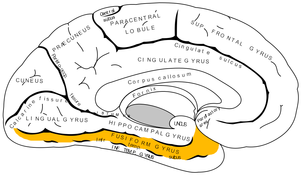
The fusiform gyrus, also known as the lateral occipitotemporal gyrus, is part of the temporal lobe and occipital lobe in Brodmann area 37. The fusiform gyrus is located between the lingual gyrus and parahippocampal gyrus above, and the inferior temporal gyrus below. Though the functionality of the fusiform gyrus is not fully understood, it has been linked with various neural pathways related to recognition. Additionally, it has been linked to various neurological phenomena such as synesthesia, dyslexia, and prosopagnosia.
Anatomically, the fusiform gyrus is the largest macro-anatomical structure within the ventral temporal cortex, which mainly includes structures involved in high-level vision. The term fusiform gyrus (lit. "spindle-shaped convolution") refers to the fact that the shape of the gyrus is wider at its centre than at its ends. This term is based on the description of the gyrus by Emil Huschke in 1854. (see also section on history). The fusiform gyrus is situated at the basal surface of the temporal and occipital lobes and is delineated by the collateral sulcus (CoS) and occipitotemporal sulcus (OTS), respectively. The OTS separates the fusiform gyrus from the inferior temporal gyrus (located laterally in respect to the fusiform gyrus) and the CoS separates the fusiform gyrus from the parahippocampal gyrus (located medially in respect to the fusiform gyrus).
The fusiform gyrus can be further delineated into a lateral and medial portion, as it is separated in its middle by the relatively shallow mid-fusiform sulcus (MFS). Thus, the lateral fusiform gyrus is delineated by the OTS laterally and the MFS medially. Likewise, the medial fusiform gyrus is delineated by the MFS laterally and the CoS medially.
Importantly, the mid-fusiform sulcus serves as a macroanatomical landmark for the fusiform face area (FFA), a functional subregion of the fusiform gyrus assumed to play a key role in processing faces.
The fusiform gyrus has a contentious history that has recently been clarified. The term was first used in 1854 by Emil Huschke from Jena, Germany, who called the fusiform gyrus a "Spindelwulst" (lit. spindle bulge). He chose this term because of the similarity that the respective cerebral gyrus bears to the shape of a spindle, or fusil, due to its wider central section. At first, researchers located the fusiform gyrus in other mammals as well, without taking into account the variations in gross organizations of other species' brains. Today, the fusiform gyrus is considered to be specific to hominoids. This is supported by research showing only three temporal gyri and no fusiform gyrus in macaques.
The first accurate definition of the mid-fusiform sulcus was coined by Gustav Retzius in 1896. He was the first to describe the sulcus sagittalis gyri fusiformis (today: mid-fusiform sulcus), and correctly determined that a sulcus divides the fusiform gyrus into lateral and medial partitions. W. Julius Mickle mentioned the mid-fusiform sulcus in 1897 and attempted to clarify the relation between temporal sulci and the fusiform gyrus, calling it the "intra-gyral sulcus of the fusiform lobule".
The exact functionality of the fusiform gyrus is still disputed, but there is relative consensus on its involvement in the following pathways:
In 2003, V. S. Ramachandran collaborated with scientists from the Salk Institute for Biological Studies in order to identify the potential role of the fusiform gyrus within the color processing pathway in the brain. Examining the relationship within the pathway specifically in cases of synesthesia, Ramachandran found that synesthetes on average have a higher density of fibers surrounding the angular gyrus. The angular gyrus is involved in higher processing of colors. The fibers relay shape information from the fusiform gyrus to the angular gyrus in order to produce the association of colors and shapes in grapheme-color synesthesia. Cross-activation between the angular and fusiform gyri has been observed in the average brain, implying that the fusiform gyrus regularly communicates with the visual pathway.
Fusiform gyrus
The fusiform gyrus, also known as the lateral occipitotemporal gyrus, is part of the temporal lobe and occipital lobe in Brodmann area 37. The fusiform gyrus is located between the lingual gyrus and parahippocampal gyrus above, and the inferior temporal gyrus below. Though the functionality of the fusiform gyrus is not fully understood, it has been linked with various neural pathways related to recognition. Additionally, it has been linked to various neurological phenomena such as synesthesia, dyslexia, and prosopagnosia.
Anatomically, the fusiform gyrus is the largest macro-anatomical structure within the ventral temporal cortex, which mainly includes structures involved in high-level vision. The term fusiform gyrus (lit. "spindle-shaped convolution") refers to the fact that the shape of the gyrus is wider at its centre than at its ends. This term is based on the description of the gyrus by Emil Huschke in 1854. (see also section on history). The fusiform gyrus is situated at the basal surface of the temporal and occipital lobes and is delineated by the collateral sulcus (CoS) and occipitotemporal sulcus (OTS), respectively. The OTS separates the fusiform gyrus from the inferior temporal gyrus (located laterally in respect to the fusiform gyrus) and the CoS separates the fusiform gyrus from the parahippocampal gyrus (located medially in respect to the fusiform gyrus).
The fusiform gyrus can be further delineated into a lateral and medial portion, as it is separated in its middle by the relatively shallow mid-fusiform sulcus (MFS). Thus, the lateral fusiform gyrus is delineated by the OTS laterally and the MFS medially. Likewise, the medial fusiform gyrus is delineated by the MFS laterally and the CoS medially.
Importantly, the mid-fusiform sulcus serves as a macroanatomical landmark for the fusiform face area (FFA), a functional subregion of the fusiform gyrus assumed to play a key role in processing faces.
The fusiform gyrus has a contentious history that has recently been clarified. The term was first used in 1854 by Emil Huschke from Jena, Germany, who called the fusiform gyrus a "Spindelwulst" (lit. spindle bulge). He chose this term because of the similarity that the respective cerebral gyrus bears to the shape of a spindle, or fusil, due to its wider central section. At first, researchers located the fusiform gyrus in other mammals as well, without taking into account the variations in gross organizations of other species' brains. Today, the fusiform gyrus is considered to be specific to hominoids. This is supported by research showing only three temporal gyri and no fusiform gyrus in macaques.
The first accurate definition of the mid-fusiform sulcus was coined by Gustav Retzius in 1896. He was the first to describe the sulcus sagittalis gyri fusiformis (today: mid-fusiform sulcus), and correctly determined that a sulcus divides the fusiform gyrus into lateral and medial partitions. W. Julius Mickle mentioned the mid-fusiform sulcus in 1897 and attempted to clarify the relation between temporal sulci and the fusiform gyrus, calling it the "intra-gyral sulcus of the fusiform lobule".
The exact functionality of the fusiform gyrus is still disputed, but there is relative consensus on its involvement in the following pathways:
In 2003, V. S. Ramachandran collaborated with scientists from the Salk Institute for Biological Studies in order to identify the potential role of the fusiform gyrus within the color processing pathway in the brain. Examining the relationship within the pathway specifically in cases of synesthesia, Ramachandran found that synesthetes on average have a higher density of fibers surrounding the angular gyrus. The angular gyrus is involved in higher processing of colors. The fibers relay shape information from the fusiform gyrus to the angular gyrus in order to produce the association of colors and shapes in grapheme-color synesthesia. Cross-activation between the angular and fusiform gyri has been observed in the average brain, implying that the fusiform gyrus regularly communicates with the visual pathway.
Recent media
Recent media