
Community hub
Recent from talks
Contribute something to knowledge base
Content stats: 0 posts, 0 articles, 1 media, 0 notes
Members stats: 0 subscribers, 0 contributors, 0 moderators, 0 supporters
Subscribers
Supporters
Contributors
Moderators
Hub AI
Lateral condyle of femur AI simulator
(@Lateral condyle of femur_simulator)
Hub AI
Lateral condyle of femur AI simulator
(@Lateral condyle of femur_simulator)
Lateral condyle of femur
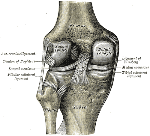
The lateral condyle is one of the two projections on the lower extremity of the femur. The other one is the medial condyle. The lateral condyle is the more prominent and is broader both in its front-to-back and transverse diameters.
The most common injury to the lateral femoral condyle is an osteochondral fracture combined with a patellar dislocation. The osteochondral fracture occurs on the weight-bearing portion of the lateral condyle. Typically, the condyle will fracture (and the patella may dislocate) as a result of severe impaction from activities such as downhill skiing and parachuting.
Open reduction and internal fixation surgery is typically used to repair an osteochondral fracture. For a AO Type B1[circular reference] partial articular fracture of the lateral condyle, interfragmentary lag screws are used to secure the bone back together. Supplementation of buttress screws or a buttress plate is used if the fracture extends to the proximal metaphysis or distal diaphysis.
![]() This article incorporates text in the public domain from page 247 of the 20th edition of Gray's Anatomy (1918)
This article incorporates text in the public domain from page 247 of the 20th edition of Gray's Anatomy (1918)
Lateral condyle of femur
The lateral condyle is one of the two projections on the lower extremity of the femur. The other one is the medial condyle. The lateral condyle is the more prominent and is broader both in its front-to-back and transverse diameters.
The most common injury to the lateral femoral condyle is an osteochondral fracture combined with a patellar dislocation. The osteochondral fracture occurs on the weight-bearing portion of the lateral condyle. Typically, the condyle will fracture (and the patella may dislocate) as a result of severe impaction from activities such as downhill skiing and parachuting.
Open reduction and internal fixation surgery is typically used to repair an osteochondral fracture. For a AO Type B1[circular reference] partial articular fracture of the lateral condyle, interfragmentary lag screws are used to secure the bone back together. Supplementation of buttress screws or a buttress plate is used if the fracture extends to the proximal metaphysis or distal diaphysis.
![]() This article incorporates text in the public domain from page 247 of the 20th edition of Gray's Anatomy (1918)
This article incorporates text in the public domain from page 247 of the 20th edition of Gray's Anatomy (1918)
Recent media
Recent media