")
Community hub
Recent from talks
Contribute something to knowledge base
Content stats: 0 posts, 0 articles, 1 media, 0 notes
Members stats: 0 subscribers, 0 contributors, 0 moderators, 0 supporters
Subscribers
Supporters
Contributors
Moderators
Hub AI
Ventricle (heart) AI simulator
(@Ventricle (heart)_simulator)
Hub AI
Ventricle (heart) AI simulator
(@Ventricle (heart)_simulator)
Ventricle (heart)
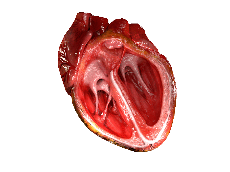
A ventricle is one of two large chambers located toward the bottom of the heart that collect and expel blood towards the peripheral beds within the body and lungs. The blood pumped by a ventricle is supplied by an atrium, an adjacent chamber in the upper heart that is smaller than a ventricle. Interventricular means between the ventricles (for example the interventricular septum), while intraventricular means within one ventricle (for example an intraventricular block).
In a four-chambered heart, such as that in humans, there are two ventricles that operate in a double circulatory system: the right ventricle pumps blood into the pulmonary circulation to the lungs, and the left ventricle pumps blood into the systemic circulation through the aorta.
Ventricles have thicker walls than atria and generate higher blood pressures. The physiological load on the ventricles requiring pumping of blood throughout the body and lungs is much greater than the pressure generated by the atria to fill the ventricles. Further, the left ventricle has thicker walls than the right because it needs to pump blood to most of the body while the right ventricle fills only the lungs.[citation needed]
On the inner walls of the ventricles are irregular muscular columns called trabeculae carneae which cover all of the inner ventricular surfaces except that of the conus arteriosus, in the right ventricle. There are three types of these muscles. The third type, the papillary muscles, give origin at their apices to the chordae tendinae which attach to the cusps of the tricuspid valve and to the mitral valve.
The mass of the left ventricle, as estimated by magnetic resonance imaging, averages 143 g ± 38.4 g, with a range of 87–224 g.
The right ventricle is equal in size to the left ventricle[citation needed] and contains roughly 85 millilitres (3 imp fl oz; 3 US fl oz) in the adult. Its upper front surface is circled and convex, and forms much of the sternocostal surface of the heart. Its under surface is flattened, forming part of the diaphragmatic surface of the heart that rests upon the diaphragm.
Its posterior wall is formed by the ventricular septum, which bulges into the right ventricle, so that a transverse section of the cavity presents a semilunar outline. Its upper and left angle forms a conical pouch, the conus arteriosus, from which the pulmonary artery arises. A tendinous band, called the tendon of the conus arteriosus, extends upward from the right atrioventricular fibrous ring and connects the posterior surface of the conus arteriosus to the aorta.[citation needed]
The left ventricle is longer and more conical in shape than the right, and on transverse section its concavity presents an oval or nearly circular outline. It forms a small part of the sternocostal surface and a considerable part of the diaphragmatic surface of the heart; it also forms the apex of the heart. The left ventricle is thicker and more muscular than the right ventricle because it pumps blood at a higher pressure.
Ventricle (heart)
A ventricle is one of two large chambers located toward the bottom of the heart that collect and expel blood towards the peripheral beds within the body and lungs. The blood pumped by a ventricle is supplied by an atrium, an adjacent chamber in the upper heart that is smaller than a ventricle. Interventricular means between the ventricles (for example the interventricular septum), while intraventricular means within one ventricle (for example an intraventricular block).
In a four-chambered heart, such as that in humans, there are two ventricles that operate in a double circulatory system: the right ventricle pumps blood into the pulmonary circulation to the lungs, and the left ventricle pumps blood into the systemic circulation through the aorta.
Ventricles have thicker walls than atria and generate higher blood pressures. The physiological load on the ventricles requiring pumping of blood throughout the body and lungs is much greater than the pressure generated by the atria to fill the ventricles. Further, the left ventricle has thicker walls than the right because it needs to pump blood to most of the body while the right ventricle fills only the lungs.[citation needed]
On the inner walls of the ventricles are irregular muscular columns called trabeculae carneae which cover all of the inner ventricular surfaces except that of the conus arteriosus, in the right ventricle. There are three types of these muscles. The third type, the papillary muscles, give origin at their apices to the chordae tendinae which attach to the cusps of the tricuspid valve and to the mitral valve.
The mass of the left ventricle, as estimated by magnetic resonance imaging, averages 143 g ± 38.4 g, with a range of 87–224 g.
The right ventricle is equal in size to the left ventricle[citation needed] and contains roughly 85 millilitres (3 imp fl oz; 3 US fl oz) in the adult. Its upper front surface is circled and convex, and forms much of the sternocostal surface of the heart. Its under surface is flattened, forming part of the diaphragmatic surface of the heart that rests upon the diaphragm.
Its posterior wall is formed by the ventricular septum, which bulges into the right ventricle, so that a transverse section of the cavity presents a semilunar outline. Its upper and left angle forms a conical pouch, the conus arteriosus, from which the pulmonary artery arises. A tendinous band, called the tendon of the conus arteriosus, extends upward from the right atrioventricular fibrous ring and connects the posterior surface of the conus arteriosus to the aorta.[citation needed]
The left ventricle is longer and more conical in shape than the right, and on transverse section its concavity presents an oval or nearly circular outline. It forms a small part of the sternocostal surface and a considerable part of the diaphragmatic surface of the heart; it also forms the apex of the heart. The left ventricle is thicker and more muscular than the right ventricle because it pumps blood at a higher pressure.
Recent media
Recent media