Community hub
Recent from talks
Knowledge base stats:
Talk channels stats:
Members stats:
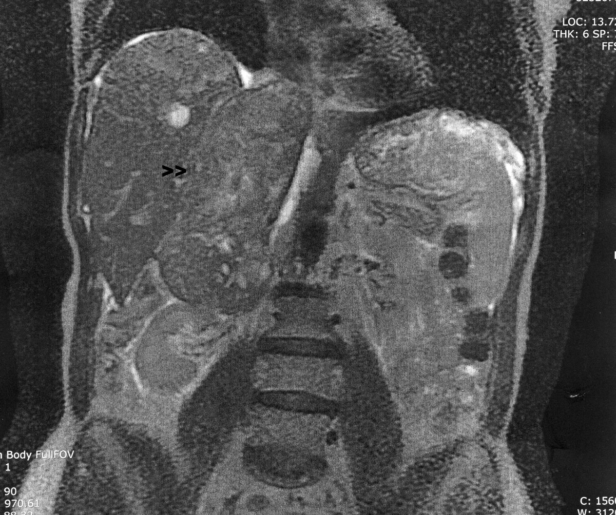
Leiomyosarcoma
A leiomyosarcoma (LMS) is a rare malignant (cancerous) smooth muscle tumor. The word is from leio- 'smooth' myo- 'muscle' and sarcoma 'tumor of connective tissue'. The stomach, bladder, uterus, blood vessels, and intestines are examples of hollow organs made up of smooth muscles where LMS can be located; however, the uterus and abdomen are the most common sites.
Although leiomyosarcomas are rare, they belong to the more common types of soft-tissue sarcoma, representing 10–20% of new cases. This type of cancer is more frequently diagnosed in adults as compared to children. When considering LMS specifically in the context of the uterus, it affects approximately 6 individuals per 1 million people in the United States each year. LMSs are resistant cancers, meaning they are generally not very responsive to chemotherapy or radiation. The best outcomes occur when the tumor tissue can be removed surgically at an early stage, while it is small and has not yet spread from the original site (it remains in situ).
Smooth muscle cells make up the involuntary muscles, which are found in most parts of the body, including the uterus, stomach and intestines, the walls of all blood vessels, and the skin. These are the areas where LMSs originate. LMSs also often develop in the retroperitoneal region which consists of the suprarenal glands, the kidney, and ureter. Just as it is not known what truly causes most sarcomas, LMSs have similarly complex karyotypes and it is suggested that because of the complexity, genomic instability might be the cause.
Uterine leiomyosarcomas come from the smooth muscle in the muscle layer of the uterus. Cutaneous leiomyosarcomas derive from the pilo-erector muscles in the skin. Gastrointestinal leiomyosarcomas might come from smooth muscle in the gastrointestinal (GI) tract, or alternatively, from a blood vessel. At most other primary sites—retroperitoneal extremity (in the abdomen, behind the intestines), truncal, abdominal organs, etc.—leiomyosarcomas appear to grow from the muscle layer of a blood vessel (the tunica media). Thus, a leiomyosarcoma can have a primary site of origin anywhere in the body from a blood vessel.
The tumors are usually hemorrhagic, soft, and microscopically marked by pleomorphism, abundant (15–30 per 10 high-power fields) abnormal mitotic figures, and coagulative tumor cell necrosis. The differential diagnosis, which includes spindle cell carcinoma, spindle cell melanoma, fibrosarcoma, malignant peripheral nerve sheath tumor, and even biphenotypic sinonasal sarcoma, is wide.
To diagnose LMS, a physical exam may be performed by one’s physician, imaging tests such as MIT, CT, and PET scans can be performed, or tissue biopsies can also be completed where the histopathology of the removed tissue sample is examined. Because LMS is a widespread disease, the symptoms vary based on the location and size of the tumor. Some of the symptoms include nausea and vomiting, palpable lumps, pain, bleeding, and unintentional weight loss.
Surgery, with as wide a margin of removal as possible, has generally been the most effective and preferred way to attack LMS. If surgical margins are narrow or not clear of tumor, however, or in some situations where tumor cells were left behind, chemotherapy or radiation has been shown to give a clear survival benefit. While LMS tends to be resistant to radiation and chemotherapy, each case is different and results can vary widely.
For metastatic (widespread) disease, chemotherapy and targeted therapies are the first choices. Chemotherapy regimens include doxorubicin/ifosfamide and doxorubicin combination/gemcitabine and docetaxel/trabectedin; pazopanib is the targeted therapy used in metastatic leiomyosarcoma as second line and is well tolerated.
Hub AI
Leiomyosarcoma AI simulator
(@Leiomyosarcoma_simulator)
Leiomyosarcoma
A leiomyosarcoma (LMS) is a rare malignant (cancerous) smooth muscle tumor. The word is from leio- 'smooth' myo- 'muscle' and sarcoma 'tumor of connective tissue'. The stomach, bladder, uterus, blood vessels, and intestines are examples of hollow organs made up of smooth muscles where LMS can be located; however, the uterus and abdomen are the most common sites.
Although leiomyosarcomas are rare, they belong to the more common types of soft-tissue sarcoma, representing 10–20% of new cases. This type of cancer is more frequently diagnosed in adults as compared to children. When considering LMS specifically in the context of the uterus, it affects approximately 6 individuals per 1 million people in the United States each year. LMSs are resistant cancers, meaning they are generally not very responsive to chemotherapy or radiation. The best outcomes occur when the tumor tissue can be removed surgically at an early stage, while it is small and has not yet spread from the original site (it remains in situ).
Smooth muscle cells make up the involuntary muscles, which are found in most parts of the body, including the uterus, stomach and intestines, the walls of all blood vessels, and the skin. These are the areas where LMSs originate. LMSs also often develop in the retroperitoneal region which consists of the suprarenal glands, the kidney, and ureter. Just as it is not known what truly causes most sarcomas, LMSs have similarly complex karyotypes and it is suggested that because of the complexity, genomic instability might be the cause.
Uterine leiomyosarcomas come from the smooth muscle in the muscle layer of the uterus. Cutaneous leiomyosarcomas derive from the pilo-erector muscles in the skin. Gastrointestinal leiomyosarcomas might come from smooth muscle in the gastrointestinal (GI) tract, or alternatively, from a blood vessel. At most other primary sites—retroperitoneal extremity (in the abdomen, behind the intestines), truncal, abdominal organs, etc.—leiomyosarcomas appear to grow from the muscle layer of a blood vessel (the tunica media). Thus, a leiomyosarcoma can have a primary site of origin anywhere in the body from a blood vessel.
The tumors are usually hemorrhagic, soft, and microscopically marked by pleomorphism, abundant (15–30 per 10 high-power fields) abnormal mitotic figures, and coagulative tumor cell necrosis. The differential diagnosis, which includes spindle cell carcinoma, spindle cell melanoma, fibrosarcoma, malignant peripheral nerve sheath tumor, and even biphenotypic sinonasal sarcoma, is wide.
To diagnose LMS, a physical exam may be performed by one’s physician, imaging tests such as MIT, CT, and PET scans can be performed, or tissue biopsies can also be completed where the histopathology of the removed tissue sample is examined. Because LMS is a widespread disease, the symptoms vary based on the location and size of the tumor. Some of the symptoms include nausea and vomiting, palpable lumps, pain, bleeding, and unintentional weight loss.
Surgery, with as wide a margin of removal as possible, has generally been the most effective and preferred way to attack LMS. If surgical margins are narrow or not clear of tumor, however, or in some situations where tumor cells were left behind, chemotherapy or radiation has been shown to give a clear survival benefit. While LMS tends to be resistant to radiation and chemotherapy, each case is different and results can vary widely.
For metastatic (widespread) disease, chemotherapy and targeted therapies are the first choices. Chemotherapy regimens include doxorubicin/ifosfamide and doxorubicin combination/gemcitabine and docetaxel/trabectedin; pazopanib is the targeted therapy used in metastatic leiomyosarcoma as second line and is well tolerated.