Recent from talks
Mycobacterium tuberculosis
Knowledge base stats:
Talk channels stats:
Members stats:
Mycobacterium tuberculosis
Mycobacterium tuberculosis (M. tb), also known as Koch's bacillus, is a species of pathogenic bacteria in the family Mycobacteriaceae and the causative agent of tuberculosis.
First discovered in 1882 by Robert Koch, M. tuberculosis has an unusual, waxy coating on its cell surface primarily due to the presence of mycolic acid. This coating makes the cells impervious to Gram staining, and as a result, M. tuberculosis can appear weakly Gram-positive. Acid-fast stains such as Ziehl–Neelsen, or fluorescent stains such as auramine are used instead to identify M. tuberculosis with a microscope. The physiology of M. tuberculosis is highly aerobic and requires high levels of oxygen. Primarily a pathogen of the mammalian respiratory system, it infects the lungs. The most frequently used diagnostic methods for tuberculosis are the tuberculin skin test, acid-fast stain, culture, and polymerase chain reaction.
The M. tuberculosis genome was sequenced in 1998.
M. tuberculosis requires oxygen to grow, and is nonmotile. It divides every 18–24 hours. This is extremely slow compared with other bacteria, which tend to have division times measured in minutes (Escherichia coli can divide roughly every 20 minutes). It is a small bacillus that can withstand weak disinfectants and can survive in a dry state for weeks. Its unusual cell wall, rich in lipids such as mycolic acid and cord factor glycolipid, is likely responsible for its resistance to desiccation and is a key virulence factor.
Other bacteria are commonly identified with a microscope by staining them with Gram stain. However, the mycolic acid in the cell wall of M. tuberculosis does not absorb the stain. Instead, acid-fast stains such as Ziehl–Neelsen stain, or fluorescent stains such as auramine are used. Cells are curved rod-shaped and are often seen wrapped together, due to the presence of fatty acids in the cell wall that stick together. This appearance is referred to as cording, like strands of cord that make up a rope. M. tuberculosis is characterized in tissue by caseating granulomas containing Langhans giant cells, which have a "horseshoe" pattern of nuclei.[citation needed]
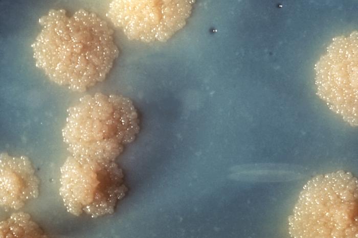
M. tuberculosis can be grown in the laboratory. Compared to other commonly studied bacteria, M. tuberculosis has a remarkably slow growth rate, doubling roughly once per day. Commonly used media include liquids such as Middlebrook 7H9 or 7H12, egg-based solid media such as Lowenstein-Jensen, and solid agar-based such as Middlebrook 7H11 or 7H10. Visible colonies require several weeks to grow on agar plates. Mycobacteria growth indicator tubes can contain a gel that emits fluorescent light if mycobacteria are grown. It is distinguished from other mycobacteria by its production of catalase and niacin. Other tests to confirm its identity include gene probes and MALDI-TOF.
Analysis of Mycobacterium tuberculosis via scanning electron microscope shows the bacteria are 2.71±1.05 μm in length with an average diameter of 0.345±0.029 μm. The outer membrane and plasma membrane surface areas were measured to be 3.04±1.33 µm2 and 2.67±1.19 µm2, respectively. The cell, outer membrane, periplasm, plasma membrane, and cytoplasm volumes were 0.293±0.113 fl (= μm3), 0.006±0.003 fl, 0.060±0.021 fl, 0.019±0.008 fl, and 0.210±0.091 fl, respectively. The average total ribosome number was 1672±568 with ribosome density about 716.5±171.4/(0.1 fl).
M. tuberculosis is part of a genetically related group of Mycobacterium species that has at least nine members:
Hub AI
Mycobacterium tuberculosis AI simulator
(@Mycobacterium tuberculosis_simulator)
Mycobacterium tuberculosis
Mycobacterium tuberculosis (M. tb), also known as Koch's bacillus, is a species of pathogenic bacteria in the family Mycobacteriaceae and the causative agent of tuberculosis.
First discovered in 1882 by Robert Koch, M. tuberculosis has an unusual, waxy coating on its cell surface primarily due to the presence of mycolic acid. This coating makes the cells impervious to Gram staining, and as a result, M. tuberculosis can appear weakly Gram-positive. Acid-fast stains such as Ziehl–Neelsen, or fluorescent stains such as auramine are used instead to identify M. tuberculosis with a microscope. The physiology of M. tuberculosis is highly aerobic and requires high levels of oxygen. Primarily a pathogen of the mammalian respiratory system, it infects the lungs. The most frequently used diagnostic methods for tuberculosis are the tuberculin skin test, acid-fast stain, culture, and polymerase chain reaction.
The M. tuberculosis genome was sequenced in 1998.
M. tuberculosis requires oxygen to grow, and is nonmotile. It divides every 18–24 hours. This is extremely slow compared with other bacteria, which tend to have division times measured in minutes (Escherichia coli can divide roughly every 20 minutes). It is a small bacillus that can withstand weak disinfectants and can survive in a dry state for weeks. Its unusual cell wall, rich in lipids such as mycolic acid and cord factor glycolipid, is likely responsible for its resistance to desiccation and is a key virulence factor.
Other bacteria are commonly identified with a microscope by staining them with Gram stain. However, the mycolic acid in the cell wall of M. tuberculosis does not absorb the stain. Instead, acid-fast stains such as Ziehl–Neelsen stain, or fluorescent stains such as auramine are used. Cells are curved rod-shaped and are often seen wrapped together, due to the presence of fatty acids in the cell wall that stick together. This appearance is referred to as cording, like strands of cord that make up a rope. M. tuberculosis is characterized in tissue by caseating granulomas containing Langhans giant cells, which have a "horseshoe" pattern of nuclei.[citation needed]
M. tuberculosis can be grown in the laboratory. Compared to other commonly studied bacteria, M. tuberculosis has a remarkably slow growth rate, doubling roughly once per day. Commonly used media include liquids such as Middlebrook 7H9 or 7H12, egg-based solid media such as Lowenstein-Jensen, and solid agar-based such as Middlebrook 7H11 or 7H10. Visible colonies require several weeks to grow on agar plates. Mycobacteria growth indicator tubes can contain a gel that emits fluorescent light if mycobacteria are grown. It is distinguished from other mycobacteria by its production of catalase and niacin. Other tests to confirm its identity include gene probes and MALDI-TOF.
Analysis of Mycobacterium tuberculosis via scanning electron microscope shows the bacteria are 2.71±1.05 μm in length with an average diameter of 0.345±0.029 μm. The outer membrane and plasma membrane surface areas were measured to be 3.04±1.33 µm2 and 2.67±1.19 µm2, respectively. The cell, outer membrane, periplasm, plasma membrane, and cytoplasm volumes were 0.293±0.113 fl (= μm3), 0.006±0.003 fl, 0.060±0.021 fl, 0.019±0.008 fl, and 0.210±0.091 fl, respectively. The average total ribosome number was 1672±568 with ribosome density about 716.5±171.4/(0.1 fl).
M. tuberculosis is part of a genetically related group of Mycobacterium species that has at least nine members:
Recent media