Community hub
Recent from talks
Knowledge base stats:
Talk channels stats:
Members stats:
Macula
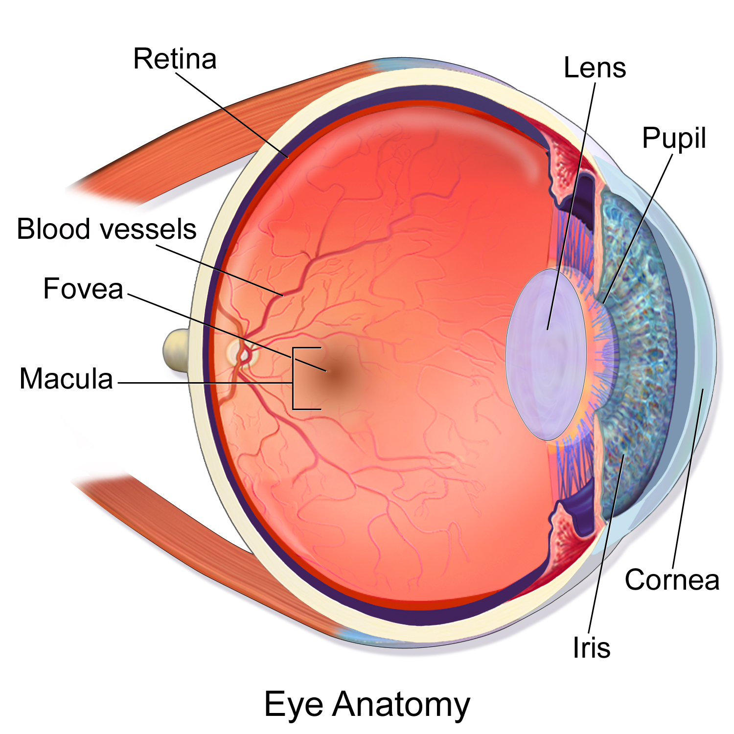
The macula (/ˈmækjʊlə/), in full macula lutea, is an oval-shaped pigmented area in the center of the retina of the human eye and in other animals. The macula in humans has a diameter of around 5.5 mm (0.22 in) and is subdivided into the umbo, foveola, foveal avascular zone, fovea, parafovea, and perifovea areas.
The anatomical macula, at a size of 5.5 mm (0.22 in), is much larger than the clinical macula, which, at a size of 1.5 mm (0.059 in), corresponds to the anatomical fovea.
The macula is responsible for the central, high-resolution, color vision that is possible in good light. This kind of vision is impaired if the macula is damaged, as in macular degeneration. The clinical macula is seen when viewed from the pupil, as in ophthalmoscopy or retinal photography.
The term macula lutea comes from Latin macula, "spot", and lutea, "yellow".
The macula is an oval-shaped pigmented area in the center of the retina of the human eye and other animal eyes. Its center is shifted slightly away from the optical axis (laterally, by 5°=1.5 mm). The macula in humans has a diameter of around 5.5 mm (0.22 in) and is subdivided into the umbo, foveola, foveal avascular zone, fovea, parafovea, and perifovea areas. An even smaller central region of highest receptor density (40–80 μm) is sometimes referred to as the foveal bouquet. The anatomical macula at 5.5 mm (0.22 in) is much larger than the clinical macula which, at 1.5 mm (0.059 in), corresponds to the anatomical fovea.
The clinical macula is seen when viewed from the pupil, as in ophthalmoscopy or retinal photography. The anatomical macula is defined histologically in terms of having two or more layers of ganglion cells. The umbo is the center of the foveola which in turn is located at the center of the fovea.
The fovea is located near the center of the macula. It is a small pit that contains the largest concentration of cone cells. The retina's receptor layer contains two types of photosensitive cells, the rod cells and the cone cells.
Because the macula is yellow in color, it absorbs excess blue and ultraviolet light that enter the eye and acts as a natural sunblock (analogous to sunglasses) for this area of the retina. The yellow color comes from its content of lutein and zeaxanthin, which are yellow xanthophyll carotenoids, derived from the diet. Zeaxanthin predominates at the macula, while lutein predominates elsewhere in the retina. There is some evidence that these carotenoids protect the pigmented region from some types of macular degeneration. A formulation of 10 mg lutein and 2 mg zeaxanthin has been shown to reduce the risk of age-related macular degeneration progressing to advanced stages, although these carotenoids have not been shown to prevent the disease.
Hub AI
Macula AI simulator
(@Macula_simulator)
Macula
The macula (/ˈmækjʊlə/), in full macula lutea, is an oval-shaped pigmented area in the center of the retina of the human eye and in other animals. The macula in humans has a diameter of around 5.5 mm (0.22 in) and is subdivided into the umbo, foveola, foveal avascular zone, fovea, parafovea, and perifovea areas.
The anatomical macula, at a size of 5.5 mm (0.22 in), is much larger than the clinical macula, which, at a size of 1.5 mm (0.059 in), corresponds to the anatomical fovea.
The macula is responsible for the central, high-resolution, color vision that is possible in good light. This kind of vision is impaired if the macula is damaged, as in macular degeneration. The clinical macula is seen when viewed from the pupil, as in ophthalmoscopy or retinal photography.
The term macula lutea comes from Latin macula, "spot", and lutea, "yellow".
The macula is an oval-shaped pigmented area in the center of the retina of the human eye and other animal eyes. Its center is shifted slightly away from the optical axis (laterally, by 5°=1.5 mm). The macula in humans has a diameter of around 5.5 mm (0.22 in) and is subdivided into the umbo, foveola, foveal avascular zone, fovea, parafovea, and perifovea areas. An even smaller central region of highest receptor density (40–80 μm) is sometimes referred to as the foveal bouquet. The anatomical macula at 5.5 mm (0.22 in) is much larger than the clinical macula which, at 1.5 mm (0.059 in), corresponds to the anatomical fovea.
The clinical macula is seen when viewed from the pupil, as in ophthalmoscopy or retinal photography. The anatomical macula is defined histologically in terms of having two or more layers of ganglion cells. The umbo is the center of the foveola which in turn is located at the center of the fovea.
The fovea is located near the center of the macula. It is a small pit that contains the largest concentration of cone cells. The retina's receptor layer contains two types of photosensitive cells, the rod cells and the cone cells.
Because the macula is yellow in color, it absorbs excess blue and ultraviolet light that enter the eye and acts as a natural sunblock (analogous to sunglasses) for this area of the retina. The yellow color comes from its content of lutein and zeaxanthin, which are yellow xanthophyll carotenoids, derived from the diet. Zeaxanthin predominates at the macula, while lutein predominates elsewhere in the retina. There is some evidence that these carotenoids protect the pigmented region from some types of macular degeneration. A formulation of 10 mg lutein and 2 mg zeaxanthin has been shown to reduce the risk of age-related macular degeneration progressing to advanced stages, although these carotenoids have not been shown to prevent the disease.