Community hub
Recent from talks
Knowledge base stats:
Talk channels stats:
Members stats:
Microsurgery
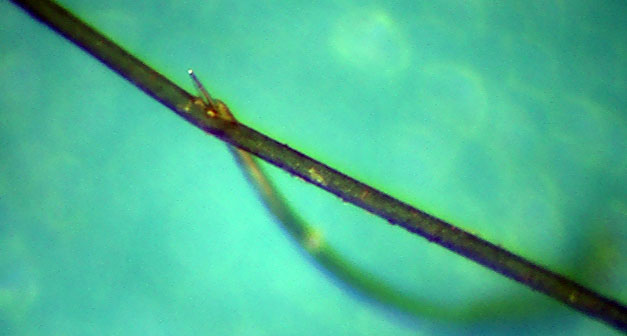
Microsurgery is a general term for surgery requiring an operating microscope. The most obvious developments have been procedures developed to allow anastomosis of successively smaller blood vessels and nerves (typically 1 mm in diameter) which have allowed transfer of tissue from one part of the body to another and re-attachment of severed parts. Microsurgical techniques are utilized by several specialties today, such as general surgery, ophthalmology, orthopedic surgery, gynecological surgery, otolaryngology, neurosurgery, oral and maxillofacial surgery, endodontic microsurgery, plastic surgery, podiatric surgery and pediatric surgery.
Otolaryngologists were the first physicians to use microsurgical techniques. A Swedish otolaryngologist, Carl-Olof Siggesson Nylén (1892–1978), was the father of microsurgery. In 1921, in the University of Stockholm, he built the first surgical microscope, a modified monocular Brinell-Leitz microscope. At first he used it for operations in animals. In November of the same year he used it to operate on a patient with chronic otitis who had a labyrinthine fistula. Nylen's microscope was soon replaced by a binocular microscope, developed in 1922 by his colleague Gunnar Holmgren (1875–1954).[citation needed]
Gradually the operating microscope began to be used for ear operations. In the 1950s many otologists began to use it in the fenestration operation, usually to perfect the opening of the fenestra in the lateral semicircular canal. The revival of the stapes mobilization operation by Rosen, in 1953, made the use of the microscope mandatory, although it was not used by the originators of the technique, Kessel (1878), Boucheron (1888) and Miot (1890). Mastoidectomies began to be performed with the surgical microscope and so were the tympanoplasty techniques that became known in the early 1950s. The stapes mobilization operation had varying results and was soon replaced by stapedectomy, first described by John Shea, Jr.; this was an operation that was always performed with the microscope.[citation needed]
Today neurosurgeons are very proud to use microscopes in their procedures. But it was not always so: many prestigious centers did not accept that idea and it had to be developed in relative isolation. In the late 1950s William House began to explore new techniques for temporal bone surgery. He developed the middle fossa approach and perfected the translabyrinthine approach and began to use these techniques to remove acoustic nerve tumors. The first neurosurgeon to make use of the surgical microscope was a Turkish emigrant, Gazi Yasargil. In 1953 he studied neurovascular surgery during work with Prof. Hugo Krayenbühl in Switzerland. His ideas interested Dr. Pete Donaghy, who invited Yasargil to his microvascular laboratory in Burlington, Vermont. After his return to Zürich in 1967 Yasargil concentrated on discovering clinical applications to their novel inventions. Publications on that topic: Micro-Vascular Surgery and Microsurgery Applied to Neurosurgery won him international recognition. His lifelong experiences with microsurgery were recapitulated in the four-volume textbook entitled simply Microneurosurgery.
Advances in the techniques and technology that popularized microsurgery began in the early 1960s to involve other medical areas. The first microvascular surgery, using a microscope to aid in the repair of blood vessels, was described by vascular surgeon, Julius H. Jacobson II of the University of Vermont in 1960. Using an operating microscope, he performed coupling of vessels as small as 1.4 mm and coined the term microsurgery. Hand surgeons at the University of Louisville, Drs. Harold Kleinert and Mort Kasdan, performed the first revascularization of a partial digital amputation in 1963.
Nakayama, a Japanese cardiothoracic surgeon, reported the first true series of microsurgical free-tissue transfers using vascularized intestinal segments to the neck for esophageal reconstruction after cancer resections using 3–4 mm vessels.
Contemporary reconstructive microsurgery was introduced by an American plastic surgeon, Dr. Harry J. Buncke. In 1964, Buncke reported a rabbit ear replantation, famously using a garage as a lab/operating theatre and home-made instruments This was the first report of successfully using blood vessels 1 millimeter in size. In 1966, Buncke used microsurgery to transplant a primate's great toe to its hand.
During the late sixties and early 1970s, plastic surgeons ushered in many new microsurgical innovations that were previously unimaginable. The first human microsurgical transplantation of the second toe to thumb was performed in February 1966 by Dr. Dong-yue Yang and Yu-dong Gu, in Shanghai, China. Great toe (big toe) to thumb was performed in April 1968 by Dr. John Cobbett, in England. In Australia work by Dr. Ian Taylor saw new techniques developed to reconstruct head and neck cancer defects with living bone from the hip or the fibula.
Hub AI
Microsurgery AI simulator
(@Microsurgery_simulator)
Microsurgery
Microsurgery is a general term for surgery requiring an operating microscope. The most obvious developments have been procedures developed to allow anastomosis of successively smaller blood vessels and nerves (typically 1 mm in diameter) which have allowed transfer of tissue from one part of the body to another and re-attachment of severed parts. Microsurgical techniques are utilized by several specialties today, such as general surgery, ophthalmology, orthopedic surgery, gynecological surgery, otolaryngology, neurosurgery, oral and maxillofacial surgery, endodontic microsurgery, plastic surgery, podiatric surgery and pediatric surgery.
Otolaryngologists were the first physicians to use microsurgical techniques. A Swedish otolaryngologist, Carl-Olof Siggesson Nylén (1892–1978), was the father of microsurgery. In 1921, in the University of Stockholm, he built the first surgical microscope, a modified monocular Brinell-Leitz microscope. At first he used it for operations in animals. In November of the same year he used it to operate on a patient with chronic otitis who had a labyrinthine fistula. Nylen's microscope was soon replaced by a binocular microscope, developed in 1922 by his colleague Gunnar Holmgren (1875–1954).[citation needed]
Gradually the operating microscope began to be used for ear operations. In the 1950s many otologists began to use it in the fenestration operation, usually to perfect the opening of the fenestra in the lateral semicircular canal. The revival of the stapes mobilization operation by Rosen, in 1953, made the use of the microscope mandatory, although it was not used by the originators of the technique, Kessel (1878), Boucheron (1888) and Miot (1890). Mastoidectomies began to be performed with the surgical microscope and so were the tympanoplasty techniques that became known in the early 1950s. The stapes mobilization operation had varying results and was soon replaced by stapedectomy, first described by John Shea, Jr.; this was an operation that was always performed with the microscope.[citation needed]
Today neurosurgeons are very proud to use microscopes in their procedures. But it was not always so: many prestigious centers did not accept that idea and it had to be developed in relative isolation. In the late 1950s William House began to explore new techniques for temporal bone surgery. He developed the middle fossa approach and perfected the translabyrinthine approach and began to use these techniques to remove acoustic nerve tumors. The first neurosurgeon to make use of the surgical microscope was a Turkish emigrant, Gazi Yasargil. In 1953 he studied neurovascular surgery during work with Prof. Hugo Krayenbühl in Switzerland. His ideas interested Dr. Pete Donaghy, who invited Yasargil to his microvascular laboratory in Burlington, Vermont. After his return to Zürich in 1967 Yasargil concentrated on discovering clinical applications to their novel inventions. Publications on that topic: Micro-Vascular Surgery and Microsurgery Applied to Neurosurgery won him international recognition. His lifelong experiences with microsurgery were recapitulated in the four-volume textbook entitled simply Microneurosurgery.
Advances in the techniques and technology that popularized microsurgery began in the early 1960s to involve other medical areas. The first microvascular surgery, using a microscope to aid in the repair of blood vessels, was described by vascular surgeon, Julius H. Jacobson II of the University of Vermont in 1960. Using an operating microscope, he performed coupling of vessels as small as 1.4 mm and coined the term microsurgery. Hand surgeons at the University of Louisville, Drs. Harold Kleinert and Mort Kasdan, performed the first revascularization of a partial digital amputation in 1963.
Nakayama, a Japanese cardiothoracic surgeon, reported the first true series of microsurgical free-tissue transfers using vascularized intestinal segments to the neck for esophageal reconstruction after cancer resections using 3–4 mm vessels.
Contemporary reconstructive microsurgery was introduced by an American plastic surgeon, Dr. Harry J. Buncke. In 1964, Buncke reported a rabbit ear replantation, famously using a garage as a lab/operating theatre and home-made instruments This was the first report of successfully using blood vessels 1 millimeter in size. In 1966, Buncke used microsurgery to transplant a primate's great toe to its hand.
During the late sixties and early 1970s, plastic surgeons ushered in many new microsurgical innovations that were previously unimaginable. The first human microsurgical transplantation of the second toe to thumb was performed in February 1966 by Dr. Dong-yue Yang and Yu-dong Gu, in Shanghai, China. Great toe (big toe) to thumb was performed in April 1968 by Dr. John Cobbett, in England. In Australia work by Dr. Ian Taylor saw new techniques developed to reconstruct head and neck cancer defects with living bone from the hip or the fibula.