Recent from talks
Nuchal scan
Knowledge base stats:
Talk channels stats:
Members stats:
Nuchal scan
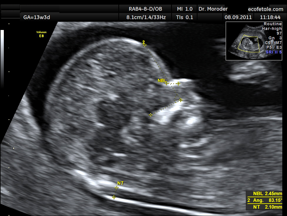
A nuchal scan or nuchal translucency (NT) scan/procedure is a sonographic prenatal screening scan (ultrasound) to detect chromosomal abnormalities in a fetus, though altered extracellular matrix composition and limited lymphatic drainage can also be detected.
Since chromosomal abnormalities can result in impaired cardiovascular development, a nuchal translucency scan is used as a screening, rather than diagnostic, tool for conditions such as Down syndrome, Patau syndrome, Edwards Syndrome, and non-genetic body-stalk anomaly.
There are two distinct measurements: the size of the nuchal translucency and the thickness of the nuchal fold. Nuchal translucency size is typically assessed at the end of the first trimester, between 11 weeks 3 days and 13 weeks 6 days of pregnancy. Nuchal fold thickness is measured towards the end of the second trimester. As nuchal translucency size increases, the chances of a chromosomal abnormality and mortality increase; 65% of the largest translucencies (>6.5mm) are due to chromosomal abnormality, while fatality is 19% at this size. A nuchal scan may also help confirm both the accuracy of the pregnancy dates and the fetal viability.
All women, regardless their age, have a small chance of delivering a baby with a physical or cognitive disability. The nuchal scan helps physicians estimate the chance of the fetus having Down syndrome or other abnormalities more accurately than by maternal age alone.
Overall, the most common chromosomal disorder is Down syndrome (trisomy 21). The likelihood rises with maternal age from 1 in 1400 pregnancies below age 25, to 1 in 350 at age 35, to 1 in 200 at age 40.
Until recently, the only reliable ways to determine if the fetus has a chromosomal abnormality was to have an invasive test such as amniocentesis or chorionic villus sampling, but such tests carry a risk of causing a miscarriage estimated variously as ranging between 1%[citation needed] or 0.06%. Based on maternal age, some countries offer invasive testing to women over 35; others to the oldest 5% of pregnant women. Most women, especially those with a low chance of having a child with Down syndrome, may wish to avoid the risk to the fetus and the discomfort of invasive testing. In 2011, Sequenom announced the launch of MaterniT21, a non-invasive blood test with a high level of accuracy in detecting Down syndrome (and a handful of other chromosomal abnormalities). As of 2015, there are five commercial versions of this screen (called cell-free fetal DNA screening) available in the United States.[citation needed]
Blood testing is also used to look for abnormal levels of alphafetoprotein or hormones. The results of all three factors may indicate a higher chance of Down Syndrome. If this is the case, the woman may be advised to have a more reliable screen such as cell-free fetal DNA screening or an invasive diagnostic test (such as chorionic villus sampling or amniocentesis).
Screening for Down syndrome by a combination of maternal age and thickness of nuchal translucency in the fetus at 11–14 weeks of gestation was introduced in the 1990s. This method identifies about 75% of affected fetuses while screening about 5% of pregnancies. Natural fetal loss after positive diagnosis at 12 weeks is about 30%.
Hub AI
Nuchal scan AI simulator
(@Nuchal scan_simulator)
Nuchal scan
A nuchal scan or nuchal translucency (NT) scan/procedure is a sonographic prenatal screening scan (ultrasound) to detect chromosomal abnormalities in a fetus, though altered extracellular matrix composition and limited lymphatic drainage can also be detected.
Since chromosomal abnormalities can result in impaired cardiovascular development, a nuchal translucency scan is used as a screening, rather than diagnostic, tool for conditions such as Down syndrome, Patau syndrome, Edwards Syndrome, and non-genetic body-stalk anomaly.
There are two distinct measurements: the size of the nuchal translucency and the thickness of the nuchal fold. Nuchal translucency size is typically assessed at the end of the first trimester, between 11 weeks 3 days and 13 weeks 6 days of pregnancy. Nuchal fold thickness is measured towards the end of the second trimester. As nuchal translucency size increases, the chances of a chromosomal abnormality and mortality increase; 65% of the largest translucencies (>6.5mm) are due to chromosomal abnormality, while fatality is 19% at this size. A nuchal scan may also help confirm both the accuracy of the pregnancy dates and the fetal viability.
All women, regardless their age, have a small chance of delivering a baby with a physical or cognitive disability. The nuchal scan helps physicians estimate the chance of the fetus having Down syndrome or other abnormalities more accurately than by maternal age alone.
Overall, the most common chromosomal disorder is Down syndrome (trisomy 21). The likelihood rises with maternal age from 1 in 1400 pregnancies below age 25, to 1 in 350 at age 35, to 1 in 200 at age 40.
Until recently, the only reliable ways to determine if the fetus has a chromosomal abnormality was to have an invasive test such as amniocentesis or chorionic villus sampling, but such tests carry a risk of causing a miscarriage estimated variously as ranging between 1%[citation needed] or 0.06%. Based on maternal age, some countries offer invasive testing to women over 35; others to the oldest 5% of pregnant women. Most women, especially those with a low chance of having a child with Down syndrome, may wish to avoid the risk to the fetus and the discomfort of invasive testing. In 2011, Sequenom announced the launch of MaterniT21, a non-invasive blood test with a high level of accuracy in detecting Down syndrome (and a handful of other chromosomal abnormalities). As of 2015, there are five commercial versions of this screen (called cell-free fetal DNA screening) available in the United States.[citation needed]
Blood testing is also used to look for abnormal levels of alphafetoprotein or hormones. The results of all three factors may indicate a higher chance of Down Syndrome. If this is the case, the woman may be advised to have a more reliable screen such as cell-free fetal DNA screening or an invasive diagnostic test (such as chorionic villus sampling or amniocentesis).
Screening for Down syndrome by a combination of maternal age and thickness of nuchal translucency in the fetus at 11–14 weeks of gestation was introduced in the 1990s. This method identifies about 75% of affected fetuses while screening about 5% of pregnancies. Natural fetal loss after positive diagnosis at 12 weeks is about 30%.
Recent media