Recent from talks
Pubis (bone)
Knowledge base stats:
Talk channels stats:
Members stats:
Pubis (bone)
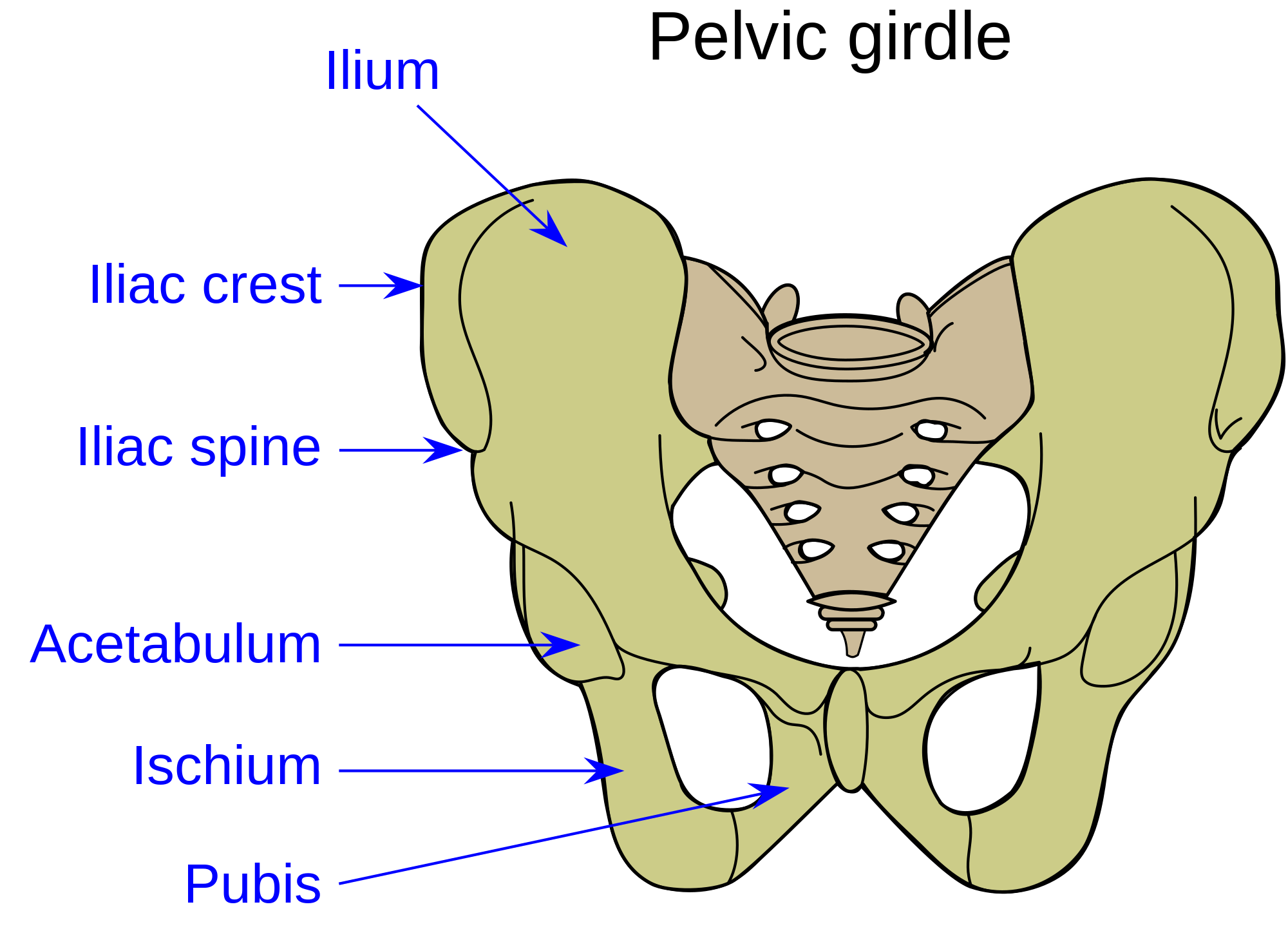
In vertebrates, the pubis or pubic bone (Latin: os pubis) forms the lower and anterior part of each side of the hip bone. The pubis is the most forward-facing (ventral and anterior) of the three bones that make up the hip bone. The left and right pubic bones are each made up of three sections; a superior ramus, an inferior ramus, and a body.
The pubic bone is made up of a body, superior ramus, and inferior ramus (Latin: branch). The left and right coxal bones join at the pubic symphysis. It is covered by a layer of fat – the mons pubis. The pubis is the lower limit of the suprapubic region. In the female, the pubis is anterior to the urethral sponge.
The body of pubis has:
The body forms the wide, strong, middle and flat part of the pubic bone. The bodies of the left and right pubic bones join at the pubic symphysis. The rough upper edge is the pubic crest, ending laterally in the pubic tubercle. This tubercle, found roughly 3 cm from the pubic symphysis, is a distinctive feature on the lower part of the abdominal wall; important when localizing the superficial inguinal ring and the femoral canal of the inguinal canal. The inner surface of the body forms part of the wall of the lesser pelvis and joints to the origin of a part of the obturator internus muscle.
The superior pubic ramus is the upper of the two rami. It forms the upper edge of the obturator foramen. It extends from the body to the median plane where it joins with the ramus of the opposite side. It consists of an inner flattened part and a narrow outer prismoid portion.
The upper border presents a prominent tubercle, the pubic tubercle (pubic spine), which projects forward; the inferior crus of the subcutaneous inguinal ring (external abdominal ring), and the inguinal ligament (Poupart's ligament) are attached to it.
Passing upward and laterally from the pubic tubercle is a well-defined ridge, forming a part of the pectineal line which marks the brim of the lesser pelvis: to it are attached a portion of the inguinal falx (conjoined tendon of obliquus internus and transversus), the lacunar ligament (Gimbernat's ligament), and the reflected inguinal ligament (triangular fascia).
Medial to the pubic tubercle is the crest, which extends from this process to the medial end of the bone. It affords attachment to the inguinal falx, and to the rectus abdominis and pyramidalis.
Hub AI
Pubis (bone) AI simulator
(@Pubis (bone)_simulator)
Pubis (bone)
In vertebrates, the pubis or pubic bone (Latin: os pubis) forms the lower and anterior part of each side of the hip bone. The pubis is the most forward-facing (ventral and anterior) of the three bones that make up the hip bone. The left and right pubic bones are each made up of three sections; a superior ramus, an inferior ramus, and a body.
The pubic bone is made up of a body, superior ramus, and inferior ramus (Latin: branch). The left and right coxal bones join at the pubic symphysis. It is covered by a layer of fat – the mons pubis. The pubis is the lower limit of the suprapubic region. In the female, the pubis is anterior to the urethral sponge.
The body of pubis has:
The body forms the wide, strong, middle and flat part of the pubic bone. The bodies of the left and right pubic bones join at the pubic symphysis. The rough upper edge is the pubic crest, ending laterally in the pubic tubercle. This tubercle, found roughly 3 cm from the pubic symphysis, is a distinctive feature on the lower part of the abdominal wall; important when localizing the superficial inguinal ring and the femoral canal of the inguinal canal. The inner surface of the body forms part of the wall of the lesser pelvis and joints to the origin of a part of the obturator internus muscle.
The superior pubic ramus is the upper of the two rami. It forms the upper edge of the obturator foramen. It extends from the body to the median plane where it joins with the ramus of the opposite side. It consists of an inner flattened part and a narrow outer prismoid portion.
The upper border presents a prominent tubercle, the pubic tubercle (pubic spine), which projects forward; the inferior crus of the subcutaneous inguinal ring (external abdominal ring), and the inguinal ligament (Poupart's ligament) are attached to it.
Passing upward and laterally from the pubic tubercle is a well-defined ridge, forming a part of the pectineal line which marks the brim of the lesser pelvis: to it are attached a portion of the inguinal falx (conjoined tendon of obliquus internus and transversus), the lacunar ligament (Gimbernat's ligament), and the reflected inguinal ligament (triangular fascia).
Medial to the pubic tubercle is the crest, which extends from this process to the medial end of the bone. It affords attachment to the inguinal falx, and to the rectus abdominis and pyramidalis.
Recent media