Recent from talks
QRS complex
Knowledge base stats:
Talk channels stats:
Members stats:
QRS complex
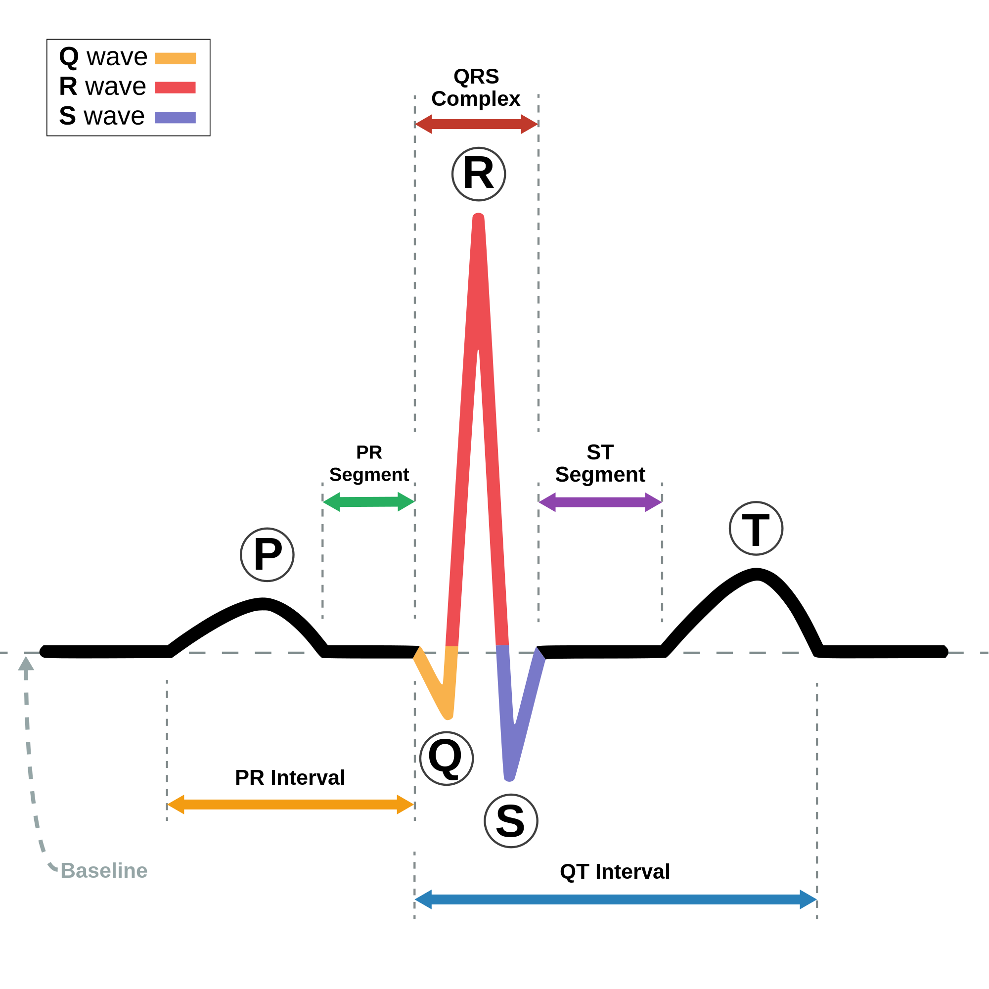
The QRS complex is the combination of three of the graphical deflections seen on a typical electrocardiogram (ECG or EKG). It is usually the central and most visually obvious part of the tracing. It corresponds to the depolarization of the right and left ventricles of the heart and contraction of the large ventricular muscles.
In adults, the QRS complex normally lasts 80 to 100 ms; in children it may be shorter. The Q, R, and S waves occur in rapid succession, do not all appear in all leads, and reflect a single event and thus are usually considered together. A Q wave is any downward deflection immediately following the P wave. An R wave follows as an upward deflection, and the S wave is any downward deflection after the R wave. The T wave follows the S wave, and in some cases, an additional U wave follows the T wave.
To measure the QRS interval start at the end of the PR interval (or beginning of the Q wave) to the end of the S wave. Normally this interval is 0.08 to 0.10 seconds. When the duration is longer it is considered a wide QRS complex.
Depolarization of the heart ventricles occurs almost simultaneously, via the bundle of His and Purkinje fibers. If they are working efficiently, the QRS complex duration in adults is 80 to 110 ms.[citation needed]
Any abnormality of conduction takes longer and causes "widened" QRS complexes, sometimes called cardiac aberrancy. In bundle branch block, there can be an abnormal second upward deflection within the QRS complex. In this case, such a second upward deflection is referred to as R′ (pronounced "R prime"). This would be described as an RSR′ pattern.
Ventricles contain more muscle mass than the atria. Therefore, the QRS complex is considerably larger than the P wave. The QRS complex is often used to determine the axis of the electrocardiogram, although it is also possible to determine a separate P wave axis.
The duration, amplitude, and morphology of the QRS complex are useful in diagnosing cardiac arrhythmias, conduction abnormalities, ventricular hypertrophy, myocardial infarction, electrolyte derangements, and other disease states.
High frequency analysis of the QRS complex may be useful for detection of coronary artery disease during an exercise stress test.
Hub AI
QRS complex AI simulator
(@QRS complex_simulator)
QRS complex
The QRS complex is the combination of three of the graphical deflections seen on a typical electrocardiogram (ECG or EKG). It is usually the central and most visually obvious part of the tracing. It corresponds to the depolarization of the right and left ventricles of the heart and contraction of the large ventricular muscles.
In adults, the QRS complex normally lasts 80 to 100 ms; in children it may be shorter. The Q, R, and S waves occur in rapid succession, do not all appear in all leads, and reflect a single event and thus are usually considered together. A Q wave is any downward deflection immediately following the P wave. An R wave follows as an upward deflection, and the S wave is any downward deflection after the R wave. The T wave follows the S wave, and in some cases, an additional U wave follows the T wave.
To measure the QRS interval start at the end of the PR interval (or beginning of the Q wave) to the end of the S wave. Normally this interval is 0.08 to 0.10 seconds. When the duration is longer it is considered a wide QRS complex.
Depolarization of the heart ventricles occurs almost simultaneously, via the bundle of His and Purkinje fibers. If they are working efficiently, the QRS complex duration in adults is 80 to 110 ms.[citation needed]
Any abnormality of conduction takes longer and causes "widened" QRS complexes, sometimes called cardiac aberrancy. In bundle branch block, there can be an abnormal second upward deflection within the QRS complex. In this case, such a second upward deflection is referred to as R′ (pronounced "R prime"). This would be described as an RSR′ pattern.
Ventricles contain more muscle mass than the atria. Therefore, the QRS complex is considerably larger than the P wave. The QRS complex is often used to determine the axis of the electrocardiogram, although it is also possible to determine a separate P wave axis.
The duration, amplitude, and morphology of the QRS complex are useful in diagnosing cardiac arrhythmias, conduction abnormalities, ventricular hypertrophy, myocardial infarction, electrolyte derangements, and other disease states.
High frequency analysis of the QRS complex may be useful for detection of coronary artery disease during an exercise stress test.
Recent media