
Community hub
Recent from talks
Contribute something to knowledge base
Content stats: 0 posts, 0 articles, 1 media, 0 notes
Members stats: 0 subscribers, 0 contributors, 0 moderators, 0 supporters
Subscribers
Supporters
Contributors
Moderators
Hub AI
Ventricular hypertrophy AI simulator
(@Ventricular hypertrophy_simulator)
Hub AI
Ventricular hypertrophy AI simulator
(@Ventricular hypertrophy_simulator)
Ventricular hypertrophy
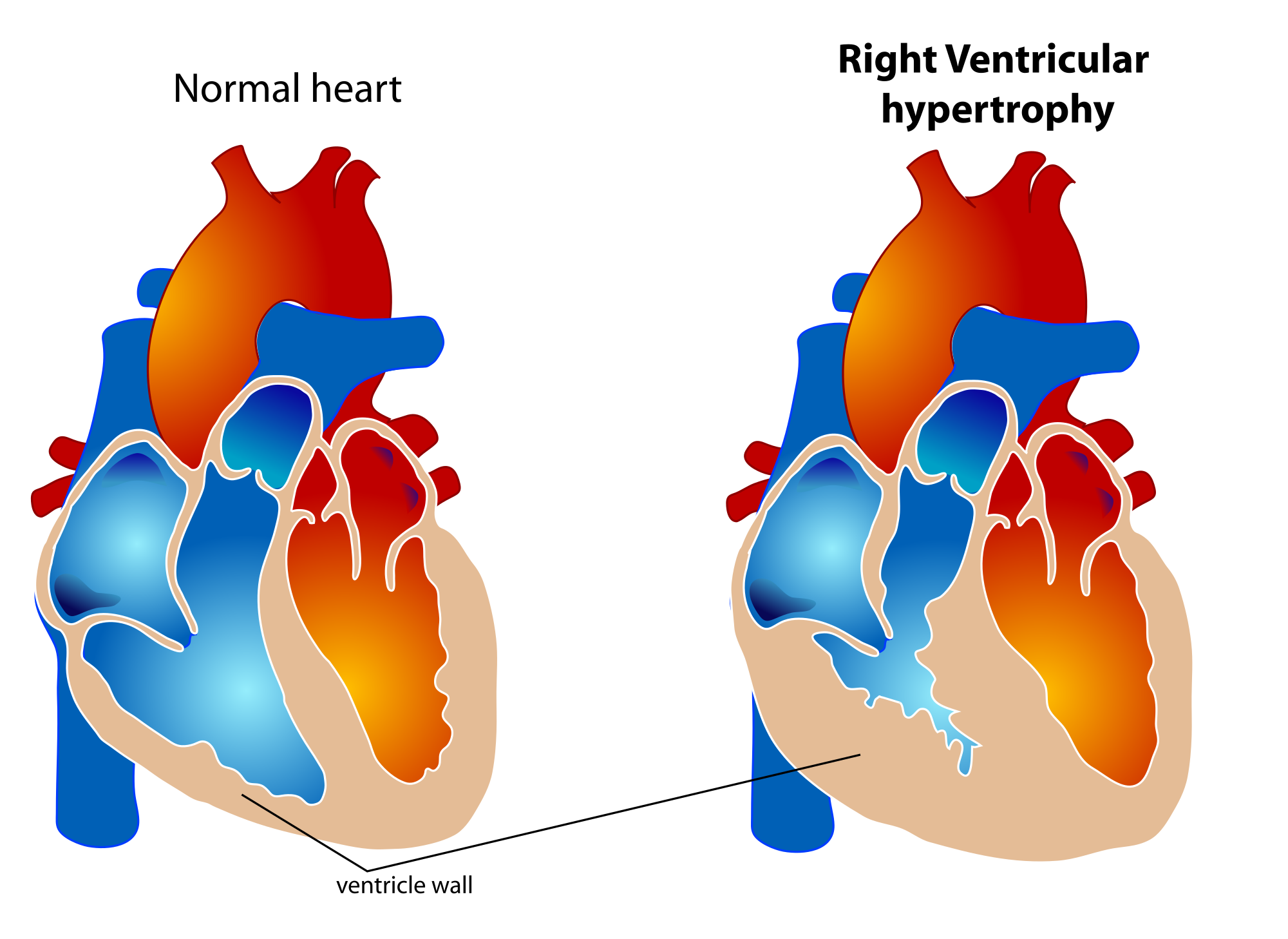
Ventricular hypertrophy (VH) is thickening of the walls of a ventricle (lower chamber) of the heart.[better source needed] Although left ventricular hypertrophy (LVH) is more common, right ventricular hypertrophy (RVH), as well as concurrent hypertrophy of both ventricles can also occur.
Ventricular hypertrophy can result from a variety of conditions, both adaptive and maladaptive. For example, it occurs in what is regarded as a physiologic, adaptive process in pregnancy in response to increased blood volume; but can also occur as a consequence of ventricular remodeling following a heart attack. Importantly, pathologic and physiologic remodeling engage different cellular pathways in the heart and result in different gross cardiac phenotypes.
In individuals with eccentric hypertrophy there may be little or no indication that hypertrophy has occurred as it is generally a healthy response to increased demands on the heart. Conversely, concentric hypertrophy can make itself known in a variety of ways. Most commonly, chest pain, either with or without exertion is present, along with shortness of breath with exertion, general fatigue, syncope, and palpitations. Overt signs of heart failure, such as edema, or shortness of breath without exertion are uncommon.[citation needed]
The ventricles are the chambers in the heart responsible for pumping blood either to the lungs (right ventricle) or to the rest of the body (left ventricle). Ventricular hypertrophy may be divided into two categories: concentric hypertrophy and eccentric hypertrophy. These adaptations are related to how the cardiomyocyte contractile units, called sarcomeres, respond to stressors such as exercise or pathology. Concentric hypertrophy is a result of pressure overload on the heart, resulting in parallel sarcomerogenesis (addition of sarcomere units parallel to existing units). Eccentric hypertrophy is related to volume overload and leads to the addition of sarcomeres in series.
Concentric hypertrophy results from various stressors to the heart including hypertension, congenital heart defects (such as Tetralogy of Fallot), valvular defects (aortic coarction or stenosis), and primary defects of the myocardium which directly cause hypertrophy (hypertrophic cardiomyopathy). The underlying commonality in these disease states is an increase in pressures that the ventricles experience. For example, in tetralogy of Fallot, the right ventricle is exposed to the high pressures of the left heart due to a defect in the septum; as a result the right ventricle undergoes hypertrophy to compensate for these increased pressures. Similarly, in systemic hypertension, the left ventricle must work harder to overcome the higher pressures of the vascular system and responds by thickening to deal with increased wall stress.
Concentric hypertrophy is characterized by an addition of sarcomeres (the contractile units of cardiac cells) in parallel. The result is an increase in thickness of the myocardium without a corresponding increase in ventricular size. This is maladaptive largely because there is not a corresponding proliferation of the vasculature supplying the myocardium, resulting in ischemic areas of the heart. Ultimately, this response can be compensatory for a duration, and allow for improved cardiac function in the face of stressors. However, this type of hypertrophy can result in a dilated ventricle which is unable to effectively pump blood, leading to heart failure. When stressors that encourage this concentric hypertrophy are reduced or eliminated (either surgically corrected in the case of cardiac defects, or hypertension is reduced from diet and exercise) it is possible for the heart to undergo 'reverse remodeling', returning to a somewhat more 'normal' state instead of progressing to a dilated, pathologic phenotype. This reversion may even go beyond muscle mass, and repair abnormalities in cardiac connective tissue.
Eccentric hypertrophy is generally regarded as healthy, or physiologic hypertrophy and is often termed "athlete's heart." It is the normal response to healthy exercise or pregnancy, which results in an increase in the heart's muscle mass and pumping ability. It is a response to 'volume-overload', either as a result of increased blood return to the heart during exercise, or a response to an actual increase in absolute blood volume as in pregnancy. This increase in pumping ability is the result of the addition of sarcomeres in series, which enables the heart to contract with greater force. This is explained by the Frank Starling mechanism, which describes the sarcomere's ability to contract with greater force as more of the elements of its contractile units become engaged. This response can be dramatic; in trained athletes have hearts that have left ventricular mass up to 60% greater than untrained subjects. Rowers, cyclists, and cross-country skiers tend to have the largest hearts, with an average left ventricular wall thickness of 1.3 centimeters, compared to 1.1 centimeters in average adults. Though eccentric hypertrophy is termed 'athlete's heart' it is typically only found in individuals who are aerobically conditioned. For example, weight lifters tend to undergo remodeling which more closely resembles concentric hypertrophy, as the heart does not experience a volume-overload, but instead responds to transient pressure overload as a consequence of increased vascular resistance from pressures exerted on arteries by sustained muscular contraction.[citation needed]
Though it is the case that eccentric hypertrophy is largely considered to be a healthy response to increased cardiac demand, it is also associated with risks. For example, in athletes with significantly increased left ventricular weight there is also a corresponding increased risk for conduction abnormalities and sudden cardiac death. Additionally, in pregnant individuals, a subpopulation progress to peripartum cardiomyopathy, characterized by a dilation of the left ventricle and a corresponding deficit in heart function. There are suggestions that this progression is partially determined by underlying metabolic derangement (diabetes) and hypertension which may result in a more maladaptive cardiac response to pregnancy. As such, though it is convenient to consider clear cut distinctions between pathologic and physiologic cardiac hypertrophy, there may be a broader range of phenotypes than may be accounted for by gross cardiac phenotypes alone.[citation needed]
Ventricular hypertrophy
Ventricular hypertrophy (VH) is thickening of the walls of a ventricle (lower chamber) of the heart.[better source needed] Although left ventricular hypertrophy (LVH) is more common, right ventricular hypertrophy (RVH), as well as concurrent hypertrophy of both ventricles can also occur.
Ventricular hypertrophy can result from a variety of conditions, both adaptive and maladaptive. For example, it occurs in what is regarded as a physiologic, adaptive process in pregnancy in response to increased blood volume; but can also occur as a consequence of ventricular remodeling following a heart attack. Importantly, pathologic and physiologic remodeling engage different cellular pathways in the heart and result in different gross cardiac phenotypes.
In individuals with eccentric hypertrophy there may be little or no indication that hypertrophy has occurred as it is generally a healthy response to increased demands on the heart. Conversely, concentric hypertrophy can make itself known in a variety of ways. Most commonly, chest pain, either with or without exertion is present, along with shortness of breath with exertion, general fatigue, syncope, and palpitations. Overt signs of heart failure, such as edema, or shortness of breath without exertion are uncommon.[citation needed]
The ventricles are the chambers in the heart responsible for pumping blood either to the lungs (right ventricle) or to the rest of the body (left ventricle). Ventricular hypertrophy may be divided into two categories: concentric hypertrophy and eccentric hypertrophy. These adaptations are related to how the cardiomyocyte contractile units, called sarcomeres, respond to stressors such as exercise or pathology. Concentric hypertrophy is a result of pressure overload on the heart, resulting in parallel sarcomerogenesis (addition of sarcomere units parallel to existing units). Eccentric hypertrophy is related to volume overload and leads to the addition of sarcomeres in series.
Concentric hypertrophy results from various stressors to the heart including hypertension, congenital heart defects (such as Tetralogy of Fallot), valvular defects (aortic coarction or stenosis), and primary defects of the myocardium which directly cause hypertrophy (hypertrophic cardiomyopathy). The underlying commonality in these disease states is an increase in pressures that the ventricles experience. For example, in tetralogy of Fallot, the right ventricle is exposed to the high pressures of the left heart due to a defect in the septum; as a result the right ventricle undergoes hypertrophy to compensate for these increased pressures. Similarly, in systemic hypertension, the left ventricle must work harder to overcome the higher pressures of the vascular system and responds by thickening to deal with increased wall stress.
Concentric hypertrophy is characterized by an addition of sarcomeres (the contractile units of cardiac cells) in parallel. The result is an increase in thickness of the myocardium without a corresponding increase in ventricular size. This is maladaptive largely because there is not a corresponding proliferation of the vasculature supplying the myocardium, resulting in ischemic areas of the heart. Ultimately, this response can be compensatory for a duration, and allow for improved cardiac function in the face of stressors. However, this type of hypertrophy can result in a dilated ventricle which is unable to effectively pump blood, leading to heart failure. When stressors that encourage this concentric hypertrophy are reduced or eliminated (either surgically corrected in the case of cardiac defects, or hypertension is reduced from diet and exercise) it is possible for the heart to undergo 'reverse remodeling', returning to a somewhat more 'normal' state instead of progressing to a dilated, pathologic phenotype. This reversion may even go beyond muscle mass, and repair abnormalities in cardiac connective tissue.
Eccentric hypertrophy is generally regarded as healthy, or physiologic hypertrophy and is often termed "athlete's heart." It is the normal response to healthy exercise or pregnancy, which results in an increase in the heart's muscle mass and pumping ability. It is a response to 'volume-overload', either as a result of increased blood return to the heart during exercise, or a response to an actual increase in absolute blood volume as in pregnancy. This increase in pumping ability is the result of the addition of sarcomeres in series, which enables the heart to contract with greater force. This is explained by the Frank Starling mechanism, which describes the sarcomere's ability to contract with greater force as more of the elements of its contractile units become engaged. This response can be dramatic; in trained athletes have hearts that have left ventricular mass up to 60% greater than untrained subjects. Rowers, cyclists, and cross-country skiers tend to have the largest hearts, with an average left ventricular wall thickness of 1.3 centimeters, compared to 1.1 centimeters in average adults. Though eccentric hypertrophy is termed 'athlete's heart' it is typically only found in individuals who are aerobically conditioned. For example, weight lifters tend to undergo remodeling which more closely resembles concentric hypertrophy, as the heart does not experience a volume-overload, but instead responds to transient pressure overload as a consequence of increased vascular resistance from pressures exerted on arteries by sustained muscular contraction.[citation needed]
Though it is the case that eccentric hypertrophy is largely considered to be a healthy response to increased cardiac demand, it is also associated with risks. For example, in athletes with significantly increased left ventricular weight there is also a corresponding increased risk for conduction abnormalities and sudden cardiac death. Additionally, in pregnant individuals, a subpopulation progress to peripartum cardiomyopathy, characterized by a dilation of the left ventricle and a corresponding deficit in heart function. There are suggestions that this progression is partially determined by underlying metabolic derangement (diabetes) and hypertension which may result in a more maladaptive cardiac response to pregnancy. As such, though it is convenient to consider clear cut distinctions between pathologic and physiologic cardiac hypertrophy, there may be a broader range of phenotypes than may be accounted for by gross cardiac phenotypes alone.[citation needed]
Recent media
Recent media