Recent from talks
Septal area
Knowledge base stats:
Talk channels stats:
Members stats:
Septal area
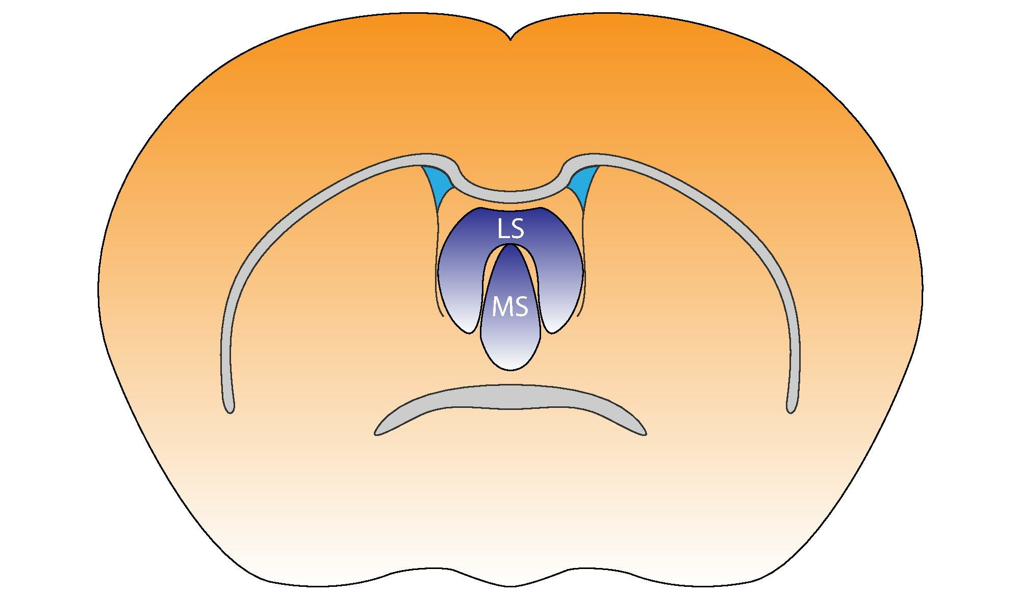
The septal area (medial olfactory area) is an area in the lower, posterior part of the medial surface of the frontal lobe, and refers to the nearby septum pellucidum. It consists of the lateral septum and medial septum.
The septal nuclei are located in this area. The septal nuclei are composed of medium-size neurons which are classified into dorsal, ventral, medial, and caudal groups. The septal nuclei receive reciprocal connections from the olfactory bulb, hippocampus, amygdala, hypothalamus, midbrain, habenula, cingulate gyrus, and thalamus. The septal nuclei are essential in generating the theta rhythm of the hippocampus.
The septal area (medial olfactory area) has no relation to the sense of smell, but it is considered a pleasure zone in animals. The septal nuclei play a role in reward and reinforcement along with the nucleus accumbens. In the 1950s, Olds & Milner showed that rats with electrodes implanted in this area will self-stimulate repeatedly (e.g., press a bar to receive electric current that stimulate the neurons). Experiments on the septal area of humans have taken place since the 1960s.
The septal area is located on the lower posterior part of the frontal lobe. The septal area refers to the nearby septum pellucidum. It is located underneath the corpus callosum and in front of the lamina terminalis. The lamina terminalis is a layer of gray matter that connects the optic chiasma and the anterior commissure. The septal nuclei in the septal area are essential in generating the theta rhythm of the hippocampus.
The dorsal septum projects to the lateral preoptic area, lateral hypothalamus, periventricular hypothalamus and midline thalamus.
Fibers from the ventral half of the septum project topographically to the hippocampal formation, thalamus, hypothalamus and midbrain. Specifically, neurons located along the midline in the vertical limb of the diagonal band of Broca project through the dorsal fornix to all CA fields of the dorsal hippocampus and adjacent subicular cortex. Other fibers from this region project through the stria medullaris to the medial and lateral habenular nuclei, the paratenial and anteromedial nucleus of the thalamus, and through the medial forebrain bundle to the pars posterior of the medial mammillary nucleus.
Cells located in the intermediolateral septum also project through the lateral part of the fimbria to all CA fields of the ventral hippocampus and adjacent subicular and entorhinal cortices. These cells also send fibers through the stria medullaris to the lateral habenular nucleus and mediodorsal thalamic nucleus. Other axons arising from these cells descend through the medial forebrain bundle to terminate in a region dorsal to the interpeduncular nucleus.
The lateral septum is a relay center for connections from the CA3 of the hippocampus to the ventral tegmental area. These connections help link reward signals with the context in which they occur.
Hub AI
Septal area AI simulator
(@Septal area_simulator)
Septal area
The septal area (medial olfactory area) is an area in the lower, posterior part of the medial surface of the frontal lobe, and refers to the nearby septum pellucidum. It consists of the lateral septum and medial septum.
The septal nuclei are located in this area. The septal nuclei are composed of medium-size neurons which are classified into dorsal, ventral, medial, and caudal groups. The septal nuclei receive reciprocal connections from the olfactory bulb, hippocampus, amygdala, hypothalamus, midbrain, habenula, cingulate gyrus, and thalamus. The septal nuclei are essential in generating the theta rhythm of the hippocampus.
The septal area (medial olfactory area) has no relation to the sense of smell, but it is considered a pleasure zone in animals. The septal nuclei play a role in reward and reinforcement along with the nucleus accumbens. In the 1950s, Olds & Milner showed that rats with electrodes implanted in this area will self-stimulate repeatedly (e.g., press a bar to receive electric current that stimulate the neurons). Experiments on the septal area of humans have taken place since the 1960s.
The septal area is located on the lower posterior part of the frontal lobe. The septal area refers to the nearby septum pellucidum. It is located underneath the corpus callosum and in front of the lamina terminalis. The lamina terminalis is a layer of gray matter that connects the optic chiasma and the anterior commissure. The septal nuclei in the septal area are essential in generating the theta rhythm of the hippocampus.
The dorsal septum projects to the lateral preoptic area, lateral hypothalamus, periventricular hypothalamus and midline thalamus.
Fibers from the ventral half of the septum project topographically to the hippocampal formation, thalamus, hypothalamus and midbrain. Specifically, neurons located along the midline in the vertical limb of the diagonal band of Broca project through the dorsal fornix to all CA fields of the dorsal hippocampus and adjacent subicular cortex. Other fibers from this region project through the stria medullaris to the medial and lateral habenular nuclei, the paratenial and anteromedial nucleus of the thalamus, and through the medial forebrain bundle to the pars posterior of the medial mammillary nucleus.
Cells located in the intermediolateral septum also project through the lateral part of the fimbria to all CA fields of the ventral hippocampus and adjacent subicular and entorhinal cortices. These cells also send fibers through the stria medullaris to the lateral habenular nucleus and mediodorsal thalamic nucleus. Other axons arising from these cells descend through the medial forebrain bundle to terminate in a region dorsal to the interpeduncular nucleus.
The lateral septum is a relay center for connections from the CA3 of the hippocampus to the ventral tegmental area. These connections help link reward signals with the context in which they occur.
Recent media