Recent from talks
Single-molecule experiment
Knowledge base stats:
Talk channels stats:
Members stats:
Single-molecule experiment
A single-molecule experiment is an experiment that investigates the properties of individual molecules. Single-molecule studies may be contrasted with measurements on an ensemble or bulk collection of molecules, where the individual behavior of molecules cannot be distinguished, and only average characteristics can be measured. Since many measurement techniques in biology, chemistry, and physics are not sensitive enough to observe single molecules, single-molecule fluorescence techniques (that have emerged since the 1990s for probing various processes on the level of individual molecules) caused a lot of excitement, since these supplied many new details on the measured processes that were not accessible in the past. Indeed, since the 1990s, many techniques for probing individual molecules have been developed.
The first single-molecule experiments were patch clamp experiments performed in the 1970s, but these were limited to studying ion channels. Today, systems investigated using single-molecule techniques include the movement of myosin on actin filaments in muscle tissue and the spectroscopic details of individual local environments in solids. Biological polymers' conformations have been measured using atomic force microscopy (AFM). Using force spectroscopy, single molecules (or pairs of interacting molecules), usually polymers, can be mechanically stretched, and their elastic response recorded in real time.
In the gas phase at ultralow pressures, single-molecule experiments have been around for decades, but in the condensed phase only since 1989 with the work by W. E. Moerner and Lothar Kador. One year later, Michel Orrit and Jacky Bernard were able to show also the detection of the absorption of single molecules by their fluorescence.
Many techniques have the ability to observe one molecule at a time, most notably mass spectrometry, where single ions are detected. In addition, one of the earliest means of detecting single molecules, came about in the field of ion channels with the development of the patch clamp technique by Erwin Neher and Bert Sakmann (who later went on to win the Nobel prize for their seminal contributions). However, the idea of measuring conductance to look at single molecules placed a serious limitation on the kind of systems which could be observed.
Fluorescence is a convenient means of observing one molecule at a time, mostly due to the sensitivity of commercial optical detectors, capable of counting single photons. However, spectroscopically, the observation of one molecule requires that the molecule is in an isolated environment and that it emits photons upon excitation, which owing to the technology to detect single photons by use of photomultiplier tubes (PMT) or avalanche photodiodes (APD), enables one to record photon emission events with great sensitivity and time resolution.
More recently, single-molecule fluorescence is the subject of intense interest for biological imaging, through the labeling of biomolecules such as proteins and nucleotides to study enzymatic function which cannot easily be studied on the bulk scale, due to subtle time-dependent movements in catalysis and structural reorganization. The most studied protein has been the class of myosin/actin enzymes found in muscle tissues. Through single-molecule techniques the step mechanism has been observed and characterized in many of these proteins.
In 1997, single-molecule detection was demonstrated with surface-enhanced Raman spectroscopy (SERS) by Katrin Kneipp, H. Kneipp, Y. Wang, L.T. Perelman and others at MIT and independently by S. Nie and S. R. Emory at Indiana University. The MIT team used non-resonance Raman excitation and surface enhancement with silver nanoclusters to detect single cresyl violet molecules, while the team at Indiana University used resonance Raman excitation and surface enhancement with silver nanoparticles to detect single rhodamine 6G molecules.
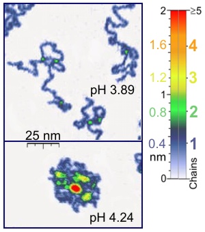
Nanomanipulators such as the atomic force microscope are also suited to single-molecule experiments of biological significance, since they work on the same length scale of most biological polymers. Besides, atomic force microscopy (AFM) is appropriate for the studies of synthetic polymer molecules. AFM provides a unique possibility of 3D visualization of polymer chains. For instance, AFM tapping mode is gentle enough for the recording of adsorbed polyelectrolyte molecules (for example, 0.4 nm thick chains of poly(2-vinylpyridine)) under liquid medium. The location of two-chain-superposition correspond in these experiments to twice the thickness of single chain (0.8 nm in the case of the mentioned example). At the application of proper scanning parameters, conformation of such molecules remain unchanged for hours that allows the performance of experiments under liquid media having various properties. Furthermore, by controlling the force between the tip and the sample high resolution images can be obtained. Optical tweezers have also been used to study and quantify DNA-protein interactions.
Hub AI
Single-molecule experiment AI simulator
(@Single-molecule experiment_simulator)
Single-molecule experiment
A single-molecule experiment is an experiment that investigates the properties of individual molecules. Single-molecule studies may be contrasted with measurements on an ensemble or bulk collection of molecules, where the individual behavior of molecules cannot be distinguished, and only average characteristics can be measured. Since many measurement techniques in biology, chemistry, and physics are not sensitive enough to observe single molecules, single-molecule fluorescence techniques (that have emerged since the 1990s for probing various processes on the level of individual molecules) caused a lot of excitement, since these supplied many new details on the measured processes that were not accessible in the past. Indeed, since the 1990s, many techniques for probing individual molecules have been developed.
The first single-molecule experiments were patch clamp experiments performed in the 1970s, but these were limited to studying ion channels. Today, systems investigated using single-molecule techniques include the movement of myosin on actin filaments in muscle tissue and the spectroscopic details of individual local environments in solids. Biological polymers' conformations have been measured using atomic force microscopy (AFM). Using force spectroscopy, single molecules (or pairs of interacting molecules), usually polymers, can be mechanically stretched, and their elastic response recorded in real time.
In the gas phase at ultralow pressures, single-molecule experiments have been around for decades, but in the condensed phase only since 1989 with the work by W. E. Moerner and Lothar Kador. One year later, Michel Orrit and Jacky Bernard were able to show also the detection of the absorption of single molecules by their fluorescence.
Many techniques have the ability to observe one molecule at a time, most notably mass spectrometry, where single ions are detected. In addition, one of the earliest means of detecting single molecules, came about in the field of ion channels with the development of the patch clamp technique by Erwin Neher and Bert Sakmann (who later went on to win the Nobel prize for their seminal contributions). However, the idea of measuring conductance to look at single molecules placed a serious limitation on the kind of systems which could be observed.
Fluorescence is a convenient means of observing one molecule at a time, mostly due to the sensitivity of commercial optical detectors, capable of counting single photons. However, spectroscopically, the observation of one molecule requires that the molecule is in an isolated environment and that it emits photons upon excitation, which owing to the technology to detect single photons by use of photomultiplier tubes (PMT) or avalanche photodiodes (APD), enables one to record photon emission events with great sensitivity and time resolution.
More recently, single-molecule fluorescence is the subject of intense interest for biological imaging, through the labeling of biomolecules such as proteins and nucleotides to study enzymatic function which cannot easily be studied on the bulk scale, due to subtle time-dependent movements in catalysis and structural reorganization. The most studied protein has been the class of myosin/actin enzymes found in muscle tissues. Through single-molecule techniques the step mechanism has been observed and characterized in many of these proteins.
In 1997, single-molecule detection was demonstrated with surface-enhanced Raman spectroscopy (SERS) by Katrin Kneipp, H. Kneipp, Y. Wang, L.T. Perelman and others at MIT and independently by S. Nie and S. R. Emory at Indiana University. The MIT team used non-resonance Raman excitation and surface enhancement with silver nanoclusters to detect single cresyl violet molecules, while the team at Indiana University used resonance Raman excitation and surface enhancement with silver nanoparticles to detect single rhodamine 6G molecules.
Nanomanipulators such as the atomic force microscope are also suited to single-molecule experiments of biological significance, since they work on the same length scale of most biological polymers. Besides, atomic force microscopy (AFM) is appropriate for the studies of synthetic polymer molecules. AFM provides a unique possibility of 3D visualization of polymer chains. For instance, AFM tapping mode is gentle enough for the recording of adsorbed polyelectrolyte molecules (for example, 0.4 nm thick chains of poly(2-vinylpyridine)) under liquid medium. The location of two-chain-superposition correspond in these experiments to twice the thickness of single chain (0.8 nm in the case of the mentioned example). At the application of proper scanning parameters, conformation of such molecules remain unchanged for hours that allows the performance of experiments under liquid media having various properties. Furthermore, by controlling the force between the tip and the sample high resolution images can be obtained. Optical tweezers have also been used to study and quantify DNA-protein interactions.
Recent media