Recent from talks
Sound localization in owls
Knowledge base stats:
Talk channels stats:
Members stats:
Sound localization in owls
Most owls are nocturnal or crepuscular birds of prey. Because they hunt at night, they must rely on non-visual senses. Experiments by Roger Payne have shown that owls are sensitive to the sounds made by their prey, not the heat or the smell. In fact, the sound cues are both necessary and sufficient for localization of mice from a distant location where they are perched. For this to work, the owls must be able to accurately localize both the azimuth and the elevation of the sound source.
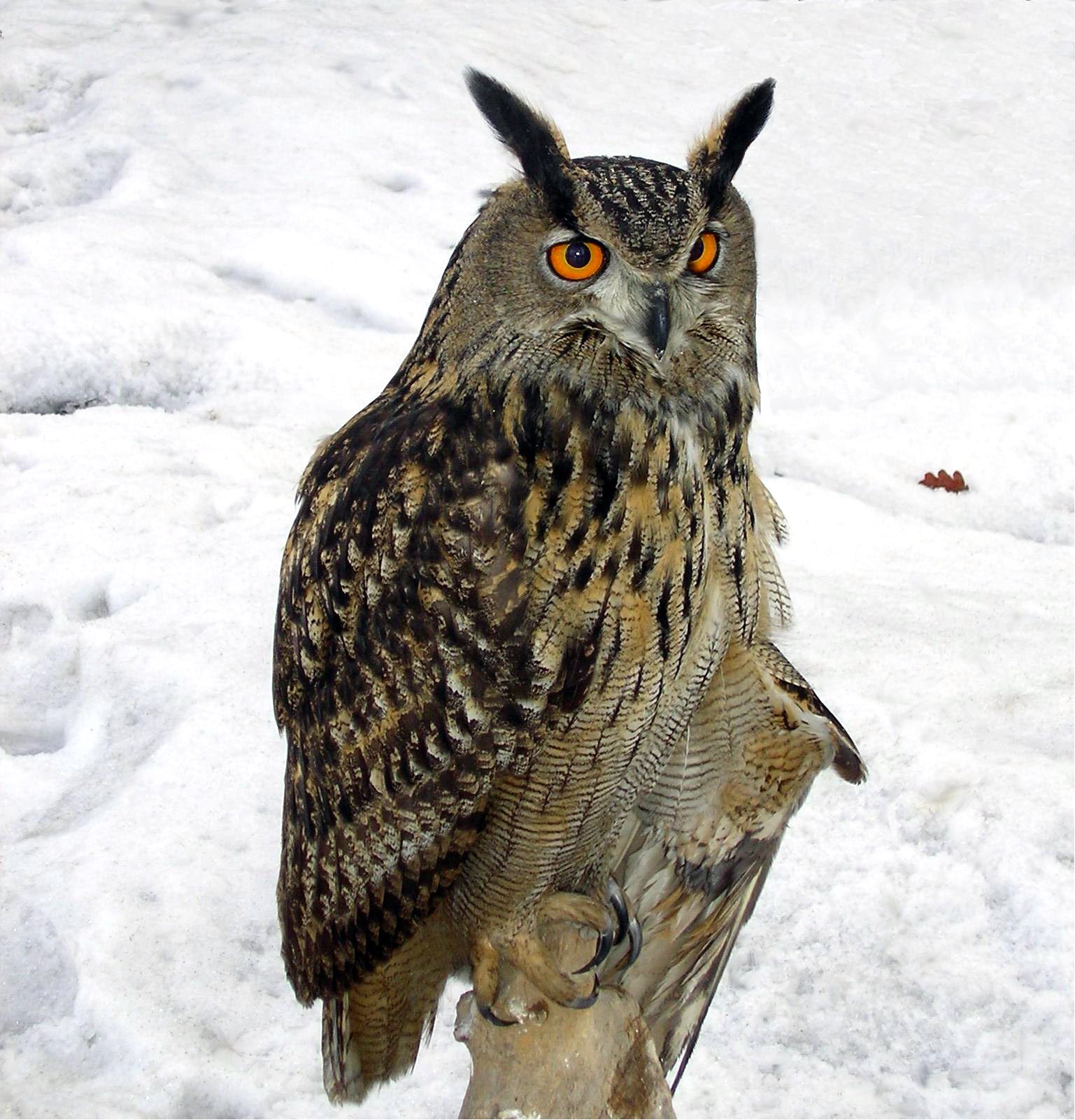
Owls are very adept nocturnal predators, hunting prey that includes small mammals, reptiles, and insects. They are able to rotate their head up to 270 degrees, lock onto prey, and launch a silent attack. Owls lock onto prey by using sound localization. Sound localization is an animal's ability to identify the origin of a sound in distance and direction. Several owl species have ears that are asymmetrical in size and location, which enhances this ability. These species include barn owls (Tyto alba), northern saw-whet owls (Aegolius acadicus), and long-eared owls (Asio otus). The barn owl (Tyto alba) is the most commonly studied for sound localization because they use similar methods to humans for interpreting interaural time differences in the horizontal plane. This species has evolved a specialized set of pathways in the brain that allow them to hear a sound and map out the possible location of the object that elicited that sound. Sound waves enter the ear via the ear canal and travel until they reach the tympanic membrane. The tympanic membrane then sends these waves through the ossicles of the middle ear and into the inner ear that includes the vestibular organ, cochlea, and auditory nerve. They are then able to use interaural time difference (ITD) and interaural level difference (ILD) to pinpoint the location and elevation of their prey.
Owls tend to have asymmetric ears, with the openings being placed just behind the eyes. The shape of the ear opening, known as the aperture, depends on the species. In some species, the opening has a valve, called an operculum, covering it. The left ear opening is typically positioned a bit higher than the right ear opening to aid with sound localization and the detection of prey, even in the dark. During flight, the left ear receives sounds from below them and the right ear receives sounds from above them. The feathers on the edge of barn owl's face creates a disc that works to trap and focus sound, similar to the outer ears of humans. The sound waves travel through the owl's ear canal until they reach the eardrum, through the ossicles, and into the inner ear so that the owl is able to perceive exactly where their prey is located. As owls depend on their ability to track prey using their super sense of hearing in order to survive, it is important to understand the structures of the ear of barn owls that work to transmit these sounds.
The inner ear of barn owls includes the vestibular organ, cochlea, and auditory nerve. The anatomy of the inner ear in barn owls was studied in an experiment where three owls were utilized and fixed at laboratories by the intravascular perfusion of 1% formaldehyde and 1.25% glutaraldehyde in a 0.1 phosphate buffer. The temporal lobes of the owls were then removed from the skulls, post-fixed in 1% osmium tetroxide, dehydrated, then embedded in Araldite to study the anatomy of the inner ear. This study revealed that the basilar papilla of barn owls has two unique features being a proliferation of lenticular cells and a thickening of the basilar membrane.
The cochlear duct of the owl contains the basilar papilla, the tectorial membrane, the tegmentum vasculum, and the macula of the lagena. The basilar papilla of the cochlea was measured to be 9.5-11.5 mm long. Proximal sensory hair cells contained mostly short hair cells along with a few intermediate hair cells, but absolutely no tall hair cells. Tall hair cells are only present on the distal half of the owl's papilla, starting at about 5 mm from the proximal end, along with some short-haired cells. The distal tip of the papilla is occupied exclusively by tall hair cells, whereas the proximal tip is occupied exclusively by lenticular cells.
Two major parts that construct the basilar membrane of the barn owl include the vestibular part and the tympanic part. The vestibular surface of the basilar membrane is covered by supporting cells and a few border cells at the inferior edge of the membrane. The basilar membrane is relatively thin toward the distal end of the papilla, but has a thick fibrous mass toward the proximal end. This dense fibrous mass measured to be about 37-57 μm in width and 8.5-11 μm in thickness among the owls utilized for this study. This mass is not to be confused with the loose fibrous mass of the tympanic part of the basilar membrane that underlies the part of the basilar membrane that is covered by sensory cells. The loose fibrous mass of the tympanic part arises at the proximal tip along with the dense fibrous mass, but the loose fibrous mass covers a greater width of the basilar membrane. The only thing that separates the vestibular and tympanic parts are thin, discontinuous cords of fibers. These cords of fibers are visible at the pendulous tympanic edge of the loose fibrous mass in the proximal part of the papilla, but are much more scattered throughout the mass distally.
Researchers have studied the embryonic development of ear asymmetry in the American barn owl (Tyto furcata pratincola) in the frame of 42 different stages of embryonic development. The experiment began by taking 19 embryos from the Institute of Biology to RWTH Aachen University. The embryos were either raised by the hen or were incubated in a Compact S84. The embryos utilized in the study were between stage 29 and stage 41 of embryonic development, with those between stages 36-39 being examined a bit more frequently than others. Micro CT-scans were used to measure the asymmetry of the ear during development, with some scans being taken using the Skyscan 1172. Aluminum filters were used to optimize image quality and phosphotungstic acid (PTA) was used to enhance soft tissue extract in the embryos. Markers were set on the embryonic heads to track differential growth. NRecon was used to reconstruct raw data, three-dimensional objects were calculated in Reconstruct, photographs were produced in Blender, and all final figures were constructed with Matlab and CorelDraw X6. The center of the ear opening, squamosum, stapes, eye lens, nose, and premaxilla were all tracked in the study. The cervical vertebra (C1) and the pituitary gland were used to define the anatomical coordinate system. Measurements of the ear-opening, surface area of the ear, and eccentricity were derived using various equations.
Through the examination of data, the development of ear asymmetry was apparent throughout the stages of embryonic development. The ears appeared slit-like at stage 29 of embryonic development and took on the ellipsoid shape at stage 32, which is also when outer ear development began. Dorso-occipital movement of the outer ear began symmetrically at stage 34, but asymmetry rapidly increased from this dorso-occipital movement by stage 37. Surface area between stages 32 and 35 was 0.4-0.5 mm2, with eccentricity between the right and left ear differing by less than 0.1 mm. Between stages 36 and 37, the surface area was 0.6-1.9 mm2. Eccentricity was no longer measured after stage 37 since the ear-opening shapes were no longer ellipsoid. The rostral margin of the ear opening started to flatten at stage 38, with the higher ear on the head having more dorso-ventral flattening. The angle of flattening margins at stage 38 differed by 18.5 degrees, but by only 16 degrees at stage 39. Ear asymmetry did not increase between stages 40 and 41, as ear asymmetry reached maximum at stage 39. The margin differed by 21 degrees at stage 40 and by 33 degrees at stage 41.
Hub AI
Sound localization in owls AI simulator
(@Sound localization in owls_simulator)
Sound localization in owls
Most owls are nocturnal or crepuscular birds of prey. Because they hunt at night, they must rely on non-visual senses. Experiments by Roger Payne have shown that owls are sensitive to the sounds made by their prey, not the heat or the smell. In fact, the sound cues are both necessary and sufficient for localization of mice from a distant location where they are perched. For this to work, the owls must be able to accurately localize both the azimuth and the elevation of the sound source.
Owls are very adept nocturnal predators, hunting prey that includes small mammals, reptiles, and insects. They are able to rotate their head up to 270 degrees, lock onto prey, and launch a silent attack. Owls lock onto prey by using sound localization. Sound localization is an animal's ability to identify the origin of a sound in distance and direction. Several owl species have ears that are asymmetrical in size and location, which enhances this ability. These species include barn owls (Tyto alba), northern saw-whet owls (Aegolius acadicus), and long-eared owls (Asio otus). The barn owl (Tyto alba) is the most commonly studied for sound localization because they use similar methods to humans for interpreting interaural time differences in the horizontal plane. This species has evolved a specialized set of pathways in the brain that allow them to hear a sound and map out the possible location of the object that elicited that sound. Sound waves enter the ear via the ear canal and travel until they reach the tympanic membrane. The tympanic membrane then sends these waves through the ossicles of the middle ear and into the inner ear that includes the vestibular organ, cochlea, and auditory nerve. They are then able to use interaural time difference (ITD) and interaural level difference (ILD) to pinpoint the location and elevation of their prey.
Owls tend to have asymmetric ears, with the openings being placed just behind the eyes. The shape of the ear opening, known as the aperture, depends on the species. In some species, the opening has a valve, called an operculum, covering it. The left ear opening is typically positioned a bit higher than the right ear opening to aid with sound localization and the detection of prey, even in the dark. During flight, the left ear receives sounds from below them and the right ear receives sounds from above them. The feathers on the edge of barn owl's face creates a disc that works to trap and focus sound, similar to the outer ears of humans. The sound waves travel through the owl's ear canal until they reach the eardrum, through the ossicles, and into the inner ear so that the owl is able to perceive exactly where their prey is located. As owls depend on their ability to track prey using their super sense of hearing in order to survive, it is important to understand the structures of the ear of barn owls that work to transmit these sounds.
The inner ear of barn owls includes the vestibular organ, cochlea, and auditory nerve. The anatomy of the inner ear in barn owls was studied in an experiment where three owls were utilized and fixed at laboratories by the intravascular perfusion of 1% formaldehyde and 1.25% glutaraldehyde in a 0.1 phosphate buffer. The temporal lobes of the owls were then removed from the skulls, post-fixed in 1% osmium tetroxide, dehydrated, then embedded in Araldite to study the anatomy of the inner ear. This study revealed that the basilar papilla of barn owls has two unique features being a proliferation of lenticular cells and a thickening of the basilar membrane.
The cochlear duct of the owl contains the basilar papilla, the tectorial membrane, the tegmentum vasculum, and the macula of the lagena. The basilar papilla of the cochlea was measured to be 9.5-11.5 mm long. Proximal sensory hair cells contained mostly short hair cells along with a few intermediate hair cells, but absolutely no tall hair cells. Tall hair cells are only present on the distal half of the owl's papilla, starting at about 5 mm from the proximal end, along with some short-haired cells. The distal tip of the papilla is occupied exclusively by tall hair cells, whereas the proximal tip is occupied exclusively by lenticular cells.
Two major parts that construct the basilar membrane of the barn owl include the vestibular part and the tympanic part. The vestibular surface of the basilar membrane is covered by supporting cells and a few border cells at the inferior edge of the membrane. The basilar membrane is relatively thin toward the distal end of the papilla, but has a thick fibrous mass toward the proximal end. This dense fibrous mass measured to be about 37-57 μm in width and 8.5-11 μm in thickness among the owls utilized for this study. This mass is not to be confused with the loose fibrous mass of the tympanic part of the basilar membrane that underlies the part of the basilar membrane that is covered by sensory cells. The loose fibrous mass of the tympanic part arises at the proximal tip along with the dense fibrous mass, but the loose fibrous mass covers a greater width of the basilar membrane. The only thing that separates the vestibular and tympanic parts are thin, discontinuous cords of fibers. These cords of fibers are visible at the pendulous tympanic edge of the loose fibrous mass in the proximal part of the papilla, but are much more scattered throughout the mass distally.
Researchers have studied the embryonic development of ear asymmetry in the American barn owl (Tyto furcata pratincola) in the frame of 42 different stages of embryonic development. The experiment began by taking 19 embryos from the Institute of Biology to RWTH Aachen University. The embryos were either raised by the hen or were incubated in a Compact S84. The embryos utilized in the study were between stage 29 and stage 41 of embryonic development, with those between stages 36-39 being examined a bit more frequently than others. Micro CT-scans were used to measure the asymmetry of the ear during development, with some scans being taken using the Skyscan 1172. Aluminum filters were used to optimize image quality and phosphotungstic acid (PTA) was used to enhance soft tissue extract in the embryos. Markers were set on the embryonic heads to track differential growth. NRecon was used to reconstruct raw data, three-dimensional objects were calculated in Reconstruct, photographs were produced in Blender, and all final figures were constructed with Matlab and CorelDraw X6. The center of the ear opening, squamosum, stapes, eye lens, nose, and premaxilla were all tracked in the study. The cervical vertebra (C1) and the pituitary gland were used to define the anatomical coordinate system. Measurements of the ear-opening, surface area of the ear, and eccentricity were derived using various equations.
Through the examination of data, the development of ear asymmetry was apparent throughout the stages of embryonic development. The ears appeared slit-like at stage 29 of embryonic development and took on the ellipsoid shape at stage 32, which is also when outer ear development began. Dorso-occipital movement of the outer ear began symmetrically at stage 34, but asymmetry rapidly increased from this dorso-occipital movement by stage 37. Surface area between stages 32 and 35 was 0.4-0.5 mm2, with eccentricity between the right and left ear differing by less than 0.1 mm. Between stages 36 and 37, the surface area was 0.6-1.9 mm2. Eccentricity was no longer measured after stage 37 since the ear-opening shapes were no longer ellipsoid. The rostral margin of the ear opening started to flatten at stage 38, with the higher ear on the head having more dorso-ventral flattening. The angle of flattening margins at stage 38 differed by 18.5 degrees, but by only 16 degrees at stage 39. Ear asymmetry did not increase between stages 40 and 41, as ear asymmetry reached maximum at stage 39. The margin differed by 21 degrees at stage 40 and by 33 degrees at stage 41.
Recent media