Hubbry Logo
CartilageCartilageMain
Open search
Cartilage
Community hub
Cartilage
logo
8 pages, 0 posts
0 subscribers
Be the first to start a discussion here.
Contribute something
Cartilage
Cartilage
from Wikipedia
Cartilage
Light micrograph of undecalcified hyaline cartilage showing chondrocytes and organelles, lacunae and matrix.
Identifiers
MeSHD002356
TA98A02.0.00.005
TA2381
Anatomical terminology

Cartilage is a resilient and smooth type of connective tissue. Semi-transparent and non-porous, it is usually covered by a tough and fibrous membrane called perichondrium. In tetrapods, it covers and protects the ends of long bones at the joints as articular cartilage,[1] and is a structural component of many body parts including the rib cage, the neck and the bronchial tubes, and the intervertebral discs. In other taxa, such as chondrichthyans and cyclostomes, it constitutes a much greater proportion of the skeleton.[2] It is not as hard and rigid as bone, but it is much stiffer and much less flexible than muscle or tendon. The matrix of cartilage is made up of glycosaminoglycans, proteoglycans, collagen fibers and, sometimes, elastin. It usually grows quicker than bone.

Because of its rigidity, cartilage often serves the purpose of holding tubes open in the body. Examples include the rings of the trachea, such as the cricoid cartilage and carina.

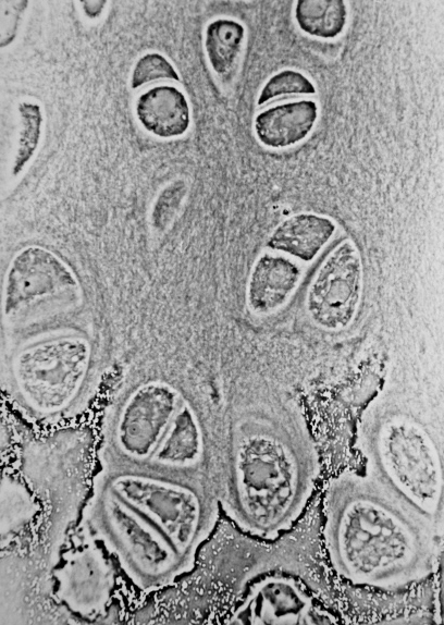
Cartilage is composed of specialized cells called chondrocytes that produce a large amount of collagenous extracellular matrix, abundant ground substance that is rich in proteoglycan and elastin fibers. Cartilage is classified into three types — elastic cartilage, hyaline cartilage, and fibrocartilage — which differ in their relative amounts of collagen and proteoglycan.

As cartilage does not contain blood vessels or nerves, it is insensitive. However, some fibrocartilage such as the meniscus of the knee has partial blood supply. Nutrition is supplied to the chondrocytes by diffusion. The compression of the articular cartilage or flexion of the elastic cartilage generates fluid flow, which assists the diffusion of nutrients to the chondrocytes. Compared to other connective tissues, cartilage has a very slow turnover of its extracellular matrix and is documented to repair at only a very slow rate relative to other tissues.

There are three different types of cartilage: elastic (A), hyaline (B), and fibrous (C). In elastic cartilage, the cells are closer together creating less intercellular space. Elastic cartilage is found in the external ear flaps and in parts of the larynx. Hyaline cartilage has fewer cells than elastic cartilage; there is more intercellular space. Hyaline cartilage is found in the nose, ears, trachea, parts of the larynx, and smaller respiratory tubes. Fibrous cartilage has the fewest cells so it has the most intercellular space. Fibrous cartilage is found in the spine and the menisci.
The physical appearance of cartilage

Structure

[edit]

Development

[edit]

In embryogenesis, the skeletal system is derived from the mesoderm germ layer. Chondrification (also known as chondrogenesis) is the process by which cartilage is formed from condensed mesenchyme tissue, which differentiates into chondroblasts and begins secreting the molecules (aggrecan and collagen type II) that form the extracellular matrix. In all vertebrates, cartilage is the main skeletal tissue in early ontogenetic stages;[3][4] in osteichthyans, many cartilaginous elements subsequently ossify through endochondral and perichondral ossification.[5]

Following the initial chondrification that occurs during embryogenesis, cartilage growth consists mostly of the maturing of immature cartilage to a more mature state. The division of cells within cartilage occurs very slowly, and thus growth in cartilage is usually not based on an increase in size or mass of the cartilage itself.[6] It has been identified that non-coding RNAs (e.g. miRNAs and long non-coding RNAs) as the most important epigenetic modulators can affect the chondrogenesis. This also justifies the non-coding RNAs' contribution in various cartilage-dependent pathological conditions such as arthritis, and so on.[7]

Articular cartilage

[edit]
Section from mouse joint showing cartilage (purple)

The articular cartilage function is dependent on the molecular composition of the extracellular matrix (ECM). The ECM consists mainly of proteoglycan and collagens. The main proteoglycan in cartilage is aggrecan, which, as its name suggests, forms large aggregates with hyaluronan and with itself.[8] These aggregates are negatively charged and hold water in the tissue. The collagen, mostly collagen type II, constrains the proteoglycans. The ECM responds to tensile and compressive forces that are experienced by the cartilage.[9] Cartilage growth thus refers to the matrix deposition, but can also refer to both the growth and remodeling of the extracellular matrix. Due to the great stress on the patellofemoral joint during resisted knee extension, the articular cartilage of the patella is among the thickest in the human body. The ECM of articular cartilage is classified into three regions: the pericellular matrix, the territorial matrix, and the interterritorial matrix.

Function

[edit]

Mechanical properties

[edit]

The mechanical properties of articular cartilage in load-bearing joints such as the knee and hip have been studied extensively at macro, micro, and nano-scales. These mechanical properties include the response of cartilage in frictional, compressive, shear and tensile loading. Cartilage is resilient and displays viscoelastic properties.[10]

Since cartilage has interstitial fluid that is free-moving, it makes the material difficult to test. One of the tests commonly used to overcome this obstacle is a confined compression test, which can be used in either a 'creep' or 'relaxation' mode.[11][12] In creep mode, the tissue displacement is measured as a function of time under a constant load, and in relaxation mode, the force is measured as a function of time under constant displacement. During this mode, the deformation of the tissue has two main regions. In the first region, the displacement is rapid due to the initial flow of fluid out of the cartilage, and in the second region, the displacement slows down to an eventual constant equilibrium value. Under the commonly used loading conditions, the equilibrium displacement can take hours to reach.

In both the creep mode and the relaxation mode of a confined compression test, a disc of cartilage is placed in an impervious, fluid-filled container and covered with a porous plate that restricts the flow of interstitial fluid to the vertical direction. This test can be used to measure the aggregate modulus of cartilage, which is typically in the range of 0.5 to 0.9 MPa for articular cartilage,[11][12][13] and the Young's Modulus, which is typically 0.45 to 0.80 MPa.[11][13] The aggregate modulus is "a measure of the stiffness of the tissue at equilibrium when all fluid flow has ceased",[11] and Young's modulus is a measure of how much a material strains (changes length) under a given stress.

The confined compression test can also be used to measure permeability, which is defined as the resistance to fluid flow through a material. Higher permeability allows for fluid to flow out of a material's matrix more rapidly, while lower permeability leads to an initial rapid fluid flow and a slow decrease to equilibrium. Typically, the permeability of articular cartilage is in the range of 10^-15 to 10^-16 m^4/Ns.[11][12] However, permeability is sensitive to loading conditions and testing location. For example, permeability varies throughout articular cartilage and tends to be highest near the joint surface and lowest near the bone (or "deep zone"). Permeability also decreases under increased loading of the tissue.

Indentation testing is an additional type of test commonly used to characterize cartilage.[11][14] Indentation testing involves using an indentor (usually <0.8 mm) to measure the displacement of the tissue under constant load. Similar to confined compression testing, it may take hours to reach equilibrium displacement. This method of testing can be used to measure the aggregate modulus, Poisson's ratio, and permeability of the tissue. Initially, there was a misconception that due to its predominantly water-based composition, cartilage had a Poisson's ratio of 0.5 and should be modeled as an incompressible material.[11] However, subsequent research has disproven this belief. The Poisson's ratio of articular cartilage has been measured to be around 0.4 or lower in humans [11][14] and ranges from 0.46–0.5 in bovine subjects.[15]

The mechanical properties of articular cartilage are largely anisotropic, test-dependent, and can be age-dependent.[11] These properties also depend on collagen-proteoglycan interactions and therefore can increase/decrease depending on the total content of water, collagen, glycoproteins, etc. For example, increased glucosaminoglycan content leads to an increase in compressive stiffness, and increased water content leads to a lower aggregate modulus.

Tendon-bone interface

[edit]

In addition to its role in load-bearing joints, cartilage serves a crucial function as a gradient material between softer tissues and bone. Mechanical gradients are crucial for your body's function, and for complex artificial structures including joint implants. Interfaces with mismatched material properties lead to areas of high stress concentration which, over the millions of loading cycles experienced by human joins over a lifetime, would eventually lead to failure. For example, the elastic modulus of human bone is roughly 20 GPa while the softer regions of cartilage can be about 0.5 to 0.9 MPa.[16][17] When there is a smooth gradient of materials properties, however, stresses are distributed evenly across the interface, which puts less wear on each individual part.

The body solves this problem with stiffer, higher modulus layers near bone, with high concentrations of mineral deposits such as hydroxyapatite. Collagen fibers (which provide mechanical stiffness in cartilage) in this region are anchored directly to bones, reducing the possible deformation. Moving closer to soft tissue into the region known as the tidemark, the density of chondrocytes increases and collagen fibers are rearranged to optimize for stress dissipation and low friction. The outermost layer near the articular surface is known as the superficial zone, which primarily serves as a lubrication region. Here cartilage is characterized by a dense extracellular matrix and is rich in proteoglycans (which dispel and reabsorb water to soften impacts) and thin collagen oriented parallel to the joint surface which have excellent shear resistant properties.[18]

Osteoarthritis and natural aging both have negative effects on cartilage as a whole as well as the proper function of the materials gradient within. The earliest changes are often in the superficial zone, the softest and most lubricating part of the tissue. Degradation of this layer can put additional stresses on deeper layers which are not designed to support the same deformations. Another common effect of aging is increased crosslinking of collagen fibers. This leads to stiffer cartilage as a whole, which again can lead to early failure as stiffer tissue is more susceptible to fatigue based failure. Aging in calcified regions also generally leads to a larger number of mineral deposits, which has a similarly undesired stiffening effect.[19] Osteoarthritis has more extreme effects and can entirely wear down cartilage, causing direct bone-to-bone contact.[20]

Frictional properties

[edit]

Lubricin, a glycoprotein abundant in cartilage and synovial fluid, plays a major role in bio-lubrication and wear protection of cartilage.[21]

Repair

[edit]

Cartilage has limited repair capabilities: Because chondrocytes are bound in lacunae, they cannot migrate to damaged areas. Therefore, cartilage damage is difficult to heal. Also, because hyaline cartilage does not have a blood supply, the deposition of new matrix is slow. Over the last years, surgeons and scientists have elaborated a series of cartilage repair procedures that help to postpone the need for joint replacement. A tear of the meniscus of the knee cartilage can often be surgically trimmed to reduce problems. Complete healing of cartilage after injury or repair procedures is hindered by cartilage-specific inflammation caused by the involvement of M1/M2 macrophages, mast cells, and their intercellular interactions.[22]

Biological engineering techniques are being developed to generate new cartilage, using a cellular "scaffolding" material and cultured cells to grow artificial cartilage.[23] Extensive researches have been conducted on freeze-thawed PVA hydrogels as a base material for such a purpose.[24] These gels have exhibited great promises in terms of biocompatibility, wear resistance, shock absorption, friction coefficient, flexibility, and lubrication, and thus are considered superior to polyethylene-based cartilages. A two-year implantation of the PVA hydrogels as artificial meniscus in rabbits showed that the gels remain intact without degradation, fracture, or loss of properties.[24]

Clinical significance

[edit]
Human skeleton with articular cartilage shown in blue

Disease

[edit]

Several diseases can affect cartilage. Chondrodystrophies are a group of diseases, characterized by the disturbance of growth and subsequent ossification of cartilage. Some common diseases that affect the cartilage are listed below.

  • Osteoarthritis: Osteoarthritis is a disease of the whole joint, however, one of the most affected tissues is the articular cartilage. The cartilage covering bones (articular cartilage—a subset of hyaline cartilage) is thinned, eventually completely wearing away, resulting in a "bone against bone" within the joint, leading to reduced motion, and pain. Osteoarthritis affects the joints exposed to high stress and is therefore considered the result of "wear and tear" rather than a true disease. It is treated by arthroplasty, the replacement of the joint by a synthetic joint often made of a stainless steel alloy (cobalt chromoly) and ultra-high-molecular-weight polyethylene. Chondroitin sulfate or glucosamine sulfate supplements, have been claimed to reduce the symptoms of osteoarthritis, but there is little good evidence to support this claim.[25] In osteoarthritis, increased expression of inflammatory cytokines and chemokines cause aberrant changes in differentiated chondrocytes function which leads to an excess of chondrocyte catabolic activity, mediated by factors including matrix metalloproteinases and aggrecanases.[26]
  • Traumatic rupture or detachment: The cartilage in the knee is frequently damaged but can be partially repaired through knee cartilage replacement therapy. Often when athletes talk of damaged "cartilage" in their knee, they are referring to a damaged meniscus (a fibrocartilage structure) and not the articular cartilage.
  • Achondroplasia: Reduced proliferation of chondrocytes in the epiphyseal plate of long bones during infancy and childhood, resulting in dwarfism.
  • Costochondritis: Inflammation of cartilage in the ribs, causing chest pain.
  • Spinal disc herniation: Asymmetrical compression of an intervertebral disc ruptures the sac-like disc, causing a herniation of its soft content. The hernia often compresses the adjacent nerves and causes back pain.
  • Relapsing polychondritis: a destruction, probably autoimmune, of cartilage, especially of the nose and ears, causing disfiguration. Death occurs by asphyxiation as the larynx loses its rigidity and collapses.

Tumors made up of cartilage tissue, either benign or malignant, can occur. They usually appear in bone, rarely in pre-existing cartilage. The benign tumors are called chondroma, the malignant ones chondrosarcoma. Tumors arising from other tissues may also produce a cartilage-like matrix, the best-known being pleomorphic adenoma of the salivary glands.

The matrix of cartilage acts as a barrier, preventing the entry of lymphocytes or diffusion of immunoglobulins. This property allows for the transplantation of cartilage from one individual to another without fear of tissue rejection.

Imaging

[edit]

Cartilage does not absorb X-rays under normal in vivo conditions, but a dye can be injected into the synovial membrane that will cause the X-rays to be absorbed by the dye. The resulting void on the radiographic film between the bone and meniscus represents the cartilage. For in vitro X-ray scans, the outer soft tissue is most likely removed, so the cartilage and air boundary are enough to contrast the presence of cartilage due to the refraction of the X-ray.[27]

Histological image of hyaline cartilage stained with haematoxylin and eosin, under polarized light

Other animals

[edit]

Cartilaginous fish

[edit]

Cartilaginous fish (Chondrichthyes) or sharks, rays and chimaeras have a skeleton composed entirely of cartilage.

Invertebrate cartilage

[edit]

Cartilage tissue can also be found among some arthropods such as horseshoe crabs, some mollusks such as marine snails and cephalopods, and some annelids like sabellid polychaetes.

Arthropods

[edit]

The most studied cartilage in arthropods is the branchial cartilage of Limulus polyphemus. It is a vesicular cell-rich cartilage due to the large, spherical and vacuolated chondrocytes with no homologies in other arthropods. Other type of cartilage found in L. polyphemus is the endosternite cartilage, a fibrous-hyaline cartilage with chondrocytes of typical morphology in a fibrous component, much more fibrous than vertebrate hyaline cartilage, with mucopolysaccharides immunoreactive against chondroitin sulfate antibodies. There are homologous tissues to the endosternite cartilage in other arthropods.[28] The embryos of Limulus polyphemus express ColA and hyaluronan in the gill cartilage and the endosternite, which indicates that these tissues are fibrillar-collagen-based cartilage. The endosternite cartilage forms close to Hh-expressing ventral nerve cords and expresses ColA and SoxE, a Sox9 analog. This is also seen in gill cartilage tissue.[29]

Mollusks

[edit]

In cephalopods, the models used for the studies of cartilage are Octopus vulgaris and Sepia officinalis. The cephalopod cranial cartilage is the invertebrate cartilage that shows more resemblance to the vertebrate hyaline cartilage. The growth is thought to take place throughout the movement of cells from the periphery to the center. The chondrocytes present different morphologies related to their position in the tissue.[28] The embryos of S. officinalis express ColAa, ColAb, and hyaluronan in the cranial cartilages and other regions of chondrogenesis. This implies that the cartilage is fibrillar-collagen-based. The S. officinalis embryo expresses hh, whose presence causes ColAa and ColAb expression and is also able to maintain proliferating cells undiferentiated. It has been observed that this species presents the expression SoxD and SoxE, analogs of the vertebrate Sox5/6 and Sox9, in the developing cartilage. The cartilage growth pattern is the same as in vertebrate cartilage.[29]

In gastropods, the interest lies in the odontophore, a cartilaginous structure that supports the radula. The most studied species regarding this particular tissue is Busycotypus canaliculatus. The odontophore is a vesicular cell rich cartilage, consisting of vacuolated cells containing myoglobin, surrounded by a low amount of extra cellular matrix containing collagen. The odontophore contains muscle cells along with the chondrocytes in the case of Lymnaea and other mollusks that graze vegetation.[28]

Sabellid polychaetes

[edit]

The sabellid polychaetes, or feather duster worms, have cartilage tissue with cellular and matrix specialization supporting their tentacles. They present two distinct extracellular matrix regions. These regions are an acellular fibrous region with a high collagen content, called cartilage-like matrix, and collagen lacking a highly cellularized core, called osteoid-like matrix. The cartilage-like matrix surrounds the osteoid-like matrix. The amount of the acellular fibrous region is variable. The model organisms used in the study of cartilage in sabellid polychaetes are Potamilla species and Myxicola infundibulum.[28]

Plants and fungi

[edit]

Vascular plants, particularly seeds, and the stems of some mushrooms, are sometimes called "cartilaginous", although they contain no cartilage.[30]

References

[edit]

Further reading

[edit]
[edit]
Revisions and contributorsEdit on WikipediaRead on Wikipedia
from Grokipedia
Cartilage is a specialized, avascular connective tissue characterized by its firmness, flexibility, and rubbery consistency, consisting primarily of chondrocytes embedded within an extracellular matrix composed of water, collagen fibers, and proteoglycans. It lacks blood vessels and nerves, deriving nutrients through diffusion from surrounding synovial fluid or perichondrium, and serves essential roles in supporting structures, cushioning joints, and facilitating smooth movement. The three primary types of cartilage—hyaline, elastic, and fibrocartilage—differ in their matrix composition and mechanical properties to suit specific anatomical needs. , the most prevalent type, features a glassy, homogeneous matrix with fine fibers and is found at the articular surfaces of long bones, in the , , and trachea, where it provides low-friction gliding and structural support. , distinguished by abundant elastic fibers that impart greater flexibility, occurs in the external (pinna), , and auditory tubes to maintain shape while allowing bending. , reinforced with dense bundles for tensile strength and shock absorption, is located in high-stress areas such as the intervertebral discs, , and menisci of the . Functionally, cartilage resists compressive forces, enhances resilience during growth, and acts as a precursor to in skeletal development, while its limited regenerative capacity makes injuries like a significant clinical concern. In adults, it maintains integrity by distributing loads and minimizing , but degeneration can lead to pain and impaired mobility.

Structure

Types

Cartilage is classified into three main types based on the composition of their and their anatomical locations: , elastic, and . Each type consists of chondrocytes housed within lacunae embedded in a specialized matrix, but they differ in fiber content and functional adaptation to specific sites. , the most abundant type, features a matrix primarily composed of fibers and proteoglycans within a highly hydrated , giving it a smooth, glassy texture. It appears pale blue-white and translucent on gross examination, with a firm yet flexible consistency. Microscopically, the matrix stains homogeneously basophilic under hematoxylin and (H&E), surrounding rounded or polygonal chondrocytes often grouped in isogenous clusters. This type is found at articular surfaces of long bones, costal cartilages connecting ribs to the , the , , trachea, and bronchi, where it provides structural support and a smooth gliding surface. Elastic cartilage resembles in its basic matrix of and proteoglycans but incorporates a dense network of branching elastic fibers, enhancing its flexibility and resilience. Grossly, it has a dull yellow hue due to these fibers and maintains a firm, pliable form. Under the with routine H&E staining, it appears similar to , showing chondrocytes in lacunae within a homogeneous matrix, though elastic fibers are prominent and darkly stained when using special elastin stains like Verhoeff's. It is located in structures requiring elasticity, such as the external ear (pinna), , and auditory (eustachian) tubes. Fibrocartilage is distinguished by its matrix, which combines elements of with dense bundles of fibers, resulting in fewer chondrocytes and a more fibrous, less hydrated structure. On gross inspection, it is white, tough, and rope-like, reflecting its high tensile strength. Microscopically, the prominent collagen bundles align in parallel rows, interspersed with small groups or linear arrangements of chondrocytes in lacunae, creating a wavy, stratified appearance under H&E . This type occurs in areas subjected to compressive and tensile forces, including the intervertebral discs (annulus fibrosus), , menisci of the , and at or insertions into .

Composition

Cartilage is primarily composed of chondrocytes embedded within an (ECM), which constitutes the bulk of its structure and imparts its unique biomechanical properties. Chondrocytes, the resident cells of cartilage, are mature, differentiated cells responsible for synthesizing and maintaining the ECM through the production of its key components, including collagens, proteoglycans, and other proteins. These cells reside in small cavities known as lacunae within the ECM, where they exhibit low metabolic activity and limited proliferative capacity in adults. Chondrocytes originate from cells during development and can differentiate into subtypes such as hypertrophic chondrocytes, which play roles in by enlarging and eventually undergoing . The ECM of cartilage is predominantly acellular, with cellularity typically ranging from 1% to 5%, and it is richly hydrated, containing 60-80% that facilitates and load distribution. The organic components of the ECM include , which are large macromolecules consisting of a core protein substituted with (GAG) chains; aggrecan is the predominant proteoglycan in , forming aggregates with hyaluronan that trap and provide compressive resistance. Collagens form the fibrous scaffold of the ECM, with being the primary isoform in hyaline and , comprising up to 50-60% of the dry weight and organized into a fine network that entraps . In fibrocartilage, predominates, contributing to its tensile strength. Non-collagenous proteins, such as link protein, stabilize proteoglycan aggregates by binding aggrecan to hyaluronan, enhancing the matrix's structural integrity. Cartilage is avascular, lacking blood vessels, which necessitates nutrient and oxygen supply via from surrounding in articular cartilage or from the in developing or non-articular cartilage. This avascular nature, combined with the post-mitotic state of mature chondrocytes, limits the tissue's regenerative capacity, as repair relies on slow processes rather than vascular-mediated . Variations in composition occur across cartilage types; for instance, incorporates elastin fibers alongside , allowing greater flexibility in structures like the . , in contrast, features high content for hydration and resilience, while has a higher proportion of and lower proteoglycans to withstand tensile forces.

Development

Cartilage development, or chondrogenesis, initiates during embryogenesis with the condensation of mesenchymal stem cells into dense aggregates known as chondrogenic foci. This process involves the recruitment and migration of mesenchymal progenitors, which upregulate cell adhesion molecules such as (NCAM) and N-cadherin to facilitate close cell-cell interactions and form precartilage condensations. The transcription factor plays a pivotal role as the master regulator of chondrogenesis, driving the expression of chondrocyte-specific genes like Col2a1 (encoding ) and promoting mesenchymal-to-chondrocyte differentiation within these foci. Chondrogenesis proceeds through distinct stages following . In the proliferative stage, chondrocytes undergo rapid to expand the cartilage anlage, synthesizing a proteoglycan-rich . This transitions to the pre-hypertrophic and hypertrophic stages, where chondrocytes enlarge significantly, express type X collagen, and prepare the matrix for mineralization through the secretion of and other factors. Finally, in the mineralization stage, the hypertrophic matrix calcifies, serving as a scaffold for vascular invasion and eventual replacement by in , though permanent cartilages like articular surfaces avoid full mineralization. Cartilage grows via two primary mechanisms during development. growth occurs internally through the division of existing chondrocytes, which secrete new matrix to expand the tissue from within, predominating in early embryonic cartilage. Appositional growth adds layers peripherally, where undifferentiated cells in the differentiate into chondroblasts that deposit matrix on the surface; in , the transforms into , contributing to formation while sustaining cartilage expansion. Postnatally, cartilage growth continues primarily at the epiphyseal plates (growth plates) in long bones, where organized zones of resting, proliferative, , and calcified drive longitudinal skeletal elongation until maturity. These plates form after secondary centers develop in the epiphyses, dividing the cartilage and maintaining growth through niches until fusion in adolescence. Several signaling pathways orchestrate cartilage formation and growth. Bone morphogenetic proteins (BMPs) promote mesenchymal condensation and chondrocyte differentiation by activating , while fibroblast growth factors (FGFs) regulate proliferation and , often synergizing with BMPs in early stages. The Wnt pathway modulates these processes in a context-dependent manner: Wnt/β-catenin signaling inhibits chondrogenesis to favor osteogenesis, whereas non-canonical pathways support chondrocyte maturation and maintenance.

Function

Mechanical Properties

Cartilage displays poroelastic behavior, manifesting as time-dependent deformation under sustained or dynamic loads, where it combines instantaneous elastic recovery with gradual viscous flow that dissipates . This dual nature enables the tissue to buffer impacts and adapt to varying physiological stresses, preventing immediate failure during activities like walking or running. The viscoelastic response is particularly evident in creep (continued deformation under constant load) and (decreasing stress under fixed strain), which arise from interactions between the solid and interstitial fluid. In compression, cartilage's load-bearing capacity follows the biphasic theory, modeling the tissue as a porous-permeable solid matrix saturated with fluid that contributes to both instantaneous and equilibrium responses. For articular cartilage, the equilibrium typically ranges from 0.5 to 1 MPa, reflecting its ability to withstand pressures up to several megapascals during daily activities. Proteoglycans within the matrix resist compressive forces primarily through osmotic hydration , generated by their negatively charged chains that attract and retain , creating a swelling that counteracts applied loads. Tensile strength in cartilage derives from the organized network, which provides resistance to stretching forces and maintains tissue integrity. In articular cartilage, tensile moduli range from 10 to 30 MPa, while exhibits higher values of 10 to 50 MPa due to denser alignment, as seen in structures like the meniscus. Shear properties complement this, with the of articular cartilage typically 0.1 to 1 MPa, varying by depth and influenced by - interactions; these enable resistance to torsional loads in joints. resistance allows cartilage to endure millions of loading cycles over a lifetime, though it diminishes with age due to matrix degradation, including reduced content and increased cross-linking, leading to stiffening and reduced energy dissipation. Biomechanical properties are assessed through methods like unconfined compression testing, where cartilage samples are subjected to controlled axial loads between platens to measure parameters such as aggregate modulus, permeability, and dynamic under oscillatory conditions. These assays reveal how exudation and matrix recoil contribute to time-dependent responses, providing insights into tissue health and degeneration.

Frictional Properties

Articular cartilage exhibits exceptionally low frictional properties, enabling smooth articulation in synovial joints with coefficients of typically ranging from 0.001 to 0.02 under physiological conditions. This low is primarily achieved through boundary lubrication mechanisms involving synovial fluid components. Glycoproteins such as lubricin (also known as proteoglycan 4 or PRG4) adsorb onto the cartilage surface, forming a protective molecular layer that minimizes direct solid-to-solid contact and reduces shear forces during sliding. Surface-active phospholipids further contribute by creating a hydrated, amphiphilic interface that enhances boundary when interacting with PRG4 and . The superficial zone of articular cartilage plays a critical role in frictional performance, characterized by tangential orientation of collagen that aligns parallel to the surface, thereby distributing shear stresses and preventing under sliding loads. This zone facilitates biphasic , a theory describing how cartilage's porous-permeable structure supports load through a combination of pressurized interstitial flow (forming a thin ) and limited direct contact between solid matrix components. pressurization within the tissue reduces effective by bearing a significant portion of the load, particularly at low sliding speeds, while boundary lubricants handle residual contacts. Cartilage's wear resistance under repetitive loading stems from the proteoglycan-rich superficial layer acting as a sacrificial boundary, which shears preferentially to protect underlying structures and maintain joint integrity over extended periods. This mechanism contributes to the longevity of synovial joints by minimizing abrasive damage and preserving viability. Experimental assessments, such as tests on intact specimens, demonstrate that the coefficient of decreases with increasing applied load, reflecting enhanced pressurization and efficiency under higher compressive states. These measurements underscore cartilage's adaptive tribological behavior, where remains remarkably low even during prolonged cyclic motion.

Tissue Interfaces

Cartilage integrates with adjacent tissues through specialized interfacial zones that enable efficient force transmission and minimize stress concentrations due to differences in mechanical properties. At the osteochondral junction, the interface between articular cartilage and subchondral , a calcified cartilage layer lies beneath the uncalcified , providing a transitional region for load distribution. The tidemark, a thin, undulating basophilic line approximately 2-5 μm thick, demarcates the boundary between the uncalcified and calcified cartilage layers, serving as a barrier while permitting exchange. This calcified layer, rich in type X and , anchors the compliant cartilage to the stiff , facilitating stress transfer during loading and preventing shear failure at the interface. The -bone interface, or , exhibits a fibrocartilaginous to accommodate the stiffness mismatch between soft / and rigid , as observed in structures like the tendons and (ACL). This comprises four zones: proper with aligned fibers, uncalcified dominated by and proteoglycans for compressive resistance, calcified with type X collagen and mineralization for enhanced rigidity, and with . In the , such as the supraspinatus insertion, these zones ensure gradual mechanical property transitions, reducing stress risers and promoting stable force transmission during motion. Similarly, the ACL features this progression, aiding in stability by distributing tensile loads without delamination. Meniscal attachments to also rely on -mediated interfaces for anchorage in the . The meniscal roots, consisting of insertional ligaments, blend circumferential fibers from the meniscus body into zones that transition to subchondral , providing robust hoop stress resistance and load transfer. These attachments, reinforced by uncalcified and calcified layers, anchor the C-shaped menisci to the tibial plateau, enabling efficient compressive and distribution during activities. Across these interfaces, biomechanical gradients in composition, particularly shifts from in uncalcified regions to type X in calcified zones and type I in , create progressive increases in stiffness and mineralization, which are essential for matching mechanical properties and averting interface failure such as under cyclic loading. These collagen variations, as detailed in cartilage composition, support the overall structural integrity at tissue boundaries.

Repair and Regeneration

Natural Mechanisms

Cartilage maintenance and minor repair rely on the intrinsic biological responses of chondrocytes, the resident cells that synthesize and turnover the (ECM). Upon injury, chondrocytes exhibit a limited proliferative response and reduced capacity for matrix synthesis, primarily due to the avascular nature of articular cartilage, which restricts nutrient delivery and cellular migration. This avascularity impairs the influx of reparative cells and growth factors, leading to an incomplete healing process where the tissue forms —a disorganized, type I collagen-rich —rather than restoring the original composed predominantly of and proteoglycans. Proteolytic remodeling is essential for ECM , involving enzymes such as matrix metalloproteinases (MMPs), particularly MMP-13, and a disintegrin and metalloproteinase with thrombospondin motifs (ADAMTS), especially ADAMTS-5, which degrade and aggrecan to facilitate turnover. These proteases maintain a balance under normal conditions, but their dysregulation can tip toward degradation. Signaling pathways mediated by insulin-like growth factor-1 (IGF-1) and transforming growth factor-β (TGF-β) promote anabolic processes, including survival and ECM production, thereby supporting cartilage ; for instance, TGF-β activates SMAD pathways to enhance synthesis. With advancing age, metabolism declines post-maturity, characterized by reduced proliferative capacity, diminished ECM synthesis, and increased , which collectively impair repair potential. Zonal variations exacerbate this: superficial zone s, which are flatter and more responsive to mechanical cues, show earlier age-related depletion and fibrillation, while deeper zone cells retain somewhat higher metabolic activity but contribute less to overall repair due to isolation from injury sites. Experimental evidence from models, such as rabbits and dogs, demonstrates that untreated full-thickness cartilage defects heal incompletely, often resulting in fibrotic tissue with inferior biomechanical properties and no restoration of architecture, underscoring the limitations of natural regeneration without external intervention.

Clinical Interventions

Microfracture and techniques involve creating small perforations in the subchondral to release bone marrow-derived cells, including mesenchymal stem cells and growth factors, which migrate to the cartilage defect site and form a repair tissue. These methods are particularly effective for small defects (less than 2 cm²), with clinical success rates of approximately 89% in terms of pain relief and functional improvement at 5 years post-procedure, though long-term survival decreases to around 68% at 10 years due to degeneration. Despite these outcomes, the repaired tissue often lacks the biomechanical durability of native , limiting applicability to larger lesions. Autologous chondrocyte implantation (ACI) entails harvesting from a non-weight-bearing cartilage site, expanding them , and reimplanting them into the defect under a periosteal flap or to promote hyaline-like cartilage regeneration. Matrix-assisted ACI (MACI) advances this by seeding the cultured onto a collagen-based scaffold, which enhances cell distribution, integration, and stability during implantation, reducing the need for periosteal harvesting. Clinical studies demonstrate MACI yields superior defect filling and symptomatic relief compared to traditional ACI, with around 70-80% of patients reporting good to excellent outcomes at 5 years, particularly for defects up to 10 cm² in the . These techniques address the limited intrinsic repair capacity of cartilage by providing a concentrated source of patient-derived cells. Stem cell therapies utilize mesenchymal stem cells (MSCs) sourced from aspirate or , injected or implanted to differentiate into chondrocytes and modulate in cartilage defects. Intra-articular MSC injections have shown safety and efficacy in phase II/III trials, with improvements in pain scores and cartilage volume on MRI in 70-80% of patients at 12-24 months, attributed to paracrine effects promoting tissue repair. In the 2020s, advancements in induced pluripotent s (iPSCs) have enabled scalable production of chondrocyte-like cells for engineering cartilage constructs, with preclinical models demonstrating stable formation and integration without tumorigenicity risks when properly differentiated. Early clinical translations of iPSC-derived therapies are underway, focusing on personalized implants for focal defects. Biomaterials and tissue engineering approaches employ hydrogels, such as those based on , to create injectable scaffolds that mimic the and support cell encapsulation for defect filling. These dynamic hydrogels facilitate diffusion and mechanical load-bearing, with clinical trials reporting enhanced cartilage regeneration and reduced in defects compared to acellular fillers. By 2025, with bioinks containing chondrocytes or MSCs has progressed to phase I/II trials, producing patient-specific osteochondral grafts that integrate with host tissue, achieving up to 90% defect coverage and improved subchondral bone repair in small cohorts. Such innovations overcome natural repair limitations by providing structural guidance for neocartilage formation. Pharmacological aids include (PRP) injections, which deliver concentrated growth factors like PDGF and TGF-β to stimulate proliferation and synthesis in early cartilage damage. Meta-analyses of randomized trials indicate PRP provides modest reduction and functional gains in mild , with effects lasting 6-12 months, though cartilage volume changes are inconsistent on imaging. Emerging gene therapies target , a master regulator of chondrogenesis, via viral vectors or nanoparticles to overexpress the gene in defect sites, enhancing MSC differentiation and repair quality in preclinical models. Early-phase clinical trials, including phase I initiated by 2025, have shown promising safety profiles for Sox9 delivery with preliminary evidence of hyaline-like tissue formation in initial studies, paving the way for combined gene-scaffold strategies.

Clinical Significance

Diseases and Disorders

(OA) is a degenerative disease characterized by the progressive loss of articular cartilage, leading to pain, stiffness, and reduced mobility. Risk factors include advanced age, , and trauma, which contribute to cartilage breakdown through mechanical stress and inflammatory processes. By 2025, OA affects an estimated 606.5 million people globally, reflecting a significant increase driven by aging populations and rising rates. Rheumatoid arthritis (RA) is an autoimmune disorder that causes chronic synovial , resulting in progressive erosion of articular cartilage and underlying bone. This pathology is primarily driven by pro-inflammatory cytokines such as tumor necrosis factor-alpha (TNF-α) and interleukin-1 (IL-1), which stimulate matrix metalloproteinases and activity, exacerbating cartilage degradation. RA typically affects multiple joints symmetrically and leads to severe functional impairment if uncontrolled. Chondromalacia patellae involves the softening and fibrillation of the on the posterior surface of the , often due to repetitive stress or misalignment of the . It is particularly prevalent among young athletes engaged in high-impact activities, such as running or jumping, where patellofemoral overload accelerates cartilage wear. Symptoms include anterior worsened by activity, potentially progressing to if unaddressed. Cartilage tumors encompass both benign and malignant forms arising from cartilaginous tissue. is a originating from , representing the second most common primary , with a predilection for the , , and proximal in adults over 40. It exhibits slow growth but can metastasize, leading to local destruction and pain. In contrast, is a benign cartilaginous outgrowth projecting from the surface, typically near growth plates, and accounts for about 35% of benign tumors; it usually presents asymptomatically in children and adolescents but may cause mechanical issues or rare . Congenital disorders affecting cartilage include , the most common form of , caused by a gain-of-function in the FGFR3 that impairs proliferation and differentiation in growth plates, resulting in shortened long bones and disproportionate stature. Epidemiological trends indicate a rising incidence of OA worldwide, partly attributable to global aging populations, with projections showing continued increases in prevalence among middle-aged and older adults.

Diagnosis and Imaging

X-ray imaging provides an indirect assessment of cartilage health primarily through measurement of space width (JSW), where narrowing indicates cartilage loss but cannot visualize the cartilage itself. This method is limited in detecting early cartilage changes, as it relies on secondary signs like subchondral alterations and fails to identify subtle degenerative processes before significant space reduction occurs. Magnetic resonance imaging (MRI) serves as the gold standard for noninvasive evaluation of cartilage due to its superior contrast and ability to directly visualize cartilage morphology and composition. Techniques such as T2 mapping assess matrix integrity by quantifying water relaxation times, which increase with disruption and increased water content in degraded cartilage. Delayed gadolinium-enhanced MRI of cartilage (dGEMRIC) evaluates content, a key indicator of early loss, by measuring T1 relaxation after contrast administration. Advancements in higher-field MRI, including and 7T systems, enhance and , enabling more precise detection of microstructural changes in cartilage. Ultrasound offers real-time imaging suitable for superficial cartilage assessment, particularly in peripheral like the or hand, where it can detect surface irregularities and thickness variations. Power Doppler complements this by identifying synovial through increased vascularity, aiding in the differentiation of active disease processes. Its portability and lack of make it valuable for dynamic evaluation during joint motion. Arthroscopy enables direct visualization of cartilage surfaces during surgical procedures, allowing for grading of defects and targeted to confirm histopathological changes. Emerging integration of (OCT) during provides high-resolution, micron-scale of subsurface cartilage , revealing early fibrillations and matrix alterations not visible macroscopically. This optical technique improves intraoperative decision-making for cartilage . Quantitative metrics from imaging, such as cartilage volume measurement via MRI segmentation, offer objective tracking of progression, with techniques like automated providing reproducible assessments of thickness and surface area. Post-2020 AI algorithms have advanced automated grading from MRI and images, achieving high accuracy in detecting and staging cartilage defects through models trained on large datasets. By 2025, AI-enhanced modalities incorporate multimodal , improving sensitivity for early cartilage compositional changes and enabling for disease trajectory.

Comparative Biology

Vertebrates

In the evolution of chordates, cartilage emerged as the foundational skeletal tissue, serving as a precursor to and enabling the development of more complex vertebral structures through processes like , where cartilage templates are gradually replaced by mineralized . This primitive cartilage matrix provided essential flexibility and support in early aquatic environments, allowing for the expansion of the head and . Even in modern bony s, cartilage retains this ancestral role, persisting as an adult tissue in regions demanding resilience over rigidity, such as the of the nose and ears, which maintains shape while permitting deformation. Across vertebrate classes, cartilage exhibits notable diversity in distribution and function, reflecting adaptations to varied lifestyles. In amphibians and reptiles, cartilaginous components remain prominent in the visceral skeleton, particularly in jaw structures like the palatoquadrate bar and Meckel's cartilage, which support feeding mechanisms and allow for kinetic skull movements. Birds, in contrast, display reduced cartilaginous elements in their skeletons, with accelerated ossification and minimized cartilage extent contributing to overall lightness essential for flight; this evolutionary trend involves fusion and resorption of skeletal parts to optimize weight without sacrificing joint functionality. The developmental pathways governing cartilage formation, or chondrogenesis, show remarkable conservation among vertebrates, primarily orchestrated by that establish positional identities along the body axis and direct mesenchymal cells toward differentiation. These genes, expressed in collinear patterns, ensure reproducible skeletal patterning from to mammals, underscoring cartilage's deep evolutionary roots. Functionally, cartilage adapts to biomechanical demands differing between terrestrial and aquatic vertebrates, with articular cartilage in land-dwelling thickened and structured for high load-bearing to counteract gravity, as seen in comparisons of long bones where terrestrial forms have proportionally thinner but denser cartilage caps. In aquatic vertebrates, cartilage often emphasizes flexibility and buoyancy-assisted support, reducing the need for extensive mineralization while maintaining shock absorption in low-gravity conditions.

Cartilaginous Fish

Cartilaginous fish, or chondrichthyans, including , rays, and chimaeras, possess a fully cartilaginous that lacks true , providing a lightweight yet robust framework adapted to aquatic life. This skeleton is reinforced through rather than , enabling flexibility and essential for predation and maneuverability in water. The prismatic calcified cartilage, a , forms a tessellated surface composed of minute, polygonal blocks known as tesserae, which are mineralized with to enhance mechanical strength without the rigidity of . These tesserae create a mosaic-like rind over the uncalcified cartilage core, distributing stress and preventing fractures during dynamic movements. Key skeletal elements in chondrichthyans include the , formed by Meckel's cartilage, which remains cartilaginous throughout and supports the robust yet flexible mandibular structure necessary for capturing prey. The cranium and vertebral column are also primarily cartilaginous, with the latter featuring calcified arches and for support while maintaining overall lightness compared to bony equivalents, which aids in and reduces energy expenditure for swimming. This reduced density—cartilage being approximately half that of —allows chondrichthyans to achieve rapid acceleration and agile turns, critical for ambush predation. Growth in the chondrichthyan occurs through continuous appositional layering, where new cartilage and mineralized tesserae are added peripherally to existing structures, without the seen in bony . Tesserae expand by accretion of layered mineralized material on their margins and surfaces, enabling lifelong skeletal enlargement and adaptation to increasing body size. This process sustains the 's integrity in adults, contrasting with the replacement of cartilage by in other gnathostomes. As basal gnathostomes, chondrichthyans represent an early divergence in jawed vertebrate evolution, with their cartilaginous and flexible jaws—supported by Meckel's cartilage—facilitating advanced predation strategies such as wide gape and rapid closure, which likely contributed to their ecological success over 400 million years. The retention of a cartilaginous framework highlights a primitive condition that prioritizes flexibility and over the weight-bearing demands of terrestrial life.

Invertebrates

In , the primary skeletal support is provided by an composed of a chitin-protein matrix, but certain internal structures display cartilage-like properties for flexibility and support. In chelicerate , such as horseshoe crabs (Limulus polyphemus), the gill books feature a cartilaginous with a sparse containing , enabling respiratory flexibility. In the leg bases of and other , the joints consist of thin, hydrated, unsclerotized chitinous that remains pliable and resilient, mimicking the flexibility of cartilage to facilitate movement without fracturing. Mollusks exhibit cartilage-like tissues adapted for feeding and . In chitons (Polyplacophora), the odontophore—a cartilaginous structure underlying the —comprises beta-chitin reinforced with mineralization, providing rigid yet flexible support for rasping food from substrates. Cephalopods possess true in the cephalic region, forming a supportive framework around the and eyes, with collagen fibers arranged in a network that allows hydrostatic pressure modulation for precise movements, including those of the chitinous . Among annelids, sabellid polychaetes, such as Sabella melanostigma, develop mucocartilage or chondroid tissue in their feeding tentacles, consisting of collagenous rods embedded in a mucoid matrix that imparts rigidity and elasticity for capturing prey in currents. These invertebrate tissues share functional homology with vertebrate cartilage in providing compressible, load-bearing support but evolved convergently, differing biochemically: and some molluskan examples rely on chitin-based matrices, while and variants are collagen-dominant.

Occurrence in Other Organisms

Plants

In plants, rigid structures resembling cartilage in function but not homology provide mechanical support, flexibility, and resistance to environmental stresses through specialized tissues composed of cell walls rather than extracellular matrices. These tissues, primarily collenchyma and sclerenchyma, enable growing organs to withstand bending, tension, and compression, analogous to cartilage's role in load-bearing and shock absorption in animals, though derived from plant-specific like , , and . Collenchyma tissue consists of living, elongated cells with unevenly thickened primary cell walls enriched in , , and , imparting flexible mechanical support to elongating stems, petioles, and leaves during active growth. These cells remain metabolically active, allowing dynamic thickening in response to mechanical stimuli, which helps prevent under wind or self-weight. A classic example is the fibrous "strings" in ( graveolens) stalks, where collenchyma bundles underlie the to reinforce the crescent-shaped petioles. Sclerenchyma fibers, in contrast, are dead cells at maturity with heavily lignified secondary walls that confer high tensile strength and rigidity, functioning similarly to by resisting pulling forces in mature parts. These elongated fibers often bundle around vascular tissues, enhancing structural integrity in stems and leaves against tensile stresses from or herbivory. Unlike collenchyma, sclerenchyma provides permanent support once deposited, contributing to the overall durability of non-growing regions. The viscoelastic properties of these supportive tissues arise from hydrated matrices of pectins and hemicelluloses embedded within frameworks, which allow elastic deformation and energy dissipation under load, aiding in wind resistance and defense against mechanical damage from herbivores. Pectins form a gel-like network that hydrates to provide , while hemicelluloses with for reversible stretching, mimicking cartilage's hydration-dependent resilience. Representative examples of rigid, cartilage-like elements include sclereids, shortened sclerenchyma cells with intensely lignified walls that create gritty textures or hard barriers; in (Pyrus spp.) fruit, brachysclereids known as stone cells form the characteristic "grit" in the pulp, deterring herbivores through abrasion. Similarly, interlocked sclereids in walnut () shells form a tough, puzzle-like 3D network that withstands compression and impact, enhancing seed protection. Plants lack true , but in growing tissues exhibit parallels as undifferentiated, proliferative units that maintain and differentiate into supportive elements, akin to chondrocyte roles in cartilage .

Fungi

In basidiomycete fungi, the cell walls of fruiting bodies, particularly in structures like mushroom stipes, consist of chitin-glucan complexes that confer elastic support and structural integrity. These complexes form a rigid inner layer, while associated beta-glucans enable hydration retention and flexibility, allowing the tissues to withstand mechanical stresses during growth and environmental exposure. Skeletal hyphae represent specialized interwoven filamentous networks observed in larger fruiting bodies of fungi such as species, providing viscoelastic properties essential for load-bearing and overall rigidity. These thick, aseptate hyphae align directionally to distribute forces, mimicking supportive frameworks through their behavior under tension and compression. These hyphal architectures play key functional roles in maintaining spore dispersal structures within fruiting bodies, ensuring stability for basidia and spore release mechanisms. Enzymatic remodeling by cell wall-modifying enzymes, including chitinases and glucanases, facilitates dynamic turnover of the matrix, similar to proteolytic processes in extracellular matrices, enabling adaptation to developmental changes and external pressures. Representative examples include edible fungi like lion's mane (), which exhibit a firm, spongy texture due to their hyphal composition. Biochemical analyses of basidiomycete fruiting bodies reveal contents typically ranging from 1% to 20% of the dry mass, contributing to this resilient quality alongside glucans.

References

Add your contribution
Related Hubs
Contribute something
User Avatar
No comments yet.