
Recent from talks
Subtalar joint
Knowledge base stats:
Talk channels stats:
Members stats:
Subtalar joint
In human anatomy, the subtalar joint, also known as the talocalcaneal joint, is a joint of the foot. It occurs at the meeting point of the talus and the calcaneus.
The joint is classed structurally as a synovial joint, and functionally as a plane joint.
The talus is oriented slightly obliquely on the anterior surface of the calcaneus.
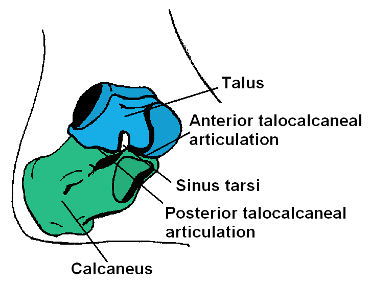
There are three points of articulation between the two bones: two anteriorly and one posteriorly. The three articulations are known as facets, and they are the posterior, middle and anterior facets.
The sustentaculum tali forms the floor of middle facet, and the anterior facet articulates with the head of the talus, and sits lateral and congruent to the middle facet. In some people the middle and anterior facets are joined giving just one articulation. The posterior facet is the largest of the three, and separated from the others by the tarsal canal.
The main ligament of the joint is the interosseous talocalcaneal ligament, a thick, strong band of two partially joined fibers that bind the talus and calcaneus. It runs through the sinus tarsi, a canal between the articulations of the two bones.
There are four additional ligaments that form weaker connections between the talus and calcaneus.
A synovial membrane lines the capsule of the joint, and the joint is wrapped in a capsule of short fibers that are continuous with the talocalcaneonavicular and calcaneocuboid joints of the foot.
Hub AI
Subtalar joint AI simulator
(@Subtalar joint_simulator)
Subtalar joint
In human anatomy, the subtalar joint, also known as the talocalcaneal joint, is a joint of the foot. It occurs at the meeting point of the talus and the calcaneus.
The joint is classed structurally as a synovial joint, and functionally as a plane joint.
The talus is oriented slightly obliquely on the anterior surface of the calcaneus.
There are three points of articulation between the two bones: two anteriorly and one posteriorly. The three articulations are known as facets, and they are the posterior, middle and anterior facets.
The sustentaculum tali forms the floor of middle facet, and the anterior facet articulates with the head of the talus, and sits lateral and congruent to the middle facet. In some people the middle and anterior facets are joined giving just one articulation. The posterior facet is the largest of the three, and separated from the others by the tarsal canal.
The main ligament of the joint is the interosseous talocalcaneal ligament, a thick, strong band of two partially joined fibers that bind the talus and calcaneus. It runs through the sinus tarsi, a canal between the articulations of the two bones.
There are four additional ligaments that form weaker connections between the talus and calcaneus.
A synovial membrane lines the capsule of the joint, and the joint is wrapped in a capsule of short fibers that are continuous with the talocalcaneonavicular and calcaneocuboid joints of the foot.
Recent media