Recent from talks
Nothing was collected or created yet.
Talus bone
View on Wikipedia| Talus bone | |
|---|---|
Anatomy of the right foot | |
Subtalar joint, viewed from an angle between lateral and frontal. | |
| Details | |
| Identifiers | |
| Latin | os talus, astragalus |
| MeSH | D013628 |
| TA98 | A02.5.10.001 |
| TA2 | 1448 |
| FMA | 9708 |
| Anatomical terms of bone | |
The talus (/ˈteɪləs/; Latin for ankle[1] or ankle bone;[2] pl.: tali), talus bone, astragalus (/əˈstræɡələs/), or ankle bone is one of the group of foot bones known as the tarsus. The tarsus forms the lower part of the ankle joint. It transmits the entire weight of the body from the lower legs to the foot.[3]
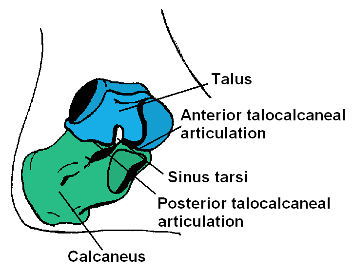
The talus has joints with the two bones of the lower leg, the tibia and thinner fibula. These leg bones have two prominences (the lateral and medial malleoli) that articulate with the talus. At the foot end, within the tarsus, the talus articulates with the calcaneus (heel bone) below, and with the curved navicular bone in front; together, these foot articulations form the ball-and-socket-shaped talocalcaneonavicular joint.
The talus is the second largest of the tarsal bones;[4] it is also one of the bones in the human body with the highest percentage of its surface area covered by articular cartilage. It is also unusual in that it has a retrograde blood supply, i.e. arterial blood enters the bone at the distal end.[citation needed]
In humans, no muscles attach to the talus, unlike most bones, and its position therefore depends on the position of the neighbouring bones.[5]
In humans
[edit]
Though irregular in shape, the talus can be subdivided into three parts.
Facing anteriorly, the head carries the articulate surface of the navicular bone, and the neck, the roughened area between the body and the head, has small vascular channels.[3]
The body features several prominent articulate surfaces: On its superior side is the trochlea tali, which is semi-cylindrical,[6] and it is flanked by the articulate facets for the two malleoli.[3] The ankle mortise, the fork-like structure of the malleoli, holds these three articulate surfaces in a steady grip, which guarantees the stability of the ankle joint. However, because the trochlea is wider in front than at the back (approximately 5–6 mm) the stability in the joint vary with the position of the foot: with the foot dorsiflexed (toes pulled upward) the ligaments of the joint are kept stretched, which guarantees the stability of the joint; but with the foot plantarflexed (as when standing on the toes) the narrower width of the trochlea causes the stability to decrease.[7] Behind the trochlea is a posterior process with a medial and a lateral tubercle separated by a groove for the tendon of the flexor hallucis longus. Exceptionally, the lateral of these tubercles forms an independent bone called os trigonum or accessory talus; it may represent the tarsale proximale intermedium. On the bone's inferior side, three articular surfaces serve for the articulation with the calcaneus, and several variously developed articular surfaces exist for the articulation with ligaments.[3]
For descriptive purposes the talus bone is divided into three sections, neck, body, and head.
Head
[edit]The talus bone of the ankle joint connects the leg to the foot.
The head of talus looks forward and medialward; its anterior articular or navicular surface is large, oval, and convex. Its inferior surface has two facets, which are best seen in the fresh condition.[8]
The medial, situated in front of the middle calcaneal facet, is convex, triangular, or semi-oval in shape, and rests on the plantar calcaneonavicular ligament; the lateral, named the anterior calcaneal articular surface, is somewhat flattened, and articulates with the facet on the upper surface of the anterior part of the calcaneus.[8]
Neck
[edit]The neck of talus is directed anteromedially, and comprises the constricted portion of the bone between the body and the oval head.[8]
Its upper and medial surfaces are rough, for the attachment of ligaments; its lateral surface is concave and is continuous below with the deep groove for the interosseous talocalcaneal ligament.[8]
Body
[edit]
The body of the talus comprises most of the volume of the talus bone (ankle bone). It presents with five surfaces; a superior, inferior, medial, lateral and a posterior:[8]
- The superior surface of the body presents, behind, a smooth trochlear surface, the trochlea, for articulation with the tibia. The trochlea is broader in front than behind, convex from before backward, slightly concave from side to side: in front it is continuous with the upper surface of the neck of the bone.
- the inferior surface presents two articular areas, the posterior and middle calcaneal surfaces, separated from one another by a deep groove, the sulcus tali. The groove runs obliquely forward and lateralward, becoming gradually broader and deeper in front: in the articulated foot it lies above a similar groove upon the upper surface of the calcaneus, and forms, with it, a canal (sinus tarsi) filled up in the fresh state by the interosseous talocalcaneal ligament. The posterior calcaneal articular surface is large and of an oval or oblong form. It articulates with the corresponding facet on the upper surface of the calcaneus, and is deeply concave in the direction of its long axis which runs forward and lateralward at an angle of about 45° with the median plane of the body. The middle calcaneal articular surface is small, oval in form and slightly convex; it articulates with the upper surface of the sustentaculum tali of the calcaneus.
- The medial surface presents at its upper part a pear-shaped articular facet for the medial malleolus, continuous above with the trochlea; below the articular surface is a rough depression for the attachment of the deep portion of the deltoid ligament of the ankle-joint.
- The lateral surface carries a large triangular facet, concave from above downward, for articulation with the lateral malleolus; its anterior half is continuous above with the trochlea; and in front of it is a rough depression for the attachment of the anterior talofibular ligament. Between the posterior half of the lateral border of the trochlea and the posterior part of the base of the fibular articular surface is a triangular facet which comes into contact with the transverse inferior tibiofibular ligament during flexion of the ankle-joint; below the base of this facet is a groove which affords attachment to the posterior talofibular ligament.
- The posterior surface is narrow, and traversed by a groove running obliquely downward and medialward, and transmitting the tendon of the Flexor hallucis longus. Lateral to the groove is a prominent tubercle, the posterior process, to which the posterior talofibular ligament is attached; this process is sometimes separated from the rest of the talus, and is then known as the os trigonum. Medial to the groove is a second smaller tubercle.
Development
[edit]During the 7th to 8th intrauterine month an ossification center is formed in the anklebone.[3]
Fracture
[edit]
The talus bone lacks a good blood supply. Because of this, healing a broken talus can take longer than most other bones. One with a broken talus may not be able to walk for many months without crutches and will further wear a walking cast or boot of some kind after that.
Talus injuries may be difficult to recognize,[9][10] and lateral process fractures in particular may be radiographically occult. If not recognized and managed appropriately, a talus fracture may result in complications and long-term morbidity. A 2015 review came to the conclusion that isolated talar body fractures may be more common than previously thought.[4]
A fractured talar body often has a displacement that is best visualised using CT imaging. In case a talus fracture is accompanied by a dislocation, restoration of articular and axial alignment is necessary to optimize ankle and hindfoot function.[9]
As dice
[edit]Dice were originally made from the talus of hoofed animals, leading to the nickname "bones" for dice. Colloquially known as "knucklebones", these are approximately tetrahedral. Modern Mongolians still use such bones as shagai for games and fortune-telling, with each piece relating to a symbolic meaning.[11]
In other animals
[edit]The talus apparently derives from the fusion of three separate bones in the feet of primitive amphibians; the tibiale, articulating with tibia, the intermedium, between the bases of the tibia and fibula, and the fourth centrale, lying in the mid-part of the tarsus. These bones are still partially separate in modern amphibians, which therefore do not have a true talus.[12]
The talus forms a considerably more flexible joint in mammals than it does in reptiles. This reaches its greatest extent in artiodactyls, where the distal surface of the bone has a smooth keel to allow greater freedom of movement of the foot, and thus increase running speed.[12]
In non-mammal amniotes, the talus is generally referred to as the astragalus.
In modern crocodiles, the astragalus bears a peg which inserts into a corresponding socket on the calcaneum, and the hinge of the ankle joint runs between the two tarsals; this condition is referred to as "croc-normal"; this "croc-normal" condition was likely ancestral for archosaurs. In dinosaurs (including modern birds) and pterosaurs, the hinge of the ankle instead is distal to the two tarsals.[13][14] Far rarer are archosaurs with a "croc-reversed" ankle joint, in which the calcaneus bears a peg whilst the astragalus bears a socket.[15]
In the theropod dinosaur lineage leading to birds, the astragalus gradually increases in size until it forms the entire proximal facet of the ankle articulation; additionally the anterior ascending process gradually extends increasingly proximally. In modern birds, the astragalus is fused with the tibia to form the tibiotarsus.[16]
Additional images
[edit]-
Talus - inferior view
-
Lateral view of the human ankle, including the talus
-
Left talus, medial surface
-
Left talus, lateral surface
See also
[edit]- Astragalomancy, a form of divination using talus bones
- Knucklebones, a dice game using astragali
- Shagai, sheep or goat talus bones used for gaming
- Squatting facets
Notes
[edit]- ^ Mosby's Medical, Nursing & Allied Health Dictionary, Fourth Edition, Mosby-Year Book Inc., 1994, p. 1526
- ^ Lewis, Charlton T.; Short, Charles (1879). "tālus". A Latin Dictionary. Clarendon Press. Archived from the original on 2021-11-20 – via the Perseus Project.
- ^ a b c d e Platzer (2004), p 216
- ^ a b Melenevsky Y, Mackey RA, Abrahams RB, Thomson NB (2015). "Talar Fractures and Dislocations: A Radiologist's Guide to Timely Diagnosis and Classification". Radiographics (Review). 35 (3): 765–79. doi:10.1148/rg.2015140156. PMID 25969933.
- ^ Bojsen-Møller, Finn; Simonsen, Erik B.; Tranum-Jensen, Jørgen (2001). Bevægeapparatets anatomi [Anatomy of the Locomotive Apparatus] (in Danish) (12th ed.). Munksgaard Danmark. p. 301. ISBN 978-87-628-0307-7.
- ^ Lee F. Rogers (1992) Radiology of skeletal trauma - Volume 2 p.1463
- ^ Thieme Atlas of Anatomy (2006), p 406
- ^ a b c d e Gray's Anatomy (1918)
- ^ a b Vallier HA (September 2015). "Fractures of the Talus: State of the Art". Journal of Orthopaedic Trauma (Review). 29 (9): 385–92. doi:10.1097/BOT.0000000000000378. PMID 26299809. S2CID 2532534.
- ^ Melenevsky Y, Mackey RA, Abrahams RB, Thomson NB (2015). "Talar Fractures and Dislocations: A Radiologist's Guide to Timely Diagnosis and Classification". Radiographics (Review). 35 (3): 765–79. doi:10.1148/rg.2015140156. PMID 25969933. Section Conclusion: "Accurately detecting, classifying, and managing talar injuries can be a challenging endeavor due to the unique anatomic characteristics of the talus and subtle radiographic findings of the injuries.", page 778.
- ^ Pegg, Carole (2001). Mongolian music, dance and oral narrative : performing diverse identities. [S.l.]: Univ. of Washington Press. p. 233. ISBN 9780295981123.
- ^ a b Romer, Alfred Sherwood; Parsons, Thomas S. (1977). The Vertebrate Body. Philadelphia, PA: Holt-Saunders International. p. 207. ISBN 0-03-910284-X.
- ^ Nesbitt, Sterling J.; Butler, Richard J.; Ezcurra, Martín D.; Barrett, Paul M.; Stocker, Michelle R.; Angielczyk, Kenneth D.; Smith, Roger M. H.; Sidor, Christian A.; Niedźwiedzki, Grzegorz; Sennikov, Andrey G.; Charig, Alan J. (April 2017). "The earliest bird-line archosaurs and the assembly of the dinosaur body plan". Nature. 544 (7651): 484–487. Bibcode:2017Natur.544..484N. doi:10.1038/nature22037. hdl:11336/49585. ISSN 1476-4687. PMID 28405026. S2CID 9095072.
- ^ Dyke, Gareth J. (1998). "Does Archosaur Phylogeny Hinge on the Ankle Joint?". Journal of Vertebrate Paleontology. 18 (3): 558–562. Bibcode:1998JVPal..18..558D. doi:10.1080/02724634.1998.10011083. ISSN 0272-4634. JSTOR 4523927.
- ^ Baczko, M. Belén von; Ezcurra, Martín D. (2013). "Ornithosuchidae: a group of Triassic archosaurs with a unique ankle joint". Geological Society, London, Special Publications. 379 (1): 187–202. Bibcode:2013GSLSP.379..187B. doi:10.1144/sp379.4. hdl:11336/41617. ISSN 0305-8719. S2CID 130687362.
- ^ Mayr, Gerald (31 October 2016). Avian evolution : the fossil record of birds and its paleobiological significance. John Wiley & Sons. ISBN 978-1-119-02076-9. OCLC 950901952.
References
[edit]
This article incorporates text in the public domain from page 266 of the 20th edition of Gray's Anatomy (1918)
- Platzer, Werner (2004). Color Atlas of Human Anatomy, Vol. 1: Locomotor System (5th ed.). Thieme. ISBN 3-13-533305-1.
- Thieme Atlas of Anatomy: General Anatomy and Musculoskeletal System. Thieme. 2006. ISBN 1-58890-419-9.
External links
[edit]- Anatomy of the talus by Maurice Laude, Laboratory of Anatomy and Organogenesis, Amiens Medical School Archived 2012-10-28 at the Wayback Machine
- Fractures of the Talus at mdmercy.com Archived 2011-07-14 at the Wayback Machine
- lljoints at The Anatomy Lesson by Wesley Norman (Georgetown University) (posterioranklejoint)
- Illustration at orthoinfo.aaos.org
Talus bone
View on GrokipediaAnatomy
Structure
The talus is an irregularly shaped tarsal bone in the human foot, characterized by its lack of muscular attachments and extensive articular surfaces, making it unique among the tarsal bones. It consists of three main components: the head, neck, and body, with the body being the largest portion. The bone's morphology supports its role as a connector between the leg and foot, with approximately 60% of its surface covered by articular cartilage.[1] The talar head is a rounded anterior projection that forms the most distal part of the bone, featuring a convex, ovoid anterior surface. This head is separated from the neck by a slight constriction and is almost entirely enveloped in hyaline cartilage on its anterior and superior aspects. Inferiorly, the head bears three articular facets—anterolateral, anteromedial, and middle—divided by ridges, contributing to its complex contour.[3][1] The neck of the talus is a constricted region connecting the head to the body, oriented obliquely in an inferomedial direction. It is narrower than both adjacent parts, measuring on average about 3.3 cm in width, and its inferior surface contains the sulcus tali, a deep groove that expands laterally into the sinus tarsi. This narrow neck renders it particularly susceptible to fractures due to its limited cross-sectional area.[1][4] The body of the talus represents the largest and most robust portion, encompassing the superior trochlea and posterior elements. It includes a lateral process on its inferolateral aspect and a posterior process that bifurcates into medial and lateral tubercles separated by a groove. The medial tubercle is smaller and projects more inferiorly, while the lateral tubercle is larger and more prominent. These tubercles and processes provide attachment points for ligaments, enhancing stability. The body's overall dimensions average approximately 5.4 cm in length, 4.1 cm in width, and 3.1 cm in height, varying slightly by sex and population.[1][3][4] The trochlea, or talar dome, forms the pulley-shaped superior surface of the body, presenting a convex, wedge-shaped convexity that is broader anteriorly (average width ~3.0 cm) than posteriorly (~2.3-2.6 cm). This anterior-posterior asymmetry contributes to the bone's tapered profile, with the trochlear surface gently curved in the sagittal plane and mildly concave in the coronal plane. Laterally and medially, the body features comma-shaped malleolar facets for interaction with the distal tibia and fibula, while the posterior aspect of the inferior surface includes a concave calcaneal articular facet.[1][4][5]Articulations and ligaments
The talus bone forms several key articulations in the foot, primarily serving as a connector between the leg and the hindfoot. The talocrural joint, also known as the ankle joint, is a synovial hinge joint where the superior trochlea of the talus articulates with the inferior surfaces of the distal tibia and the medial malleolus of the fibula, allowing for dorsiflexion and plantarflexion movements.[1] This articulation is reinforced by the surrounding ligaments and transmits forces from the lower leg to the foot.[1] Inferiorly, the talus articulates with the calcaneus at the subtalar joint, a compound synovial joint comprising posterior, middle, and anterior facets; the posterior and middle facets on the talus specifically interface with corresponding surfaces on the calcaneus, facilitating inversion and eversion of the foot.[1] The head of the talus articulates anteriorly with the navicular bone, forming part of the transverse tarsal joint (also called the Chopart joint), which connects the hindfoot to the midfoot and contributes to the foot's adaptability during weight-bearing.[1] These articulations collectively enable the talus to link the ankle to the subtalar and midtarsal regions without direct muscular attachments, relying instead on ligamentous stability.[1] The ligaments associated with the talus provide crucial medial and lateral support to these joints. On the medial side, the deltoid ligament, a strong triangular complex, originates from the medial malleolus of the tibia and fans out to attach to the talus, calcaneus, and navicular; it consists of superficial components (such as the tibionavicular and tibiocalcaneal ligaments) and deeper posterior tibiotalar fascicles, collectively resisting eversion and talar abduction.[6] Laterally, the collateral ligament complex includes three primary bands: the anterior talofibular ligament (ATFL), which extends from the anterior inferior lateral malleolus of the fibula to the anterior aspect of the talar neck, limiting anterior talar displacement and inversion during plantarflexion; the calcaneofibular ligament (CFL), running from the lateral malleolus to the lateral calcaneus, stabilizing both the talocrural and subtalar joints in neutral and dorsiflexed positions; and the posterior talofibular ligament (PTFL), connecting the posterior lateral malleolus to the posterior talus, providing posterior stability and resisting excessive dorsiflexion.[6][7] The sinus tarsi and tarsal canal play integral roles in ligament passage and joint stabilization. The sinus tarsi, a lateral bony depression between the talus and calcaneus, contains the cervical and interosseous talocalcaneal ligaments, which reinforce the subtalar joint and provide proprioceptive feedback.[1] Extending medially from the sinus tarsi, the tarsal canal is a narrower osseous tunnel that accommodates the interosseous talocalcaneal ligament and branches of the posterior tibial artery, further securing the posterior aspect of the subtalar joint against shear forces.[1]Blood supply and innervation
The talus bone receives its arterial blood supply primarily from three major sources: the posterior tibial artery, the anterior tibial artery (also known as the dorsalis pedis artery), and the peroneal artery. The posterior tibial artery contributes through branches that supply the medial and lateral tubercles via the posterior tubercle and forms the tarsal canal artery, which provides the dominant supply to the talar body, including a deltoid branch for the medial third. The anterior tibial artery supplies the superomedial aspect of the talar neck via medial tarsal branches, while the peroneal artery anastomoses with the dorsalis pedis to form the tarsal sinus artery, which together with the tarsal canal artery supplies the inferolateral talar neck. These arteries enter the talus through vascular foramina on its non-articular surfaces, with key branches including the sinus tarsi artery and deltoid branches.[1][8][9] Venous drainage of the talus follows the arterial supply, primarily via the peroneal and posterior tibial veins, which converge into the venous plexuses of the foot and ankle.[1] Intraosseous circulation within the talus is limited due to its extensive articular cartilage coverage, which encompasses approximately 60-70% of its surface, making the bone heavily reliant on extraosseous vascular supply with minimal anastomoses between the major arterial branches. This precarious intraosseous network contributes to the talus's vulnerability to ischemia, particularly when extraosseous vessels are compromised.[1][9] Innervation to the talus is predominantly sensory, derived from branches of the deep peroneal nerve (supplying the dorsal aspect), the tibial nerve (including posterior tibial branches for the medial side), and the sural nerve (for lateral sensory feedback), with no significant motor innervation due to the absence of muscular attachments. Additional contributions come from the saphenous nerve medially and branches of the superficial peroneal nerve for the dorsolateral and lateral regions.[1][10] The talar neck represents a critical watershed area with tenuous blood supply, rendering it particularly susceptible to ischemia and avascular necrosis following disruption of the extraosseous vessels.[1][9]Function
Role in locomotion
The talus bone serves as a critical intermediary in the lower limb, transmitting body weight from the tibia to the foot bones during the stance phase of locomotion. Positioned at the ankle joint, it receives the vertical forces generated by body mass and redistributes them across the tarsal bones, enabling efficient weight-bearing while maintaining structural integrity. This transmission is essential for activities such as walking and standing, where the talus acts as a pivot to transfer up to several times the body's weight without compromising mobility.[11] In facilitating ankle movements, the talus enables dorsiflexion, ranging from 0 to 20 degrees, which lifts the foot upward to clear obstacles during the swing phase, and plantarflexion, up to 50 degrees, which points the foot downward for propulsion. At the subtalar joint, the talus articulates with the calcaneus to permit inversion and eversion, allowing the foot to adapt to uneven terrain by tilting medially or laterally, thus enhancing balance and directional changes during locomotion. These motions collectively ensure smooth transitions between phases of movement, integrating the talus's role in both the talocrural and subtalar joints.[12][13] During the gait cycle, the talus contributes to heel strike absorption by cushioning initial ground contact through controlled dorsiflexion and eversion, dissipating impact forces to prevent jarring. In the push-off phase, it supports propulsion via plantarflexion and inversion, generating forward momentum as the body advances. This integration optimizes energy efficiency and stride length in walking and running.[14] The talus also provides stability in upright posture by forming a key component of the medial longitudinal arch, which supports the foot's vaulted structure and distributes weight evenly to maintain equilibrium without excessive strain on surrounding tissues. This arch configuration, with the talus as its keystone, resists collapse under gravitational load, facilitating prolonged standing and bipedal posture.[15][16]Biomechanics
The talus serves as a pivotal structure in the lower limb's load transmission pathway, articulating with the tibia and fibula to transfer compressive forces from the body to the foot. During normal walking, the ankle joint complex, with the talus at its core, experiences peak loads of approximately five times body weight, escalating to up to thirteen times body weight during activities like running. The trochlear surface of the talus, characterized by its wedge-shaped morphology—wider anteriorly than posteriorly—facilitates this transmission while resisting anterior translation, thereby maintaining joint stability under dynamic loading. This design ensures efficient force distribution to the subtalar and transverse tarsal joints, with the tibiotalar articulation handling about 83% of the axial load and the talar dome supporting 77–90% of the tibial-talar interface forces.[17][18][17] Joint congruence in the talocrural articulation varies with position, influencing load distribution and minimizing stress concentrations. In the neutral position, the contact area between the talar trochlea and tibial plafond is relatively limited, typically around 4–5 cm² under physiological loads, representing less than 50% of the available cartilage-covered surface to allow for mobility. This area expands significantly during dorsiflexion, reaching up to 7.3 cm² at 20° of dorsiflexion, which enhances congruence by engaging more of the broader anterior trochlea within the mortise, thereby optimizing force dissipation across the joint. Such positional changes in contact area are critical for accommodating the ankle's multiplanar demands without excessive wear.[19][20] Stress distribution across the talar surfaces reflects these biomechanical adaptations, with compressive forces concentrated on the trochlear dome during weight-bearing. Under static loads equivalent to twice body weight (approximately 1.5 kN), mean contact pressures on the talocrural joint reach 9.9 MPa, with higher values observed in plantarflexion due to reduced contact area compared to dorsiflexion. These stresses are unevenly distributed, often peaking anterolaterally in neutral positions, underscoring the talus's role in buffering peak forces through its geometric and material resilience.[17] Motion kinematics of the talus involve coupled interactions between the talocrural and subtalar joints, enabling triplanar foot motion. For instance, talocrural dorsiflexion or plantarflexion in the sagittal plane induces corresponding eversion or inversion at the subtalar joint, with the subtalar contribution accounting for about 20% of the total ankle complex's sagittal range of motion (typically 65–75° overall). A representative coupling ratio shows that 10° of talocrural motion may elicit 2–5° of subtalar motion, depending on the plane and loading, facilitating adaptive pronation and supination during gait. The cortical bone of the talus, with an apparent density of 1.2–1.8 g/cm³ and a modulus of elasticity of 15–20 GPa, provides the necessary stiffness to withstand these kinematic demands without deformation.[17][21][22][23]Development and variations
Ossification and growth
The talus bone originates embryologically from the somatic layer of the lateral plate mesoderm, which contributes to the formation of the limb skeleton during early development.[24] The lower limb bud emerges around the fourth week of gestation, with mesenchymal condensation leading to the cartilage anlage of the talus by the seventh week, establishing its precartilaginous model prior to ossification.[25] This endochondral process follows the typical pattern for tarsal bones, where the initial hyaline cartilage template defines the bone's future morphology. The primary ossification center for the talus appears in the body of the bone during the sixth to eighth month of fetal life, typically around seven months in utero, marking the onset of endochondral ossification.[26] Unlike long bones, the talus lacks secondary ossification centers and epiphyseal growth plates; instead, postnatal growth occurs primarily through periosteal apposition, where new bone is deposited on the outer surfaces to increase size and shape the bone progressively.[27] By three months postnatal, approximately 55% of the talus is ossified, with continued expansion leading to largely complete ossification of the main body by around six years of age.[27] Ossification and growth of the talus complete by puberty, with the posterior process potentially involving a separate center (os trigonum) that fuses between ages 7-10 in females and 9-12 in males, though this is not universal.[27] Sex differences are evident throughout development, with females exhibiting earlier and more advanced ossification centers in the talus and other tarsal bones compared to males at equivalent gestational or postnatal ages, reflecting broader skeletal dimorphism.[28]Anatomical variations
The os trigonum represents one of the most common anatomical variations of the talus, manifesting as an accessory ossicle derived from the unfused ossification center of the posterolateral tubercle. This variant occurs in 7-25% of individuals, with higher detection rates on imaging such as CT scans where prevalence reaches up to 30% in asymptomatic populations.[29][30] Although typically benign, it can become symptomatic in activities involving repetitive plantar flexion, particularly among ballet dancers, where it contributes to posterior ankle impingement through compression of adjacent soft tissues.[31][32] Variations in the lateral talar tubercle include elongation, termed Stieda's process, and less commonly bifid configurations, which alter the posterior contour and may impinge on the sinus tarsi, potentially leading to lateral ankle discomfort.[31][33] These forms arise from incomplete fusion during development and have a reported prevalence of 1-15% depending on the population studied, with elongated variants more frequently associated with mechanical overload in dynamic ankle movements.[34] Trochlear ridge anomalies encompass deviations in the shape of the talar dome, such as flat or excessively domed profiles, which affect joint congruence and stability. A flat trochlea reduces the depth of the articular surface and has been linked to diminished tibiotalar stability, increasing susceptibility to subluxation.[35] Size asymmetries between left and right tali reflect fluctuating asymmetry in skeletal development.[36] Racial variations further influence dimensions, with studies indicating longer talar lengths in African populations compared to Caucasians or Asians, aiding in population affinity assessments.[37] Recent investigations have expanded understanding of these variants. A 2024 atlas based on over 900 tali documented more than 15 distinct morphological variants, including facet and tubercle anomalies, establishing a standardized nomenclature to facilitate clinical and bioanthropological applications.[38]Clinical significance
Fractures and injuries
Talus fractures are relatively uncommon, accounting for less than 1% of all fractures and 3% to 6% of foot fractures.[9] These injuries typically result from high-energy trauma, such as motor vehicle accidents or falls from height, which impose axial loads on the dorsiflexed foot, or from lower-energy mechanisms like repetitive stress in activities involving jumping, as seen in athletes.[9][39] The talus's precarious blood supply, detailed in anatomical descriptions, heightens the risk of complications when these vascular structures are disrupted.[9] Classification systems guide management, with the Hawkins classification (modified by Canale) applied to talar neck fractures, the most common type: Type I involves nondisplaced fractures; Type II features subtalar subluxation or dislocation; Type III includes both subtalar and ankle joint involvement; and Type IV extends to talonavicular dislocation.[9] For talar body fractures, the Sneppen classification delineates types based on fracture plane, such as coronal or sagittal shear, or comminuted crush patterns.[9] Diagnosis relies on imaging, where computed tomography (CT) is the gold standard with superior accuracy over plain X-rays, which have approximately 60% sensitivity for detecting and classifying talar injuries, as shown in a 2025 meta-analysis.[40] Complications are frequent due to the talus's limited vascularity, with avascular necrosis occurring in 0-15% of Hawkins Type I nondisplaced talar neck fractures, 20-50% of displaced neck fractures (rising to over 90% in Types III and IV) from disruption of retinacular vessels.[9][41][42] Nonunion affects 10-20% of cases, particularly in comminuted body fractures, while malunion can lead to joint incongruity.[9] Treatment for nondisplaced fractures, particularly Hawkins Type I talar neck fractures, typically involves conservative (non-operative) treatment with below-knee cast immobilization for 8-12 weeks (non-weight-bearing for the initial 6 weeks), followed by gradual progressive weight-bearing. Displaced fractures require open reduction and internal fixation (ORIF) using screws to restore alignment; severe comminuted or Hawkins Type IV injuries may necessitate primary arthrodesis to prevent collapse.[9][39][41] Outcomes for Hawkins Type I fractures are generally favorable, with high fracture union rates, a low risk of avascular necrosis (0-15%), and good to excellent functional results in approximately 65-90% of cases, though some patients may experience post-traumatic arthrosis, restricted joint motion, or mild pain.[42][41] Outcomes vary by fracture severity and timeliness of intervention, with post-traumatic arthritis developing in 40-60% of patients, predominantly affecting the subtalar and tibiotalar joints, and leading to chronic pain and stiffness.[43] Rehabilitation emphasizes early motion preservation through protected weight-bearing protocols to mitigate stiffness, though functional scores like the AOFAS hindfoot scale often remain moderate (around 70-80 points) in displaced cases.[9][39]Other disorders
Osteochondritis dissecans (OCD) of the talus primarily affects the talar dome and represents approximately 4% of all osteochondral lesions.[44] These lesions involve focal disruption of the subchondral bone and overlying articular cartilage, often leading to pain, swelling, and mechanical symptoms such as catching or locking in the ankle joint. The condition is classified into stages I through IV using the Berndt-Harty system, which assesses lesion stability and fragmentation: stage I indicates subchondral compression, stage II shows incomplete fracture, stage III denotes a nondisplaced fragment, and stage IV involves a displaced or loose fragment.[45] Early detection via MRI is crucial, as progression can result in cartilage degeneration and osteoarthritis if untreated. Avascular necrosis (AVN) of the talus, also known as osteonecrosis, arises from disrupted blood supply and can be idiopathic or post-traumatic, with the latter often following injuries that compromise the talus's retrograde vascularity.[46] The Ficat and Arlet staging system is applied to talar AVN, progressing from stage I (preradiographic necrosis detectable by MRI) to stage IV (advanced collapse with secondary arthritis). Approximately 15-30% of cases progress to subchondral collapse, particularly in shoulder-type lesions involving the talar neck or body, leading to pain, stiffness, and joint deformity.[47] Management focuses on preserving bone viability in early stages through non-weight-bearing protocols, though advanced collapse often necessitates surgical intervention like core decompression. Talar coalition refers to anomalous bony, cartilaginous, or fibrous union between the talus and adjacent tarsal bones, most commonly the talocalcaneal joint, with a prevalence of 1-2% in the general population.[48] This congenital condition restricts subtalar motion, causing rigid flatfoot, recurrent ankle sprains, and peroneal spasm due to compensatory muscle hyperactivity. Talocalcaneal coalitions, accounting for about 37% of tarsal coalitions, often manifest in adolescence as the coalition ossifies, leading to hindfoot valgus and potential secondary osteoarthritis. Diagnosis relies on imaging features like the C-sign on lateral radiographs, with treatment ranging from orthotics to resection for symptomatic cases. Tumors of the talus are rare, comprising less than 3% of primary bone neoplasms, with benign lesions predominating. Chondroblastoma, a benign cartilaginous tumor arising from epiphyseal chondroblasts, occurs in 1-3% of all bone tumors and involves the talus in about 4% of its cases, typically presenting with pain and swelling in young patients aged 10-20 years.[49] These lytic lesions with chondroid matrix may mimic infection or AVN radiographically. Intraosseous lipomas, benign fatty tumors accounting for 0.1-2.5% of bone tumors, can also affect the talus, often appearing as well-defined cystic lesions with fat attenuation on MRI and rarely causing symptoms unless fractured. Malignant tumors like osteosarcoma are exceptionally uncommon in the talus. Infections involving the talus, such as post-injury osteomyelitis, occur when bacteria invade bone following trauma, leading to bone destruction, abscess formation, and chronic pain if not promptly treated with antibiotics and debridement.[50] Gouty arthropathy can affect the talus through urate crystal deposition, forming tophi that erode subchondral bone and induce osteochondral lesions, mimicking trauma or necrosis with acute inflammatory flares and joint effusion.[51] These tophaceous deposits, seen in advanced hyperuricemia, may cause cystic changes or pathologic fractures, emphasizing the need for serum uric acid monitoring in atypical ankle pathologies.Comparative and historical aspects
In other animals
In quadrupedal mammals adapted for cursorial locomotion, such as horses, the talus exhibits an elongated morphology that facilitates efficient propulsion and stability during high-speed, straight-line movement. This adaptation supports the animal's ability to cover large distances rapidly, with the talus forming a key component of the hock joint alongside the calcaneus and other tarsal bones.[52] In equids, the talus contributes to the overall rigidity of the tarsus, enhancing force transmission from the hindlimb to the ground.[53] Among primates, the talus shows notable variations tied to locomotor styles, with arboreal species like gibbons displaying a reduced overall size and a more mobile talar head to accommodate flexible foot movements during brachiation and leaping. This morphology, characterized by lower curvature in the talar head and sustentaculum facets, promotes transverse tarsal joint mobility and elastic energy storage in the foot, aiding in agile arboreal navigation.[54] In contrast, more terrestrial primates exhibit talar features suited to weight-bearing, though gibbons' design emphasizes dorsiflexion and inversion for grasping branches.[55] In birds, the talus, or astragalus, is evolutionarily incorporated into the proximal tarsals that fuse with the tibia to form the tibiotarsus, while the distal tarsals merge with the metatarsals to create the tarsometatarsus, an elongated bone analogous to the mammalian tarsus for weight support during perching or walking. This fusion pattern enhances lightweight leg structure for flight in most species. In flightless birds, such as ostriches and emus, the tarsometatarsus and associated structures become robustly developed rather than vestigial, with increased cross-sectional thickness and length to support graviportal terrestrial locomotion and high-speed running.[56][57] Evolutionary trends in the talus trace back to basal amniotes, where the astragalus and calcaneum originated as separate elements but underwent fusion in the proximal tarsus for enhanced terrestrial stability; in reptiles like mesosaurs, the astragalus forms via early ontogenetic fusion of the intermedium, tibiale, and proximal centralia, while the calcaneum derives from the fibulare and remains distinct. This separation persists and diversifies in mammals, allowing greater ankle mobility compared to the more rigid reptilian configurations, reflecting adaptations from sprawling to upright postures.[58][59] Veterinarily, talus fractures hold significant importance in performance equines like racehorses, where stress-induced injuries predominate due to repetitive high-impact loading. Sagittal or incomplete fractures, often arising from maladaptive bone remodeling under compression, shear, and torsional stresses, are rare but can cause severe lameness and career-ending damage, particularly in the proximal trochlear groove.[60][61] Early detection via MRI or CT is crucial, as these microfractures may underlie catastrophic failures and require prolonged rest or arthroscopic intervention for recovery.[62]Etymology and use as dice
The term "talus" originates from the Latin word talus, which denoted the ankle or anklebone and was also applied to dice fashioned from such bones.[63][64] This nomenclature reflects the bone's historical dual role in anatomy and gaming. The Greek equivalent, astragalos, similarly referred to the ankle bone of animals like sheep or goats and extended to knucklebones used as rudimentary dice, highlighting a shared Indo-European linguistic root tied to both skeletal structure and play.[65] Astragali, the knucklebones derived from the talus of sheep, goats, or cattle, served as precursors to modern dice due to their natural irregular shape, featuring four relatively flat sides suitable for landing unpredictably.[66] These sides were often marked with values such as 1, 3, 4, and 6, providing inherent randomness without needing carving, unlike later cubic forms. In ancient Rome, the game of tali involved throwing four such bones, with outcomes scored based on the upward-facing sides; the highest throw, Venus, occurred when all four showed different sides (1, 3, 4, and 6), while the lowest, Canis, featured all 1s.[67][68] Beyond gaming, astragali held ritual importance, particularly in divination practices known as astragalomancy. In ancient Greece, oracles cast five knucklebones—each valued at 1, 3, 4, or 6—to interpret outcomes from 32 possible combinations, often invoking deities like Apollo for guidance.[69][70] This method persisted into Roman and medieval periods for fortune-telling, especially among women, before evolving into more standardized board games.[71] By the 14th century, knucklebones largely gave way to six-sided cubic dice in Europe, which offered greater fairness through symmetry and uniform numbering from 1 to 6, marking a shift from organic to manufactured gaming tools.[72] Astragali appear frequently in archaeological contexts, underscoring their cultural ubiquity; for instance, ivory examples were interred in Tutankhamun's tomb around 1323 BCE, likely for use in the afterlife.[73] In Greek mythology, the talus indirectly evokes vulnerability through the Achilles' heel motif, where the hero's unprotected tendon insertion at the heel symbolizes a fatal weakness, though the bone itself was not the precise target.[74] Today, the term "talus" endures in architecture to describe the sloping base of a fortified wall, thicker at the bottom for stability, a usage derived from the bone's sloped contours.[75][76]References
- https://en.wiktionary.org/wiki/talus