Hubbry Logo
ABO blood group systemABO blood group systemMain
Open search
ABO blood group system
Community hub
ABO blood group system
logo
8 pages, 0 posts
0 subscribers
Be the first to start a discussion here.
ABO blood group system
from Wikipedia

ABO blood group antigens present on red blood cells and IgM antibodies present in the serum

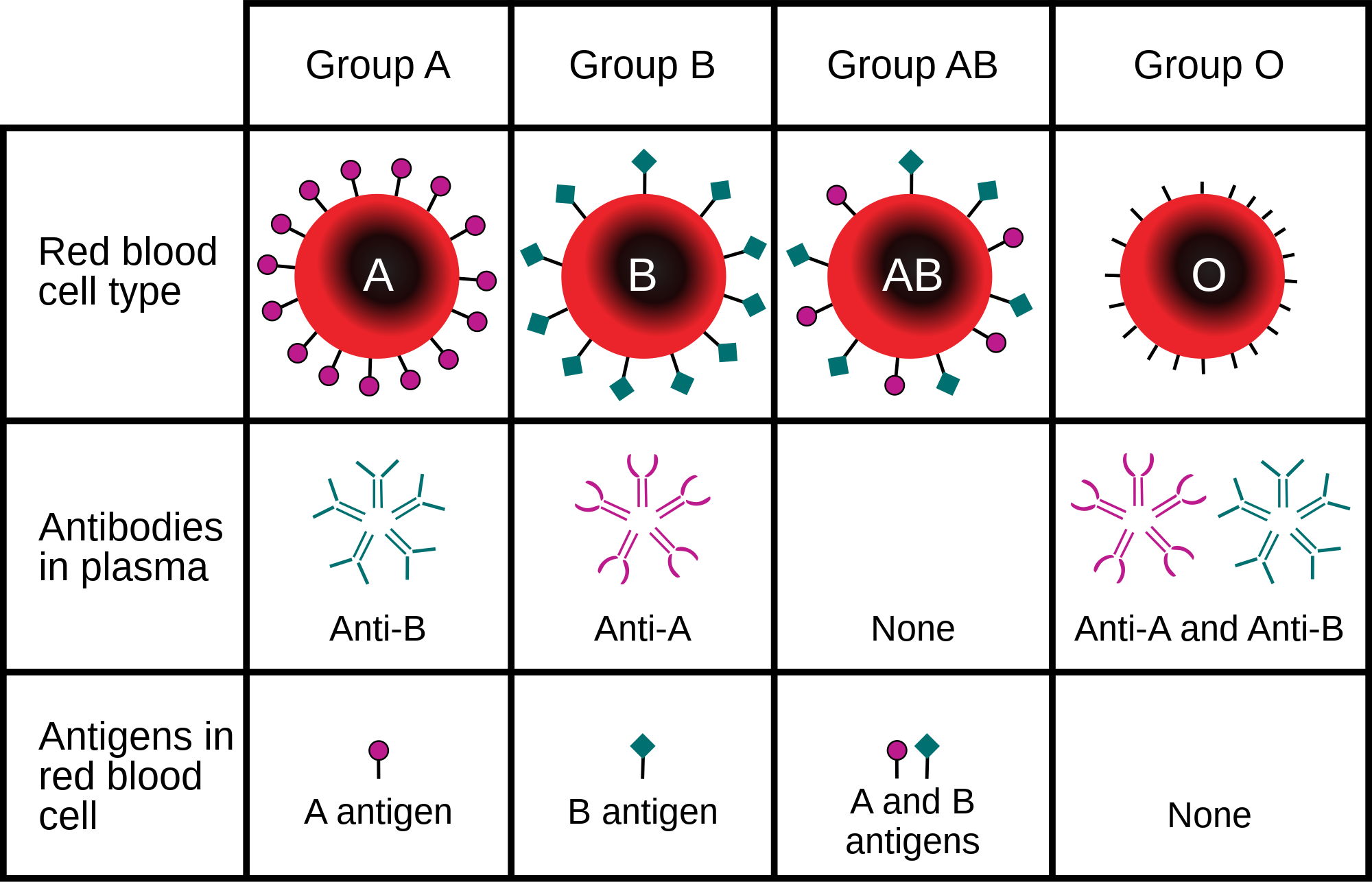
The ABO blood group system is used to denote the presence of one, both, or neither of the A and B antigens on erythrocytes (red blood cells).[1] For human blood transfusions, it is the most important of the 48 different blood type (or group) classification systems currently recognized by the International Society of Blood Transfusions (ISBT) as of June 2025.[2][3] A mismatch in this serotype (or in various others) can cause a potentially fatal adverse reaction after a transfusion, or an unwanted immune response to an organ transplant.[4] Such mismatches are rare in modern medicine. The associated anti-A and anti-B antibodies are usually IgM antibodies, produced in the first years of life by sensitization to environmental substances such as food, bacteria, and viruses.

The ABO blood types were discovered by Karl Landsteiner in 1901; he received the Nobel Prize in Physiology or Medicine in 1930 for this discovery.[5] ABO blood types are also present in other primates such as apes, monkeys and Old World monkeys.[6]

History

[edit]

Discovery

[edit]

The ABO blood types were first discovered by an Austrian physician, Karl Landsteiner, working at the Pathological-Anatomical Institute of the University of Vienna (now Medical University of Vienna). In 1900, he found that red blood cells would clump together (agglutinate) when mixed in test tubes with sera from different persons, and that some human blood also agglutinated with animal blood.[7] He wrote a two-sentence footnote:

The serum of healthy human beings not only agglutinates animal red cells, but also often those of human origin, from other individuals. It remains to be seen whether this appearance is related to inborn differences between individuals or it is the result of some damage of bacterial kind.[8]

This was the first evidence that blood variations exist in humans — it was believed that all humans have similar blood. The next year, in 1901, he made a definitive observation that blood serum of an individual would agglutinate with only those of certain individuals. Based on this he classified human blood into three groups, namely group A, group B, and group C. He defined that group A blood agglutinates with group B, but never with its own type. Similarly, group B blood agglutinates with group A. Group C blood is different in that it agglutinates with both A and B.[9]

This was the discovery of blood groups for which Landsteiner was awarded the Nobel Prize in Physiology or Medicine in 1930. In his paper, he referred to the specific blood group interactions as isoagglutination, and also introduced the concept of agglutinins (antibodies), which is the actual basis of antigen-antibody reaction in the ABO system.[10] He asserted:

[It] may be said that there exist at least two different types of agglutinins, one in A, another one in B, and both together in C. The red blood cells are inert to the agglutinins which are present in the same serum.[9]

Thus, he discovered two antigens (agglutinogens A and B) and two antibodies (agglutinins — anti-A and anti-B). His third group (C) indicated absence of both A and B antigens, but contains anti-A and anti-B.[10] The following year, his students Adriano Sturli and Alfred von Decastello discovered the fourth type (but not naming it, and simply referred to it as "no particular type").[11][12]

Ukraine marine uniform imprint, showing the wearer's blood type as "B (III) Rh+"

In 1910, Ludwik Hirszfeld and Emil Freiherr von Dungern introduced the term 0 (null) for the group Landsteiner designated as C, and AB for the type discovered by Sturli and von Decastello. They were also the first to explain the genetic inheritance of the blood groups.[13][14]

Classification systems

[edit]
Jan Janský, who invented type I, II, III, IV system

Czech serologist Jan Janský independently introduced blood type classification in 1907 in a local journal.[15] He used the Roman numerical I, II, III, and IV (corresponding to modern O, A, B, and AB). Unknown to Janský, an American physician William L. Moss devised a slightly different classification using the same numerical;[16] his I, II, III, and IV corresponding to modern AB, A, B, and O.[12]

These two systems created confusion and potential danger in medical practice. Moss's system was adopted in Britain, France, and US, while Janský's was preferred in most European countries and some parts of US. To resolve the chaos, the American Association of Immunologists, the Society of American Bacteriologists, and the Association of Pathologists and Bacteriologists made a joint recommendation in 1921 that the Jansky classification be adopted based on priority.[17] But it was not followed particularly where Moss's system had been used.[18]

In 1927, Landsteiner had moved to the Rockefeller Institute for Medical Research in New York. As a member of a committee of the National Research Council concerned with blood grouping, he suggested to substitute Janský's and Moss's systems with the letters O, A, B, and AB. (There was another confusion on the use of figure 0 for German null as introduced by Hirszfeld and von Dungern, because others used the letter O for ohne, meaning without or zero; Landsteiner chose the latter.[18]) This classification was adopted by the National Research Council and became variously known as the National Research Council classification, the International classification, and most popularly the "new" Landsteiner classification. The new system was gradually accepted and by the early 1950s, it was universally followed.[19]

Other developments

[edit]

The first practical use of blood typing in transfusion was by an American physician Reuben Ottenberg in 1907. Large-scale application began during the First World War (1914–1915) when citric acid began to be used for blood clot prevention.[10] Felix Bernstein demonstrated the correct blood group inheritance pattern of multiple alleles at one locus in 1924.[20] Watkins and Morgan, in England, discovered that the ABO epitopes were conferred by sugars, to be specific, N-acetylgalactosamine for the A-type and galactose for the B-type.[21][22][23] After much published literature claiming that the ABH substances were all attached to glycosphingolipids, Finne et al. (1978) found that the human erythrocyte glycoproteins contain polylactosamine chains[24] that contains ABH substances attached and represent the majority of the antigens.[25][26][27] The main glycoproteins carrying the ABH antigens were identified to be the Band 3 and Band 4.5 proteins and glycophorin.[28] Later, Yamamoto's group showed the precise glycosyl transferase set that confers the A, B and O epitopes.[29]

Genetics

[edit]
A and B are codominant, giving the AB phenotype.
Punnett square of the possible genotypes and phenotypes of children given genotypes and phenotypes of their mother (rows) and father (columns) shaded by phenotype

Blood groups are inherited from both parents. The ABO blood type is controlled by a single gene (the ABO gene) with three types of alleles inferred from classical genetics: i, IA, and IB. The I designation stands for isoagglutinogen, another term for antigen.[31] The gene encodes a glycosyltransferase—that is, an enzyme that modifies the carbohydrate content of the red blood cell antigens. The gene is located on the long arm of the ninth chromosome (9q34).[32]

The IA allele gives type A, IB gives type B, and i gives type O. As both IA and IB are dominant over i, only ii people have type O blood. Individuals with IAIA or IAi have type A blood, and individuals with IBIB or IBi have type B. IAIB people have both phenotypes, because A and B express a special dominance relationship: codominance, which means that type A and B parents can have an AB child. A couple with type A and type B can also have a type O child if they are both heterozygous (IBi and IAi). The cis-AB phenotype has a single enzyme that creates both A and B antigens. The resulting red blood cells do not usually express A or B antigen at the same level that would be expected on common group A1 or B red blood cells, which can help solve the problem of an apparently genetically impossible blood group.[33]

Blood group inheritance
Blood type O A B AB
Genotype ii (OO) IAi (AO) IAIA (AA) IBi (BO) IBIB (BB) IAIB (AB)
O ii (OO) O
OO OO OO OO
O or A
AO OO AO OO
A
AO AO AO AO
O or B
BO OO BO OO
B
BO BO BO BO
A or B
AO BO AO BO
A IAi (AO) O or A
AO AO OO OO
O or A
AA AO AO OO
A
AA AA AO AO
O, A, B or AB
AB AO BO OO
B or AB
AB AB BO BO
A, B or AB
AA AB AO BO
IAIA (AA) A
AO AO AO AO
A
AA AO AA AO
A
AA AA AA AA
A or AB
AB AO AB AO
AB
AB AB AB AB
A or AB
AA AB AA AB
B IBi (BO) O or B
BO BO OO OO
O, A, B or AB
AB BO AO OO
A or AB
AB AB AO AO
O or B
BB BO BO OO
B
BB BB BO BO
A, B or AB
AB BB AO BO
IBIB (BB) B
BO BO BO BO
B or AB
AB BO AB BO
AB
AB AB AB AB
B
BB BO BB BO
B
BB BB BB BB
B or AB
AB BB AB BB
AB IAIB (AB) A or B
AO AO BO BO
A, B or AB
AA AO AB BO
A or AB
AA AA AB AB
A, B or AB
AB AO BB BO
B or AB
AB AB BB BB
A, B, or AB
AA AB AB BB

Individuals with the rare Bombay phenotype (hh) produce antibodies against the A, B, and O groups and can only receive transfusions from other hh individuals. The table above summarizes the various blood groups that children may inherit from their parents.[34][35] Genotypes are shown in the second column and in small print for the offspring: AO and AA both test as type A; BO and BB test as type B. The four possibilities represent the combinations obtained when one allele is taken from each parent; each has a 25% chance, but some occur more than once. The text above them summarizes the outcomes.

Blood group inheritance by phenotype only
Blood type O A B AB
O O O or A O or B A or B
A O or A O or A O, A, B or AB A, B or AB
B O or B O, A, B or AB O or B A, B or AB
AB A or B A, B or AB A, B or AB A, B or AB

Historically, ABO blood tests were used in paternity testing, but in 1957 only 50% of American men falsely accused were able to use them as evidence against paternity.[36] Occasionally, the blood types of children are not consistent with expectations—for example, a type O child can be born to an AB parent—due to rare situations, such as Bombay phenotype and cis AB.[37]

Subgroups

[edit]

The A blood type contains about 20 subgroups, of which A1 and A2 are the most common (over 99%). A1 makes up about 80% of all A-type blood, with A2 making up almost all of the rest.[38] These two subgroups are not always interchangeable as far as transfusion is concerned, as some A2 individuals produce antibodies against the A1 antigen. Complications can sometimes arise in rare cases when typing the blood.[38]

With the development of DNA sequencing, it has been possible to identify a much larger number of alleles at the ABO locus, each of which can be categorized as A, B, or O in terms of the reaction to transfusion, but which can be distinguished by variations in the DNA sequence. There are six common alleles in white individuals of the ABO gene that produce one's blood type:[39][40]

A B O
A101 (A1)
A201 (A2)
B101 (B1) O01 (O1)
O02 (O1v)
O03 (O2)

The same study also identified 18 rare alleles, which generally have a weaker glycosylation activity. People with weak alleles of A can sometimes express anti-A antibodies, though these are usually not clinically significant as they do not stably interact with the antigen at body temperature.[41]

Cis AB is another rare variant, in which A and B antigens are transmitted together from a single parent.[42]

Distribution and evolutionary history

[edit]

The distribution of the blood groups A, B, O and AB varies across the world according to the population. There are also variations in blood type distribution within human subpopulations.[citation needed]

In the UK, the distribution of blood type frequencies through the population still shows some correlation to the distribution of placenames and to the successive invasions and migrations including Celts, Norsemen, Danes, Anglo-Saxons, and Normans who contributed the morphemes to the placenames and the genes to the population. The native Celts tended to have more type O blood, while the other populations tended to have more type A.[43]

The two common O alleles, O01 and O02, share their first 261 nucleotides with the group A allele A01.[44] However, unlike the group A allele, a guanosine base is subsequently deleted. A premature stop codon results from this frame-shift mutation. This variant is found worldwide, and likely predates human migration from Africa. The O01 allele is considered to predate the O02 allele.[citation needed]

Some evolutionary biologists theorize that there are four main lineages of the ABO gene and that mutations creating type O have occurred at least three times in humans.[45] From oldest to youngest, these lineages comprise the following alleles: A101/A201/O09, B101, O02 and O01. The continued presence of the O alleles is hypothesized to be the result of balancing selection.[45] Both theories contradict the previously held theory that type O blood evolved first.[citation needed]

Origin theories

[edit]

It is possible that food and environmental antigens (bacterial, viral, or plant antigens) have epitopes similar enough to A and B glycoprotein antigens. The antibodies created against these environmental antigens in the first years of life can cross-react with ABO-incompatible red blood cells that it comes in contact with during blood transfusion later in life. Anti-A antibodies are hypothesized to originate from immune response towards influenza virus, whose epitopes are similar enough to the α-D-N-galactosamine on the A glycoprotein to be able to elicit a cross-reaction. Anti-B antibodies are hypothesized to originate from antibodies produced against Gram-negative bacteria, such as E. coli, cross-reacting with the α-D-galactose on the B glycoprotein.[46]

However, it is more likely that the force driving evolution of allele diversity is simply negative frequency-dependent selection; cells with rare variants of membrane antigens are more easily distinguished by the immune system from pathogens carrying antigens from other hosts. Thus, individuals possessing rare types are better equipped to detect pathogens. The high within-population diversity observed in human populations would, then, be a consequence of natural selection on individuals.[47]

Clinical relevance

[edit]

The carbohydrate molecules on the surfaces of red blood cells have roles in cell membrane integrity, cell adhesion, membrane transportation of molecules, and acting as receptors for extracellular ligands, and enzymes. ABO antigens are found having similar roles on epithelial cells as well as red blood cells.[48][49]

Bleeding and thrombosis (von Willebrand factor)

[edit]

The ABO antigen is also expressed on the von Willebrand factor (vWF) glycoprotein,[50] which participates in hemostasis (control of bleeding). In fact, having type O blood predisposes to bleeding,[51] as 30% of the total genetic variation observed in plasma vWF is explained by the effect of the ABO blood group,[52] and individuals with group O blood normally have significantly lower plasma levels of vWF (and Factor VIII) than do non-O individuals.[53][54] In addition, vWF is degraded more rapidly due to the higher prevalence of blood group O with the Cys1584 variant of vWF (an amino acid polymorphism in VWF):[55] the gene for ADAMTS13 (vWF-cleaving protease) maps to human chromosome 9 band q34.2, the same locus as ABO blood type. Higher levels of vWF are more common amongst people who have had ischemic stroke (from blood clotting) for the first time. The results of this study found that the occurrence was not affected by ADAMTS13 polymorphism, and the only significant genetic factor was the person's blood group.[56]

ABO(H) blood group antigens are also carried by other hemostatically relevant glycoproteins, such as platelet glycoprotein Ibα, which is a ligand for vWF on platelets.[57] The significance of ABO(H) antigen expression on these other hemostatic glycoproteins is not fully defined, but may also be relevant for bleeding and thrombosis.

ABO hemolytic disease of the newborn

[edit]

ABO blood group incompatibilities between the mother and child do not usually cause hemolytic disease of the newborn (HDN) because antibodies to the ABO blood groups are usually of the IgM type, which do not cross the placenta. However, in an O-type mother, IgG ABO antibodies are produced and the baby can potentially develop ABO hemolytic disease of the newborn.[58]

Clinical applications

[edit]

In human cells, the ABO alleles and their encoded glycosyltransferases have been described in several oncologic conditions.[59] Using anti-GTA/GTB monoclonal antibodies, it was demonstrated that a loss of these enzymes was correlated to malignant bladder and oral epithelia.[60][61] Furthermore, the expression of ABO blood group antigens in normal human tissues is dependent the type of differentiation of the epithelium. In most human carcinomas, including oral carcinoma, a significant event as part of the underlying mechanism is decreased expression of the A and B antigens.[62] Several studies have observed that a relative down-regulation of GTA and GTB occurs in oral carcinomas in association with tumor development.[62][63] More recently, a genome wide association study (GWAS) has identified variants in the ABO locus associated with susceptibility to pancreatic cancer.[64] In addition, another large GWAS study has associated ABO-histo blood groups as well as FUT2 secretor status with the presence in the intestinal microbiome of specific bacterial species. In this case the association was with Bacteroides and Faecalibacterium spp. Bacteroides of the same OTU (operational taxonomic unit) have been shown to be associated with inflammatory bowel disease,[65][66] thus the study suggests an important role for the ABO histo-blood group antigens as candidates for direct modulation of the human microbiome in health and disease.[67]

Clinical marker

[edit]

A multi-locus genetic risk score study based on a combination of 27 loci, including the ABO gene, identified individuals at increased risk for both incident and recurrent coronary artery disease events, as well as an enhanced clinical benefit from statin therapy. The study was based on a community cohort study (the Malmo Diet and Cancer study) and four additional randomized controlled trials of primary prevention cohorts (JUPITER and ASCOT) and secondary prevention cohorts (CARE and PROVE IT-TIMI 22).[68]

Alteration of ABO antigens for transfusion

[edit]

In April 2007, an international team of researchers announced in the journal Nature Biotechnology an inexpensive and efficient way to convert types A, B, and AB blood into type O.[69] This is done by using glycosidase enzymes from specific bacteria to strip the blood group antigens from red blood cells. The removal of A and B antigens still does not address the problem of the Rh blood group antigen on the blood cells of Rh positive individuals, and so blood from Rh negative donors must be used. The modified blood is named "enzyme converted to O" (ECO blood) but despite the early success of converting B- to O-type RBCs and clinical trials without adverse effects transfusing into A- and O-type patients,[70] the technology has not yet become clinical practice.[71]

Another approach to the blood antigen problem is the manufacture of artificial blood, which could act as a substitute in emergencies.[72]

Pseudoscience

[edit]

In Japan and other parts of East Asia, there is a popular belief in blood type personality theory, which claims that blood types predict or influence personality. This claim is not scientifically based, and there is scientific consensus that no such link exists; the scientific community considers it a pseudoscience and a superstition.[73]

The belief originated in the 1930s, when it was introduced as part of Japan's eugenics program.[74] Its popularity faded following Japan's defeat in World War 2 and Japanese support for eugenics faltered, but it resurfaced in the 1970s by a journalist named Masahiko Nomi. Despite its status as a pseudoscience, it remains widely popular throughout East Asia.[75]

Other popular ideas are blood type-specific dietary needs, that group A causes severe hangovers, that group O is associated with better teeth, and that those with group A2 have the highest IQ scores. As with blood type personality theory, these and other popular ideas lack scientific evidence, and many are discredited or pseudoscientific.[76]

See also

[edit]

References

[edit]

Further reading

[edit]
[edit]
Revisions and contributorsEdit on WikipediaRead on Wikipedia
from Grokipedia
The ABO blood group system is a method of classifying human blood into four principal types—A, B, AB, and O—based on the presence or absence of carbohydrate antigens known as A and B on the surface of red blood cells, along with corresponding antibodies in the plasma. Discovered by Austrian immunologist Karl Landsteiner in 1900 through experiments mixing red blood cells and sera from colleagues, which revealed agglutination patterns defining the A, B, and C (later renamed O) groups, with AB identified shortly after in 1902, this system revolutionized transfusion medicine by preventing fatal hemolytic reactions from incompatible blood. Landsteiner's work earned him the Nobel Prize in Physiology or Medicine in 1930, underscoring the system's foundational role in immunology and hematology. Genetically, the ABO blood groups are determined by a single gene on chromosome 9q34, featuring three main alleles: A, B, and O, where A and B are codominant and O is recessive, resulting in phenotypes such as AA or AO for type A, BB or BO for type B, AB for type AB, and OO for type O. The A and B antigens are synthesized by glycosyltransferase enzymes encoded by the A and B alleles, which add specific sugar molecules (N-acetylgalactosamine for A and galactose for B) to the H antigen precursor on red blood cells, while the O allele produces an inactive enzyme leading to no A or B addition. Plasma antibodies—anti-B in type A individuals, anti-A in type B, none in AB, and both in O—develop naturally early in life due to exposure to environmental antigens, enabling immune recognition and response to foreign blood types. Clinically, the ABO system is paramount for safe blood transfusions, , and , as incompatible matches can trigger acute hemolytic transfusion reactions via antibody-mediated destruction of donor cells. Type O red blood cells, lacking A or B antigens, serve as universal donors for red cell transfusions (especially O-negative), while AB individuals are universal recipients due to the absence of anti-A and anti-B antibodies; however, plasma compatibility follows the inverse pattern. In pregnancy, maternal-fetal ABO incompatibility may cause mild , though it is far less severe than Rh incompatibility. Beyond , ABO types show global population variations influenced by evolutionary pressures like infectious diseases—such as higher type O prevalence in regions with history—and associations with conditions including and certain cancers.

Overview

Antigens and Antibodies

The ABO blood group system is defined by antigens expressed primarily on the surface of blood cells, as well as on other cells and in secretions. The foundational structure is the , a precursor chain consisting of a residue linked α-1,2 to the terminal of a precursor glycan (such as type 1 or type 2 chains). This is synthesized by α-1,2-fucosyltransferases encoded by the FUT1 gene on erythrocytes and the FUT2 gene in secretory tissues, enabling the expression of ABO antigens in a tissue-specific manner. Individuals with the rare Bombay phenotype, lacking functional FUT1, do not express on blood cells and thus cannot form A or B antigens. The A antigen is formed when an N-acetylgalactosaminyltransferase (A transferase), encoded by the A allele of the ABO gene, transfers an N-acetylgalactosamine (GalNAc) residue from UDP-GalNAc to the terminal galactose of the H antigen in an α-1,3 linkage, resulting in the structure GalNAcα1-3(Fucα1-2)Gal-. This modification creates the immunodominant A epitope recognized by the immune system. Similarly, the B antigen arises from a galactosyltransferase (B transferase), encoded by the B allele, which adds a galactose (Gal) residue from UDP-Gal to the same position on the H antigen, forming Galα1-3(Fucα1-2)Gal-. In group AB individuals, both transferases are active, leading to co-expression of A and B antigens on the same or different glycan chains. Corresponding to these antigens, naturally occurring antibodies are present in human plasma, primarily of the IgM isotype, which bind specifically to non-self ABO structures. Anti-A antibodies, which react with A and AB red cells, are found in individuals of blood groups B and O, while anti-B antibodies, reactive with B and AB cells, occur in groups A and O; group O individuals produce both anti-A and anti-B (collectively anti-AB), which react with A, B, and AB cells. These antibodies are predominantly IgM pentamers that agglutinate red cells most efficiently at temperatures below body temperature (cold-reacting, optimal at 4–22°C), though some IgG subclasses can react at 37°C. Unlike alloantibodies to other blood group antigens, ABO antibodies arise spontaneously in infancy without prior exposure to foreign blood, driven by immune with similar carbohydrate epitopes on common gut bacteria such as and species.

Blood Types and Compatibility

The ABO blood group system classifies human blood into four main types—A, B, AB, and O—based on the presence or absence of A and B antigens on the surface of red blood cells (RBCs). Individuals with A have A antigens on their RBCs and produce anti-B antibodies in their plasma. Those with B possess B antigens and anti-A antibodies. AB features both A and B antigens with no corresponding anti-A or anti-B antibodies, while type O lacks both A and B antigens but contains both anti-A and anti-B antibodies. These antigen-antibody profiles determine compatibility for blood transfusions, as transfusing incompatible blood can trigger agglutination and hemolysis due to antibody binding to foreign antigens. In the ABO system alone, type O blood serves as the universal donor because it lacks A and B antigens, allowing it to be safely transfused to recipients of any ABO type without eliciting an anti-A or anti-B response. Conversely, type AB blood acts as the universal recipient, as the absence of anti-A and anti-B antibodies permits it to accept RBCs from any ABO donor. (Note: Rh factor compatibility is addressed separately and influences overall transfusion safety.) The following table summarizes ABO compatibility for RBC transfusions, indicating which donor types are suitable for each recipient type:
RecipientCompatible Donors
AA, O
BB, O
ABAB, A, B, O
OO
A rare exception is the Bombay phenotype (also known as Oh), resulting from a homozygous recessive hh at the H locus, which prevents synthesis of the —the precursor required for A and B expression. Individuals with this lack H, A, and B antigens on their RBCs, causing them to appear as type O in standard ABO typing; however, they produce potent anti-H antibodies that make them incompatible with type O blood, requiring Bombay-specific donors for transfusions.

History

Discovery

In 1900, Austrian pathologist and immunologist began investigating agglutinins—substances in blood serum that cause clumping of red blood cells—amid efforts to understand why early blood transfusions often failed disastrously. In 1900, while investigating agglutinins in blood serum, he noted in a brief footnote that some normal human sera agglutinated red blood cells from other people, indicating physiological variations in blood composition. Building on this observation, Landsteiner conducted deliberate experiments in 1900–1901 at the University of Vienna, using blood samples from himself and five colleagues. He separated the red blood cells (erythrocytes) from the serum of each donor, then systematically mixed cells from one with serum from another, observing reactions under a microscope. These tests revealed specific patterns of agglutination: serum from certain donors clumped cells from some but not others, allowing him to define three distinct groups—A, B, and C—based on the presence or absence of agglutinins and agglutinogens (antigens) on the cells. For instance, serum from group A donors agglutinated group B cells but not group A or C cells, while group C serum agglutinated both A and B cells. Landsteiner published these findings in a seminal 1901 paper in Wiener klinische Wochenschrift, proposing that these groups explained transfusion incompatibilities. In 1902, two of Landsteiner's associates, Alfredo von Decastello and Adriano Sturli, extended the work by testing additional samples and identifying a fourth group, AB (originally without a letter designation), whose cells lacked agglutination by A or B sera but whose serum failed to agglutinate any cells. This completed the initial framework of four groups: A, B, AB, and C (later redesignated O, from the German ohne meaning "without," to reflect the absence of A or B antigens). Early nomenclature showed variations; Landsteiner primarily used letters A, B, and C for the cell groups, while sera were classified by their agglutinating specificity, sometimes denoted as types I through IV in contemporaneous descriptions to distinguish anti-A, anti-B, and combined activities. Landsteiner's identification of the ABO system through these foundational experiments earned him sole recognition with the in or in 1930, honoring "his discovery of human blood groups" and its implications for safe medical practices. Landsteiner's discovery of the ABO blood groups is considered a major scientific discovery in medicine and immunology, enabling safe blood transfusions by explaining the causes of fatal reactions in incompatible transfusions. Similarly, Ilya Mechnikov (also known as Élie Metchnikoff)'s discovery of phagocytosis in 1882, which demonstrated how certain white blood cells engulf and destroy harmful microorganisms, established a key mechanism of innate cellular immunity. This breakthrough earned him the shared Nobel Prize in Physiology or Medicine in 1908 with Paul Ehrlich for their work on immunity. Both discoveries are foundational to modern immunology and transfusion medicine.

Classification Development

Following Karl Landsteiner's initial identification of three blood groups in 1901, which he designated as A, B, and C based on reactions, the evolved rapidly to standardize terminology across scientific communities. In 1910, Emil von Dungern and Ludwik Hirszfeld proposed the modern ABO system, renaming Landsteiner's group C as O (for "ohne," meaning without agglutinable substance in German) and introducing AB for the combined type, establishing the terms A, B, AB, and O that became universally adopted. This shift addressed inconsistencies in earlier classifications, such as Jan Janský's 1907 use of (I for O, II for A, III for B, IV for AB) and William Moss's 1910 variant (I for AB, II for B, III for A, IV for O), facilitating clearer communication in transfusion research. By the early 1910s, recognition of subgroups within the A type emerged, with von Dungern and Hirszfeld describing in 1911 the distinction between A1 (strongly reactive with anti-A sera) and A2 (weaker reactivity), based on serological observations of variable agglutination strengths among group A individuals. This early identification highlighted heterogeneity in antigen expression, though full genetic mechanisms were not yet elucidated, and similar variations in B subgroups were noted shortly thereafter in the 1920s. Serological typing methods solidified during the 1910s and 1920s, with forward typing (testing red blood cells against anti-A and anti-B sera to detect antigens) and reverse typing (testing serum against known A and B cells to detect antibodies) becoming established protocols by the mid-1920s to ensure accurate ABO determination and minimize transfusion risks. The integration of the Rh factor into ABO classification occurred in the 1940s following its discovery in 1940 by Landsteiner and Alexander Wiener, who identified the Rh antigen in rhesus experiments that explained hemolytic disease in newborns. By the mid-1940s, routine blood typing combined ABO with Rh (positive or negative based on D presence), forming the ABO-Rh system essential for safe transfusions, as evidenced by widespread adoption in clinical practice post-World War II.

Key Scientific Advances

In the mid-20th century, significant progress was made in elucidating the biochemical nature of ABO antigens. During the 1950s, researchers Winifred M. Watkins and Walter T. J. Morgan demonstrated that the ABO blood group antigens are carbohydrate structures attached to glycoproteins and glycolipids on cell surfaces, marking a pivotal shift from earlier protein-based hypotheses to a glycan-focused understanding. Their work involved inhibition studies using plant and , revealing that specific sugar residues, such as for A and for B, confer antigenic specificity. Advancing into the 1970s, genetic mapping efforts linked the ABO locus to human . In 1976, a study using hybrids and family pedigrees assigned the ABO:Np:AK1 linkage group to the long arm of (9q34), providing the first chromosomal localization and facilitating subsequent genomic studies. This assignment was confirmed through recombination analysis, establishing a of approximately 15 centimorgans between ABO and the nail-patella syndrome locus. A landmark molecular breakthrough occurred in 1990 with the cloning of the ABO gene. Fumi-ichiro Yamamoto and colleagues isolated and sequenced cDNAs encoding the A and B glycosyltransferases from human gastric cancer cell lines, revealing that the A and B alleles differ by only four amino acid substitutions in the enzyme's active site, which determine substrate specificity for adding distinct sugar moieties. This discovery not only explained the molecular basis of ABO polymorphism but also identified the O allele as resulting from a frameshift mutation leading to a truncated, nonfunctional protein. Recent years have seen the identification of novel ABO alleles through advanced sequencing technologies. In 2025, a study reported five new variant alleles—ABOA1.952A, ABOA1.973C, ABOA1.28+5A, ABOA2.01.407A, and ABO*B1.860T—detected in individuals with discrepant serological phenotypes, each involving single changes that attenuate antigen expression. These findings, analyzed via next-generation sequencing, highlight ongoing in the ABO system and underscore the need for updated allele databases in .

Genetics and Inheritance

ABO Locus and Alleles

The ABO gene is located on the long arm of human chromosome 9 at the cytogenetic band 9q34.1-q34.2, spanning approximately 18-20 kb of genomic DNA with seven exons. This gene encodes a glycosyltransferase enzyme that determines the ABO blood group antigens by modifying precursor structures on cell surfaces. The ABO locus features three principal alleles—A, B, and O—that give rise to the corresponding blood types through distinct enzymatic activities. The A allele encodes an α-1,3-N-acetylgalactosaminyltransferase, which transfers N-acetylgalactosamine (GalNAc) in an α(1,3)-linkage to the terminal galactose of the H antigen, forming the A antigen. The B allele encodes an α-1,3-galactosyltransferase, which adds galactose (Gal) in an α(1,3)-linkage to the same H antigen precursor, producing the B antigen. In contrast, the O allele is nonfunctional due to a single nucleotide deletion (guanine at position 261) that causes a frameshift mutation, leading to a premature stop codon and an inactive enzyme incapable of modifying the H antigen. ABO inheritance follows Mendelian principles at this single autosomal locus, which exhibits multiple allelism with over 200 known variants beyond the primary A, B, and O forms. The A and B alleles are codominant, meaning both are fully expressed in heterozygotes, while the O allele is recessive to both A and B. Recent reviews from 2020 to 2025, including a 2020 update and a 2025 StatPearls overview, affirm this established model of ABO inheritance as autosomal Mendelian with A and B alleles codominant (AB expresses both) and O recessive (OO expresses neither), with no major changes to the basic structure or inheritance. Common genotypes include I^A I^A and I^A i for blood type A, I^B I^B and I^B i for type B, I^A I^B for type AB, and i i for type O, where i denotes the O allele. When one parent has blood type O (genotype ii), they contribute only an O allele (i). The child's possible ABO blood types depend on the other parent's blood type and the allele contributed: if the other parent has type A blood, the child can have type A or O; if type B, the child can have type B or O; if type AB, the child can have type A or B; if type O, the child can have only type O. The child cannot have type AB, as that phenotype requires one A allele and one B allele, which the type O parent cannot provide.

Subgroups and Variants

The ABO blood group system includes several subgroups and variants that deviate from the standard A, B, O, and AB phenotypes due to allelic variations affecting antigen expression levels. These arise primarily from mutations in the ABO gene that alter the activity or specificity of the glycosyltransferase enzyme responsible for adding N-acetylgalactosamine (for A) or galactose (for B) to the H antigen precursor. Subgroups are classified based on serological reactivity strength, with weaker expressions often linked to reduced enzyme efficiency. Within the A blood group, the two most common subgroups are A1 and A2, accounting for over 99% of A phenotypes. A1 exhibits strong antigen expression, reacting robustly with anti-A lectins like Dolichos biflorus, while A2 shows weaker reactivity due to approximately 20% of the antigen sites being unmodified H substance. A2 comprises about 20% of all A types globally, though frequencies vary by population, such as 19.25% in some South Asian cohorts. Genetically, the A2 phenotype results from a single nucleotide polymorphism (c.703G>A) in exon 7 of the ABO gene, causing an amino acid substitution (p.Met235Ile) that reduces transferase activity to 1-5% of A1 levels without abolishing it entirely. This variant is inherited as an allele distinct from the standard A1 (ABO*A1.01). B subgroups are rarer than A subgroups, occurring in less than 1% of B phenotypes, and include B3 and Bx, characterized by diminished B antigen density on red blood cells. The B3 subgroup displays mixed-field agglutination (weak to 2+ strength) with anti-B and anti-AB reagents, reflecting heterogeneous antigen expression, and has a reported frequency of about 1 in 7,297 among B donors in some populations. Bx, even weaker, shows minimal or no direct agglutination but may produce naturally occurring anti-B in plasma, with frequencies around 1 in 29,190 B donors. These phenotypes stem from various missense mutations or splice site alterations in the ABO gene that impair enzyme function, such as those reducing catalytic efficiency in exon 7. Rare variants further diversify the ABO system, with the cis-AB being a notable example where a single encodes a chimeric capable of synthesizing both A and B antigens, often at reduced levels. This results in a weak AB-like that can mimic acquired B or other discrepancies, and it predominates in East Asian populations, with over 70 described to date. Another unique feature is the potential for anti-A1 production in some cis-AB individuals if paired with an A1 . In 2025, five novel variant were identified through serological and molecular analysis of discrepant samples: ABOA1.952A and ABOA1.973C (weak A1 ), ABOA1.28+5A (splice site variant causing attenuated A expression), ABOA2.01.407A (modified A2), and ABO*B1.860T (weak B). These discoveries highlight ongoing , often involving intronic or exonic changes that subtly alter stability or activity. Detection of these subgroups and variants typically begins with serological discrepancies in forward and reverse , followed by advanced techniques. Adsorption-elution tests involve incubating cells with specific antisera to adsorb antibodies, washing, and eluting them at higher temperatures to confirm weak presence, particularly useful for subgroups like Ax, Bx, or cis-AB where direct fails. Molecular , using PCR-sequence-specific primers or next-generation sequencing, identifies causative mutations by amplifying and analyzing ABO exons 6 and 7, enabling precise assignment and family studies. These methods are essential in to avoid hemolytic reactions from mistyping.

Evolutionary Origins and Distribution

The ABO blood group system exhibits significant variation in allele frequencies across global populations, reflecting historical migrations and . Globally, group O is the most prevalent, occurring in approximately 46% of individuals, while AB is the rarest at around 4%. Regional differences are pronounced: group A reaches its highest frequency in , where it accounts for about 40-45% of the , whereas group B predominates in parts of with frequencies exceeding 25%. These patterns underscore the system's role as a marker of structure. The evolutionary origins of the ABO system trace back to a common ancestor of primates, predating the divergence between humans and chimpanzees by at least 20 million years. Phylogenetic analyses of ABO gene sequences across primate species reveal that the A and B alleles represent a trans-species polymorphism, maintained through deep evolutionary time without fixation in any lineage. This ancient polymorphism suggests that the functional diversity of ABO antigens conferred selective advantages long before the emergence of modern humans. The persistence of ABO polymorphism is attributed to balancing selection, likely driven by differential resistance to pathogens. Individuals with blood group O exhibit reduced rosetting of Plasmodium falciparum-infected erythrocytes, conferring protection against severe malaria compared to non-O types. Conversely, non-O blood groups (A, B, and AB) are associated with milder cholera outcomes, as Vibrio cholerae toxins bind less effectively to their surface antigens, potentially sparing A and B individuals from severe dehydration. These pathogen-specific advantages have maintained allelic diversity despite varying disease pressures across human history. Recent genomic studies utilizing have reinforced these ancient origins, analyzing ABO loci in Eurasian Homo sapiens and remains dating from 120,000 to 20,000 years ago. These investigations reveal high ABO diversity in early modern humans post-Out-of-Africa migrations, with frequencies shifting rapidly in response to local environments, consistent with pathogen-driven selection. For instance, analyses of pre-agricultural skeletons confirm the presence of O-dominant patterns in some ancient populations, aligning with contemporary distributions in indigenous groups. Such findings from 2023-2025 highlight the system's stability over millennia while illuminating migration-linked variations.

Biochemistry

Antigen Biosynthesis

Recent reviews from 2020–2025, including the 2025 StatPearls overview, affirm the established model of ABO antigen structure and biosynthesis, with no major changes: the A and B antigens are oligosaccharides on red blood cell surfaces, built from the H antigen precursor by glycosyltransferases encoded by the ABO gene on chromosome 9; the A allele adds N-acetylgalactosamine to H, the B allele adds galactose, and the O allele produces no functional enzyme. The ABO blood group antigens are complex structures synthesized via sequential enzymatic additions to precursor chains on glycoproteins and glycolipids. These precursors primarily consist of type 1 chains, characterized by a Galβ1-3GlcNAcβ1-R motif, which predominate in secretory fluids and mucosal tissues, and type 2 chains, with a Galβ1-4GlcNAcβ1-R structure, which are more common on red blood cells and vascular endothelia. Both chain types serve as substrates for the initial step that establishes the foundation for all ABO antigens. The begins with the formation of the , the essential precursor for A and B s, through the action of α1,2-fucosyltransferases. These enzymes, encoded by the FUT1 and FUT2 genes, catalyze the transfer of L-fucose from GDP-L-fucose to the terminal β-galactose residue of the precursor chain via an α1,2-glycosidic linkage. FUT1 predominantly modifies type 2 chains in erythroid lineages to produce H type 2 , while FUT2 acts on type 1 chains in secretory epithelia to generate H type 1 . The reaction proceeds as follows: Precursor (Galβ1-3/4GlcNAc-R)+GDP-FucFUT1/2H antigen (Fucα1-2Galβ1-3/4GlcNAc-R)+GDP\text{Precursor (Galβ1-3/4GlcNAc-R)} + \text{GDP-Fuc} \xrightarrow{\text{FUT1/2}} \text{H antigen (Fucα1-2Galβ1-3/4GlcNAc-R)} + \text{GDP}
Add your contribution
Related Hubs
User Avatar
No comments yet.