Hubbry Logo
Red blood cellRed blood cellMain
Open search
Red blood cell
Community hub
Red blood cell
logo
8 pages, 0 posts
0 subscribers
Be the first to start a discussion here.
Red blood cell
Red blood cell
from Wikipedia

Red blood cells
3D rendering of human red blood cells (c. 6–8 μm in diameter)
Details
FunctionOxygen transport
Identifiers
AcronymRBC
MeSHD004912
THH2.00.04.1.01001
FMA62845
Anatomical terms of microanatomy

Red blood cells (RBCs), referred to as erythrocytes (from Ancient Greek erythros 'red' and kytos 'hollow vessel', with -cyte translated as 'cell' in modern usage) in academia and medical publishing, also known as red cells,[1] erythroid cells, and rarely haematids, are the most common type of blood cell and the vertebrate's principal means of delivering oxygen (O2) to the body tissues—via blood flow through the circulatory system.[2] Erythrocytes take up oxygen in the lungs, or in fish the gills, and release it into tissues while squeezing through the body's capillaries.

The cytoplasm of a red blood cell is rich in hemoglobin (Hb), an iron-containing biomolecule that can bind oxygen and is responsible for the red color of the cells and the blood. Each human red blood cell contains approximately 270 million hemoglobin molecules.[3] The cell membrane is composed of proteins and lipids, and this structure provides properties essential for physiological cell function such as deformability and stability of the blood cell while traversing the circulatory system and specifically the capillary network.

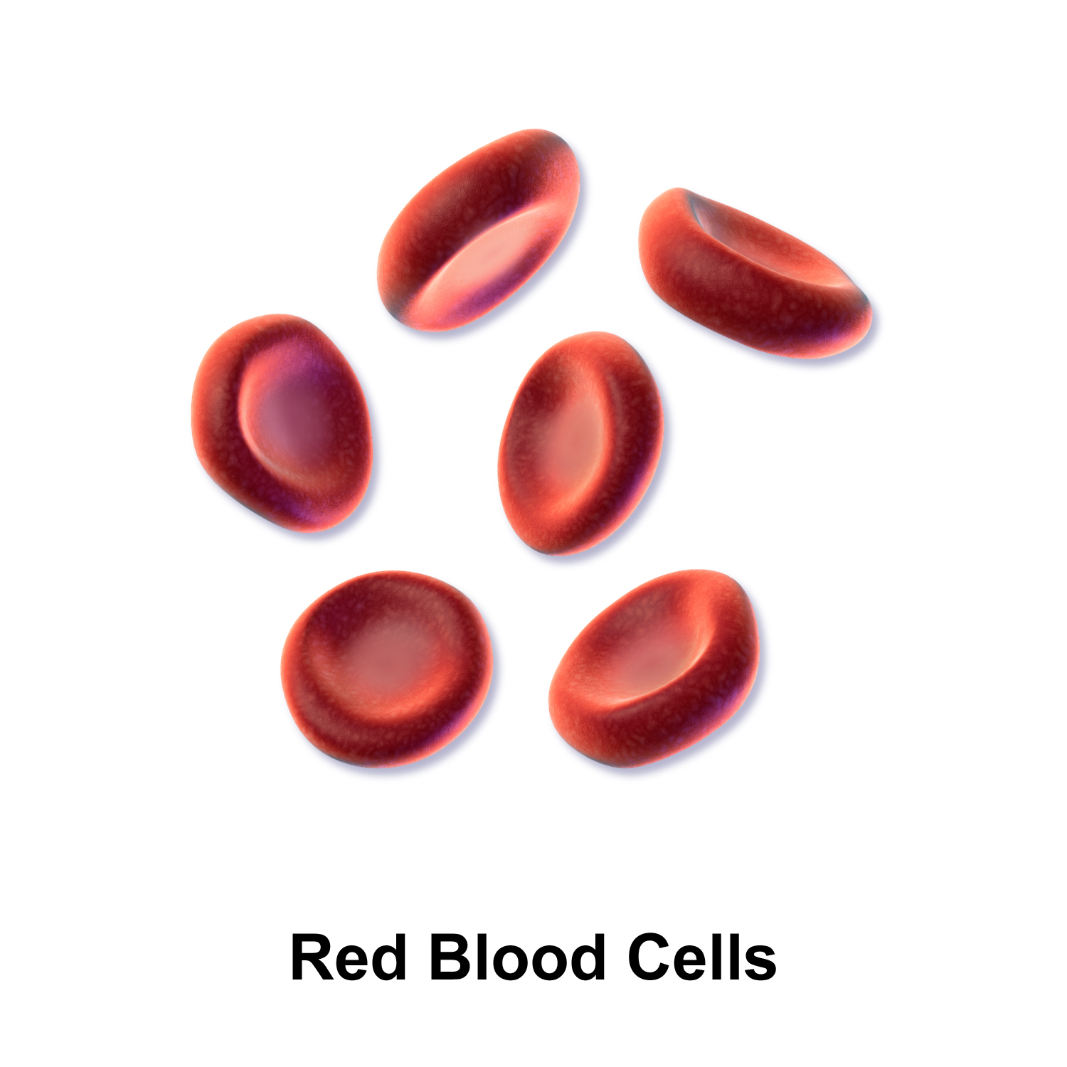
In humans, mature red blood cells are flexible biconcave disks. They lack a cell nucleus (which is expelled during development) and organelles, to accommodate maximum space for hemoglobin; they can be viewed as sacks of hemoglobin, with a plasma membrane as the sack. Approximately 2.4 million new erythrocytes are produced per second in human adults.[4] The cells develop in the bone marrow and circulate for about 100–120 days in the body before their components are recycled by macrophages. Each circulation takes about 60 seconds (one minute).[5] Approximately 84% of the cells in the human body are the 20–30 trillion red blood cells.[6][7][8][9] Nearly half of the blood's volume (40% to 45%) is red blood cells.

Packed red blood cells are red blood cells that have been donated, processed, and stored in a blood bank for blood transfusion.

Structure

[edit]

Vertebrates

[edit]
There is an immense size variation in vertebrate red blood cells, as well as a correlation between cell and nucleus size. Mammalian red blood cells, which do not contain nuclei, are considerably smaller than those of most other vertebrates.[10]
Mature red blood cells of birds have a nucleus, however in the blood of adult females of penguin Pygoscelis papua enucleated red blood cells (B) have been observed, but with very low frequency.

The vast majority of vertebrates, including mammals and humans, have red blood cells. These erythrocytes are cells present in blood to transport oxygen. The only known vertebrates without red blood cells are the crocodile icefish (family Channichthyidae); they live in very oxygen-rich cold water and transport oxygen freely dissolved in their blood.[11] While they no longer use hemoglobin, remnants of hemoglobin genes can be found in their genome.[12]

Vertebrate red blood cells consist mainly of hemoglobin, a complex metalloprotein containing heme groups whose iron atoms temporarily bind to oxygen molecules (O2) in the lungs or gills and release them throughout the body. Oxygen can easily diffuse through the erythrocyte's cell membrane. Hemoglobin in the red blood cells also carries some of the waste product carbon dioxide back from the tissues; most waste carbon dioxide, however, is transported back to the pulmonary capillaries of the lungs as bicarbonate (HCO3) dissolved in the blood plasma. Myoglobin, a compound related to hemoglobin, acts to store oxygen in muscle cells.[13]

The color of red blood cells is due to the heme group of hemoglobin. The blood plasma alone is straw-colored, but the red blood cells change color depending on the state of the hemoglobin: when combined with oxygen the resulting oxyhemoglobin is scarlet, and when oxygen has been released the resulting deoxyhemoglobin is of a dark red burgundy color. However, blood can appear bluish when seen through the vessel wall and skin.[14] Pulse oximetry takes advantage of the hemoglobin color change to directly measure the arterial blood oxygen saturation using colorimetric techniques. Hemoglobin also has a very high affinity for carbon monoxide, forming carboxyhemoglobin which is a very bright red in color. Flushed, confused patients with a saturation reading of 100% on pulse oximetry are sometimes found to be suffering from carbon monoxide poisoning.[citation needed]

Having oxygen-carrying proteins inside specialized cells (as opposed to oxygen carriers being dissolved in body fluid) was an important step in the evolution of vertebrates as it allows for less viscous blood, higher concentrations of oxygen, and better diffusion of oxygen from the blood to the tissues. The size of red blood cells varies widely among vertebrate species; red blood cell width is on average about 25% larger than capillary diameter, and it has been hypothesized that this improves the oxygen transfer from red blood cells to tissues.[15]

Mammals

[edit]
Typical mammalian red blood cells: (a) seen from surface; (b) in profile, forming rouleaux; (c) rendered spherical by water; (d) rendered crenate (shrunken and spiky) by salt. (c) and (d) do not normally occur in the body. The last two shapes are due to water being transported into, and out of, the cells, by osmosis.

The red blood cells of mammals are typically shaped as biconcave disks: flattened and depressed in the center, with a dumbbell-shaped cross section, and a torus-shaped rim on the edge of the disk. This shape allows for a high surface-area-to-volume (SA/V) ratio to facilitate diffusion of gases.[16] However, there are some exceptions concerning shape in the artiodactyl order (even-toed ungulates including cattle, deer, and their relatives), which displays a wide variety of bizarre red blood cell morphologies: small and highly ovaloid cells in llamas and camels (family Camelidae), tiny spherical cells in mouse deer (family Tragulidae), and cells which assume fusiform, lanceolate, crescentic, and irregularly polygonal and other angular forms in red deer and wapiti (family Cervidae). Members of this order have clearly evolved a mode of red blood cell development substantially different from the mammalian norm.[10][17] Overall, mammalian red blood cells are remarkably flexible and deformable so as to squeeze through tiny capillaries, as well as to maximize their apposing surface by assuming a cigar shape, where they efficiently release their oxygen load.[18]

Red blood cells in mammals are unique amongst vertebrates as most species do not have nuclei when mature.[19] They do have nuclei during early phases of erythropoiesis, but extrude them during development as they mature; this provides more space for hemoglobin. The red blood cells without nuclei, called reticulocytes, subsequently lose all other cellular organelles such as their mitochondria, Golgi apparatus and endoplasmic reticulum.

The spleen acts as a reservoir of red blood cells, but this effect is somewhat limited in humans. In some other mammals such as dogs and horses, the spleen sequesters large numbers of red blood cells, which are dumped into the blood during times of exertion stress, yielding a higher oxygen transport capacity.

Scanning electron micrograph of blood cells. From left to right: human red blood cell, thrombocyte (platelet), leukocyte.

Human

[edit]
Two drops of blood are shown with a bright red oxygenated drop on the left and a darker red deoxygenated drop on the right.
Animation of a typical human red blood cell cycle in the circulatory system. This animation occurs at a faster rate (~20 seconds of the average 60-second cycle) and shows the red blood cell deforming as it enters capillaries, as well as the bars changing color as the cell alternates in states of oxygenation along the circulatory system.

A typical human red blood cell has a disk diameter of approximately 6.2–8.2 μm[20] and a maximum thickness of 2–2.5 μm and a minimum thickness in the centre of 0.8–1 μm, being much smaller than most other human cells. These cells have an average volume of about 90 fL[21] with a surface area of about 136 μm2, and can swell up to a sphere shape containing 150 fL, without membrane distension.

Adult humans have roughly 20–30 trillion red blood cells at any given time, constituting approximately 70% of all cells by number.[22] Women have about 4–5 million red blood cells per microliter (cubic millimeter) of blood and men about 5–6 million; people living at high altitudes with low oxygen tension will have more. Red blood cells are thus much more common than the other blood particles: there are about 4,000–11,000 white blood cells and about 150,000–400,000 platelets per microliter.

Human red blood cells take on average 60 seconds to complete one cycle of circulation.[5][9][23]

The blood's red color is due to the spectral properties of the hemic iron ions in hemoglobin. Each hemoglobin molecule carries four heme groups; hemoglobin constitutes about a third of the total cell volume. Hemoglobin is responsible for the transport of more than 98% of the oxygen in the body (the remaining oxygen is carried dissolved in the blood plasma). The red blood cells of an average adult human male store collectively about 2.5 grams of iron, representing about 65% of the total iron contained in the body.[24][25]

Microstructure

[edit]

Nucleus

[edit]

Red blood cells in mammals are anucleate when mature, meaning that they lack a cell nucleus. In comparison, the red blood cells of other vertebrates have nuclei; the only known exceptions are salamanders of the family Plethodontidae, where five different clades has evolved various degrees of enucleated red blood cells (most evolved in some species of the genus Batrachoseps), and fish of the genus Maurolicus.[26][27][28]

The elimination of the nucleus in vertebrate red blood cells has been offered as an explanation for the subsequent accumulation of non-coding DNA in the genome.[17] The argument runs as follows: Efficient gas transport requires red blood cells to pass through very narrow capillaries, and this constrains their size. In the absence of nuclear elimination, the accumulation of repeat sequences is constrained by the volume occupied by the nucleus, which increases with genome size.

Nucleated red blood cells in mammals consist of two forms: normoblasts, which are normal erythropoietic precursors to mature red blood cells, and megaloblasts, which are abnormally large precursors that occur in megaloblastic anemias.

Membrane composition

[edit]

Red blood cells are deformable, flexible, are able to adhere to other cells, and are able to interface with immune cells. Their membrane plays many roles in this. These functions are highly dependent on the membrane composition. The red blood cell membrane is composed of 3 layers: the glycocalyx on the exterior, which is rich in carbohydrates; the lipid bilayer which contains many transmembrane proteins, besides its lipidic main constituents; and the membrane skeleton, a structural network of proteins located on the inner surface of the lipid bilayer. Half of the membrane mass in human and most mammalian red blood cells are proteins. The other half are lipids, namely phospholipids and cholesterol.[29]

Membrane lipids

[edit]
The most common red blood cell membrane lipids, schematically disposed as they are distributed on the bilayer. Relative abundances are not at scale.

The red blood cell membrane comprises a typical lipid bilayer, similar to what can be found in virtually all human cells. Simply put, this lipid bilayer is composed of cholesterol and phospholipids in equal proportions by weight. The lipid composition is important as it defines many physical properties such as membrane permeability and fluidity. Additionally, the activity of many membrane proteins is regulated by interactions with lipids in the bilayer.

Unlike cholesterol, which is evenly distributed between the inner and outer leaflets, the 5 major phospholipids are asymmetrically disposed, as shown below:

Outer monolayer

Inner monolayer

This asymmetric phospholipid distribution among the bilayer is the result of the function of several energy-dependent and energy-independent phospholipid transport proteins. Proteins called "Flippases" move phospholipids from the outer to the inner monolayer, while others called "floppases" do the opposite operation, against a concentration gradient in an energy-dependent manner. Additionally, there are also "scramblase" proteins that move phospholipids in both directions at the same time, down their concentration gradients in an energy-independent manner. There is still considerable debate ongoing regarding the identity of these membrane maintenance proteins in the red cell membrane.

The maintenance of an asymmetric phospholipid distribution in the bilayer (such as an exclusive localization of PS and PIs in the inner monolayer) is critical for the cell integrity and function due to several reasons:

  • Macrophages recognize and phagocytose red cells that expose PS at their outer surface. Thus the confinement of PS in the inner monolayer is essential if the cell is to survive its frequent encounters with macrophages of the reticuloendothelial system, especially in the spleen.
  • Premature destruction of thallassemic and sickle red cells has been linked to disruptions of lipid asymmetry leading to exposure of PS on the outer monolayer.
  • An exposure of PS can potentiate adhesion of red cells to vascular endothelial cells, effectively preventing normal transit through the microvasculature. Thus it is important that PS is maintained only in the inner leaflet of the bilayer to ensure normal blood flow in microcirculation.
  • Both PS and phosphatidylinositol 4,5-bisphosphate (PIP2) can regulate membrane mechanical function, due to their interactions with skeletal proteins such as spectrin and protein 4.1R. Recent studies have shown that binding of spectrin to PS promotes membrane mechanical stability. PIP2 enhances the binding of protein band 4.1R to glycophorin C but decreases its interaction with protein band 3, and thereby may modulate the linkage of the bilayer to the membrane skeleton.

The presence of specialized structures named "lipid rafts" in the red blood cell membrane have been described by recent studies. These are structures enriched in cholesterol and sphingolipids associated with specific membrane proteins, namely flotillins, STOMatins (band 7), G-proteins, and β-adrenergic receptors. Lipid rafts that have been implicated in cell signaling events in nonerythroid cells have been shown in erythroid cells to mediate β2-adregenic receptor signaling and increase cAMP levels, and thus regulating entry of malarial parasites into normal red cells.[30][31]

Membrane proteins

[edit]
Red blood cell membrane proteins separated by SDS-PAGE and silverstained[32]

The proteins of the membrane skeleton are responsible for the deformability, flexibility and durability of the red blood cell, enabling it to squeeze through capillaries less than half the diameter of the red blood cell (7–8 μm) and recovering the discoid shape as soon as these cells stop receiving compressive forces, in a similar fashion to an object made of rubber.

There are currently more than 50 known membrane proteins, which can exist in a few hundred up to a million copies per red blood cell. Approximately 25 of these membrane proteins carry the various blood group antigens, such as the A, B and Rh antigens, among many others. These membrane proteins can perform a wide diversity of functions, such as transporting ions and molecules across the red cell membrane, adhesion and interaction with other cells such as endothelial cells, as signaling receptors, as well as other currently unknown functions. The blood types of humans are due to variations in surface glycoproteins of red blood cells. Disorders of the proteins in these membranes are associated with many disorders, such as hereditary spherocytosis, hereditary elliptocytosis, hereditary stomatocytosis, and paroxysmal nocturnal hemoglobinuria.[29][30]

The red blood cell membrane proteins organized according to their function:

Red blood cell membrane major proteins

Transport

Cell adhesion

Structural role – The following membrane proteins establish linkages with skeletal proteins and may play an important role in regulating cohesion between the lipid bilayer and membrane skeleton, likely enabling the red cell to maintain its favorable membrane surface area by preventing the membrane from collapsing (vesiculating).

  • Ankyrin-based macromolecular complex – proteins linking the bilayer to the membrane skeleton through the interaction of their cytoplasmic domains with Ankyrin.
    • Band 3 – also assembles various glycolytic enzymes, the presumptive CO2 transporter, and carbonic anhydrase into a macromolecular complex termed a "metabolon", which may play a key role in regulating red cell metabolism and ion and gas transport function.
    • RHAG – also involved in transport, defines associated unusual blood group phenotype Rhmod.
  • Protein 4.1R-based macromolecular complex – proteins interacting with Protein 4.1R.
    • Protein 4.1R – weak expression of Gerbich antigens;
    • Glycophorin C and D – glycoprotein, defines Gerbich Blood Group;
    • XK – defines the Kell Blood Group and the Mcleod unusual phenotype (lack of Kx antigen and greatly reduced expression of Kell antigens);
    • RhD/RhCE – defines Rh Blood Group and the associated unusual blood group phenotype Rhnull;
    • Duffy protein – has been proposed to be associated with chemokine clearance;[35]
    • Adducin – interaction with band 3;
    • Dematin- interaction with the Glut1 glucose transporter.

[29][30]

Surface electrostatic potential

[edit]

The zeta potential is an electrochemical property of cell surfaces that is determined by the net electrical charge of molecules exposed at the surface of cell membranes of the cell. The normal zeta potential of the red blood cell is −15.7 millivolts (mV).[36] Much of this potential appears to be contributed by the exposed sialic acid residues in the membrane: their removal results in zeta potential of −6.06 mV.

Function

[edit]

Role in CO2 transport

[edit]

Recall that respiration, as illustrated schematically here with a unit of carbohydrate, produces about as many molecules of carbon dioxide, CO2, as it consumes of oxygen, O2.[37]

Thus, the function of the circulatory system is as much about the transport of carbon dioxide as about the transport of oxygen. As stated elsewhere in this article, most of the carbon dioxide in the blood is in the form of bicarbonate ion. The bicarbonate provides a critical pH buffer.[38] Thus, unlike hemoglobin for O2 transport, there is a physiological advantage to not having a specific CO2 transporter molecule.

Red blood cells, nevertheless, play a key role in the CO2 transport process, for two reasons. First, because, besides hemoglobin, they contain a large number of copies of the enzyme carbonic anhydrase on the inside of their cell membrane.[39] Carbonic anhydrase, as its name suggests, acts as a catalyst of the exchange between carbonic acid and carbon dioxide (which is the anhydride of carbonic acid). Because it is a catalyst, it can affect many CO2 molecules, so it performs its essential role without needing as many copies as are needed for O2 transport by hemoglobin. In the presence of this catalyst carbon dioxide and carbonic acid reach an equilibrium very rapidly, while the red cells are still moving through the capillary. Thus it is the RBC that ensures that most of the CO2 is transported as bicarbonate.[40][41] At physiological pH the equilibrium strongly favors carbonic acid, which is mostly dissociated into bicarbonate ion.[42]

The H+ ions released by this rapid reaction within RBC, while still in the capillary, act to reduce the oxygen binding affinity of hemoglobin, the Bohr effect.

The second major contribution of RBC to carbon dioxide transport is that carbon dioxide directly reacts with globin protein components of hemoglobin to form carbaminohemoglobin compounds. As oxygen is released in the tissues, more CO2 binds to hemoglobin, and as oxygen binds in the lung, it displaces the hemoglobin bound CO2, this is called the Haldane effect. Despite the fact that only a small amount of the CO2 in blood is bound to hemoglobin in venous blood, a greater proportion of the change in CO2 content between venous and arterial blood comes from the change in this bound CO2.[43] That is, there is always an abundance of bicarbonate in blood, both venous and arterial, because of its aforementioned role as a pH buffer.

In summary, carbon dioxide produced by cellular respiration diffuses very rapidly to areas of lower concentration, specifically into nearby capillaries.[44][45] When it diffuses into a RBC, CO2 is rapidly converted by the carbonic anhydrase found on the inside of the RBC membrane into bicarbonate ion. The bicarbonate ions in turn leave the RBC in exchange for chloride ions from the plasma, facilitated by the band 3 anion transport protein colocated in the RBC membrane. The bicarbonate ion does not diffuse back out of the capillary, but is carried to the lung. In the lung the lower partial pressure of carbon dioxide in the alveoli causes carbon dioxide to diffuse rapidly from the capillary into the alveoli. The carbonic anhydrase in the red cells keeps the bicarbonate ion in equilibrium with carbon dioxide. So as carbon dioxide leaves the capillary, and CO2 is displaced by O2 on hemoglobin, sufficient bicarbonate ion converts rapidly to carbon dioxide to maintain the equilibrium.[39][46][47][48]

Secondary functions

[edit]

When red blood cells undergo shear stress in constricted vessels, they release ATP, which causes the vessel walls to relax and dilate so as to promote normal blood flow.[49]

When their hemoglobin molecules are deoxygenated, red blood cells release S-Nitrosothiols, which also act to dilate blood vessels,[50] thus directing more blood to areas of the body depleted of oxygen.

Red blood cells can also synthesize nitric oxide enzymatically, using L-arginine as substrate, as do endothelial cells.[51] Exposure of red blood cells to physiological levels of shear stress activates nitric oxide synthase and export of nitric oxide,[52] which may contribute to the regulation of vascular tonus.

Red blood cells can also produce hydrogen sulfide, a signalling gas that acts to relax vessel walls. It is believed that the cardioprotective effects of garlic are due to red blood cells converting its sulfur compounds into hydrogen sulfide.[53]

Red blood cells also play a part in the body's immune response: when lysed by pathogens such as bacteria, their hemoglobin releases free radicals, which break down the pathogen's cell wall and membrane, killing it.[54][55]

Cellular processes

[edit]

As a result of not containing mitochondria, red blood cells use none of the oxygen they transport; instead they produce the energy carrier ATP by the glycolysis of glucose and lactic acid fermentation on the resulting pyruvate.[56][57] Furthermore, the pentose phosphate pathway plays an important role in red blood cells; see glucose-6-phosphate dehydrogenase deficiency for more information.

As red blood cells contain no nucleus, protein biosynthesis is currently assumed to be absent in these cells.

Because of the lack of nuclei and organelles, mature red blood cells do not contain DNA and cannot synthesize any RNA (although it does contain RNAs),[58][59] and consequently cannot divide and have limited repair capabilities.[60] The inability to carry out protein synthesis means that no virus can evolve to target mammalian red blood cells.[61] However, infection with parvoviruses (such as human parvovirus B19) can affect erythroid precursors while they still have DNA, as recognized by the presence of giant pronormoblasts with viral particles and inclusion bodies, thus temporarily depleting the blood of reticulocytes and causing anemia.[62]

Life cycle

[edit]

Human red blood cells are produced through a process named erythropoiesis, developing from committed stem cells to mature red blood cells in about 7 days. When matured, in a healthy individual these cells live in blood circulation for about 100 to 120 days (and 80 to 90 days in a full term infant).[63] At the end of their lifespan, they are removed from circulation. In many chronic diseases, the lifespan of the red blood cells is reduced.

Creation

[edit]

Erythropoiesis is the process by which new red blood cells are produced; it lasts about 7 days. Through this process red blood cells are continuously produced in the red bone marrow of large bones. (In the embryo, the liver is the main site of red blood cell production.) The production can be stimulated by the hormone erythropoietin (EPO), synthesised by the kidney. Just before and after leaving the bone marrow, the developing cells are known as reticulocytes; these constitute about 1% of circulating red blood cells.

Functional lifetime

[edit]

The functional lifetime of a red blood cell is about 100–120 days, during which time the red blood cells are continually moved by the blood flow push (in arteries), pull (in veins) and a combination of the two as they squeeze through microvessels such as capillaries. They are also recycled in the bone marrow.[64]

Senescence

[edit]

The aging red blood cell undergoes changes in its plasma membrane, making it susceptible to selective recognition by macrophages and subsequent phagocytosis in the mononuclear phagocyte system (spleen, liver and lymph nodes), thus removing old and defective cells and continually purging the blood. This process is termed eryptosis, red blood cell programmed death.[65] This process normally occurs at the same rate of production by erythropoiesis, balancing the total circulating red blood cell count. Eryptosis is increased in a wide variety of diseases including sepsis, haemolytic uremic syndrome, malaria, sickle cell anemia, beta-thalassemia, glucose-6-phosphate dehydrogenase deficiency, phosphate depletion, iron deficiency and Wilson's disease. Eryptosis can be elicited by osmotic shock, oxidative stress, and energy depletion, as well as by a wide variety of endogenous mediators and xenobiotics. Excessive eryptosis is observed in red blood cells lacking the cGMP-dependent protein kinase type I or the AMP-activated protein kinase AMPK. Inhibitors of eryptosis include erythropoietin, nitric oxide, catecholamines and high concentrations of urea.

Much of the resulting breakdown products are recirculated in the body. The heme constituent of hemoglobin are broken down into iron (Fe3+) and biliverdin. The biliverdin is reduced to bilirubin, which is released into the plasma and recirculated to the liver bound to albumin. The iron is released into the plasma to be recirculated by a carrier protein called transferrin. Almost all red blood cells are removed in this manner from the circulation before they are old enough to hemolyze. Hemolyzed hemoglobin is bound to a protein in plasma called haptoglobin, which is not excreted by the kidney.[66]

Clinical significance

[edit]

Disease

[edit]
Affected by Sickle-cell disease, red blood cells alter shape and threaten to damage internal organs.

Blood diseases involving the red blood cells include:

  • Anemias (or anaemias) are diseases characterized by low oxygen transport capacity of the blood, because of low red cell count or some abnormality of the red blood cells or the hemoglobin.
  • Iron deficiency anemia is the most common anemia; it occurs when the dietary intake or absorption of iron is insufficient, and hemoglobin, which contains iron, cannot be formed.
  • Sickle-cell disease is a genetic disease that results in abnormal hemoglobin molecules. When these release their oxygen load in the tissues, they become insoluble, leading to mis-shaped red blood cells. These sickle shaped red cells are less deformable and viscoelastic, meaning that they have become rigid and can cause blood vessel blockage, pain, strokes, and other tissue damage.
  • Thalassemia is a genetic disease that results in the production of an abnormal ratio of hemoglobin subunits.
  • Hereditary spherocytosis syndromes are a group of inherited disorders characterized by defects in the red blood cell's cell membrane, causing the cells to be small, sphere-shaped, and fragile instead of donut-shaped and flexible. These abnormal red blood cells are destroyed by the spleen. Several other hereditary disorders of the red blood cell membrane are known.[67]
Effect of osmotic pressure on blood cells
Micrographs of the effects of osmotic pressure
  • Hemolysis is the general term for excessive breakdown of red blood cells. It can have several causes and can result in hemolytic anemia.
  • The malaria parasite spends part of its life-cycle in red blood cells, feeds on their hemoglobin and then breaks them apart, causing fever. Both sickle-cell disease and thalassemia are more common in malaria areas, because these mutations convey some protection against the parasite.
  • Polycythemias (or erythrocytoses) are diseases characterized by a surplus of red blood cells. The increased viscosity of the blood can cause a number of symptoms.
  • In polycythemia vera the increased number of red blood cells results from an abnormality in the bone marrow.

Transfusion

[edit]

Red blood cells may be given as part of a blood transfusion. Blood may be donated from another person, or stored by the recipient at an earlier date. Donated blood usually requires screening to ensure that donors do not contain risk factors for the presence of blood-borne diseases, or will not suffer themselves by giving blood. Blood is usually collected and tested for common or serious blood-borne diseases including Hepatitis B, Hepatitis C and HIV. The blood type (A, B, AB, or O) or the blood product is identified and matched with the recipient's blood to minimise the likelihood of acute hemolytic transfusion reaction, a type of transfusion reaction. This relates to the presence of antigens on the cell's surface. After this process, the blood is stored, and within a short duration is used. Blood can be given as a whole product or the red blood cells separated as packed red blood cells.

Blood is often transfused when there is known anaemia, active bleeding, or when there is an expectation of serious blood loss, such as prior to an operation. Before blood is given, a small sample of the recipient's blood is tested with the transfusion in a process known as cross-matching.

In 2008 it was reported that human embryonic stem cells had been successfully coaxed into becoming red blood cells in the lab. The difficult step was to induce the cells to eject their nucleus; this was achieved by growing the cells on stromal cells from the bone marrow. It is hoped that these artificial red blood cells can eventually be used for blood transfusions.[68]

A human trial is conducted in 2022, using blood cultured from stem cells obtained from donor blood.[69]

Tests

[edit]
Variations of red blood cell shape, overall termed poikilocytosis

Several blood tests involve red blood cells. These include a RBC count (the number of red blood cells per volume of blood), calculation of the hematocrit (percentage of blood volume occupied by red blood cells), and the erythrocyte sedimentation rate. The blood type needs to be determined to prepare for a blood transfusion or an organ transplantation.

Many diseases involving red blood cells are diagnosed with a blood film (or peripheral blood smear), where a thin layer of blood is smeared on a microscope slide. This may reveal poikilocytosis, which are variations in red blood cell shape. When red blood cells sometimes occur as a stack, flat side next to flat side. This is known as rouleaux formation, and it occurs more often if the levels of certain serum proteins are elevated, as for instance during inflammation.

Separation and blood doping

[edit]

Red blood cells can be obtained from whole blood by centrifugation, which separates the cells from the blood plasma in a process known as blood fractionation. Packed red blood cells, which are made in this way from whole blood with the plasma removed, are used in transfusion medicine.[70] During plasma donation, the red blood cells are pumped back into the body right away and only the plasma is collected.

Some athletes have tried to improve their performance by blood doping: first about 1 litre of their blood is extracted, then the red blood cells are isolated, frozen and stored, to be reinjected shortly before the competition. (Red blood cells can be conserved for 5 weeks at −79 °C or −110 °F, or over 10 years using cryoprotectants[71]) This practice is hard to detect but may endanger the human cardiovascular system which is not equipped to deal with blood of the resulting higher viscosity. Another method of blood doping involves injection with erythropoietin to stimulate production of red blood cells. Both practices are banned by the World Anti-Doping Agency.

History

[edit]

The first person to describe red blood cells was the young Dutch biologist Jan Swammerdam, who had used an early microscope in 1658 to study the blood of a frog.[72] Unaware of this work, Anton van Leeuwenhoek provided another microscopic description in 1674, this time providing a more precise description of red blood cells, even approximating their size, "25,000 times smaller than a fine grain of sand".

In the 1740s, Vincenzo Menghini in Bologna was able to demonstrate the presence of iron by passing magnets over the powder or ash remaining from heated red blood cells.

In 1901, Karl Landsteiner published his discovery of the three main blood groups—A, B, and C (which he later renamed to O). Landsteiner described the regular patterns in which reactions occurred when serum was mixed with red blood cells, thus identifying compatible and conflicting combinations between these blood groups. A year later Alfred von Decastello and Adriano Sturli, two colleagues of Landsteiner, identified a fourth blood group—AB.

In 1959, by use of X-ray crystallography, Max Perutz was able to unravel the structure of hemoglobin, the red blood cell protein that carries oxygen.[73]

The oldest intact red blood cells ever discovered were found in Ötzi the Iceman, a natural mummy of a man who died around 3255 BCE. These cells were discovered in May 2012.[74]

See also

[edit]

References

[edit]
[edit]
Revisions and contributorsEdit on WikipediaRead on Wikipedia
from Grokipedia
Red blood cells (RBCs), also known as erythrocytes, are the most abundant cells in blood, comprising approximately 40-45% of and numbering 4-6 million per cubic millimeter, with their primary function being the transport of oxygen from the lungs to body tissues via and the return of to the lungs for . These cells are uniquely adapted for their role, featuring a biconcave discoid that provides a large surface area-to-volume for efficient and flexibility to navigate narrow capillaries. Lacking a nucleus and most organelles in their mature form, mature RBCs measure about 7.5 to 8.7 μm in diameter and 1.7 to 2.2 μm in thickness at the rim, allowing them to maximize internal space for , the iron-containing protein that binds up to four oxygen molecules per molecule. RBCs are produced through , a process in which hematopoietic stem cells in the differentiate into proerythroblasts and progress through stages including basophilic erythroblasts, polychromatophilic erythroblasts, orthochromatic erythroblasts, and reticulocytes, culminating in the expulsion of the nucleus to form mature erythrocytes; this entire maturation takes about 5-7 days and is tightly regulated by , a secreted primarily by the kidneys in response to low oxygen levels. Newly formed RBCs enter circulation and typically survive for around 120 days before being removed by macrophages in the , liver, and , during which time their is broken down, with iron recycled for new RBC production. Disruptions in RBC structure, production, or function can lead to disorders such as , where insufficient oxygen delivery causes fatigue and pallor, or conditions like , characterized by abnormally shaped cells that impair blood flow.

Structure

Vertebrates

Red blood cells, or erythrocytes, exhibit significant structural diversity across vertebrate classes, reflecting adaptations to varying physiological and environmental demands. In non-mammalian vertebrates, including , amphibians, reptiles, and birds, erythrocytes are typically nucleated, containing a nucleus and various organelles that support cellular functions beyond oxygen transport. In contrast, mammalian erythrocytes are anucleate, having extruded their nucleus during maturation, a trait unique to this class among vertebrates and present in all mammalian lineages, including monotremes, marsupials, and placentals. This enucleation is believed to have evolved to enhance metabolic efficiency and flexibility for high-energy lifestyles. The of erythrocytes also varies systematically across groups. Non-mammalian erythrocytes are generally oval or elliptical, accommodating the central nucleus and providing a more rigid structure compared to their mammalian counterparts. Mammalian erythrocytes, lacking a nucleus, adopt a biconcave disc morphology that increases surface area for and facilitates passage through narrow capillaries. These shape differences contribute to evolutionary optimizations in circulation, with the elliptical form in lower vertebrates suiting slower metabolic rates and larger body sizes in some ectothermic species. Erythrocyte size shows pronounced variation, correlating with metabolic demands and phylogenetic position. erythrocytes are notably large, often ranging from 10 to 70 μm in diameter, enabling extended circulation in low-oxygen aquatic environments. In comparison, mammalian erythrocytes are smaller, typically 5-8 μm, as seen in humans, allowing for higher cell counts and efficient oxygen delivery in endothermic systems. Reptilian and avian erythrocytes fall between these extremes, with sizes increasing from birds (around 10-15 μm) to reptiles, reflecting a gradient from endothermy to ectothermy. erythrocytes can be similarly large or larger in some species, adapted to variable oxygen levels in water. These structural features have evolved to optimize oxygen delivery in diverse habitats, from aquatic to aerial environments. The retention of nuclei in non-mammalian vertebrates permits ongoing protein synthesis, allowing erythrocytes to respond dynamically to stressors like hypoxia through de novo production of proteins and metabolites. For instance, avian erythrocytes, which are nucleated and elliptical, support the intense metabolic requirements of flight by maintaining organelles for energy production and potentially contributing to immune functions, adaptations that enhance endurance in oxygen-demanding aerial locomotion.

Mammals

Mammalian red blood cells (RBCs) are characterized by enucleation, a during terminal where the nucleus and most organelles, including mitochondria and ribosomes, are extruded, resulting in mature cells that lack these structures. This adaptation maximizes intracellular space for , enhancing oxygen-carrying capacity, while increasing cell deformability to navigate the efficiently. In contrast to nucleated RBCs in other vertebrates, this enucleation enables mammalian RBCs to prioritize gas over protein synthesis or energy production via . The typical mammalian RBC adopts a biconcave discoid shape, which optimizes the surface-to-volume ratio for rapid gas across the . This morphology features a diameter of approximately 6-8 μm, with a thickness of about 2 μm at the rim and 1 μm at the central depression. The biconcave form facilitates efficient oxygen loading in the lungs and unloading in tissues by promoting and minimizing diffusion distances. Flexibility is a hallmark of mammalian RBCs, conferred by a spectrin-based cytoskeleton that forms a dynamic meshwork beneath the plasma membrane, linking filaments and other proteins to maintain structural integrity under . This network allows RBCs to deform reversibly, passing through microcapillaries as narrow as 3 μm in diameter—smaller than their resting size—without rupture, ensuring unobstructed . RBC morphology varies across mammalian species, reflecting physiological adaptations; for instance, mouse RBCs are smaller, with a diameter of about 6 μm, suited to their high metabolic rate and compact vasculature. In contrast, camel RBCs are larger, measuring approximately 8-9 μm in length with an oval shape rather than biconcave discoid, which resists swelling and maintains function during dehydration by tolerating up to 25% body water loss without hemolysis. Llamas, as high-altitude camelids, exhibit similar oval RBCs that enhance deformability under hypoxic conditions; recent studies on camelid RBCs show reduced deformability indices (e.g., 0.024 in camels vs. 0.215 in humans) but improved resilience in low-oxygen environments, supporting oxygen delivery at elevations over 4,000 meters.

Human

Human red blood cells, or erythrocytes, exhibit precise morphological characteristics that optimize their function in circulation. The average measures approximately 7.8 μm, with a of 90-100 fL and a surface area of about 140 μm². The biconcave discoid shape of human erythrocytes enhances deformability, allowing passage through narrow capillaries, with the membrane's ranging from 10-50 kPa as measured by . This configuration optimizes the surface area to volume ratio at approximately 1.5 μm⁻¹, facilitating efficient oxygen diffusion while minimizing energy expenditure for shape maintenance. SAV1.5μm1\frac{SA}{V} \approx 1.5 \, \mu\mathrm{m}^{-1} Recent 2024 advances in super-resolution imaging have revealed nanoscale structural details on the erythrocyte surface, including protein distributions that contribute to immune evasion mechanisms such as CD47-mediated "don't eat me" signaling.

Microstructure

Nucleus and Organelles

In non-mammalian vertebrates, such as fish, birds, and non-avian reptiles, mature red blood cells retain a nucleus and contain organelles including mitochondria. This nuclear presence enables DNA replication and RNA transcription, supporting ongoing gene expression and protein synthesis within these cells. Additionally, the mitochondria facilitate aerobic respiration, allowing efficient ATP production to meet the cells' energy demands during circulation. In contrast, mammalian red blood cells, including those in humans, undergo enucleation as a hallmark of terminal , extruding the nucleus during the transition from orthochromatic erythroblasts to reticulocytes. This process also results in the clearance of organelles such as mitochondria, ribosomes, and the Golgi apparatus, leaving mature cells devoid of these structures. Consequently, mammalian red blood cells depend entirely on proteins and enzymes pre-synthesized in precursor stages, as they lack the machinery for new transcription or . The nuclear extrusion in reticulocytes is an asymmetric process involving actomyosin contractions, where polarized assembly and disassembly, along with non-muscle IIB, drive the nucleus toward the cell periphery and facilitate its expulsion into a pyrenocyte that is subsequently phagocytosed. The absence of a nucleus and organelles profoundly impacts function: metabolic activity is severely limited to anaerobic glycolysis for ATP generation, as no mitochondrial is possible. Without nuclear oversight or ribosomal capacity for protein repair and turnover, these cells cannot mitigate cumulative damage from or mechanical shear, thereby restricting their lifespan to approximately 120 days in humans. Recent 2025 research has revealed that human mature red blood cells may retain trace nuclear remnants, including DNA fragments and RNA species, detectable through sequencing and microscopic techniques, which could contribute to immune recognition and accelerated clearance.

Membrane Composition

The red blood cell (RBC) membrane features a lipid bilayer that accounts for approximately 40% of the membrane's mass, with the lipid fraction consisting of roughly 50% phospholipids and 40% cholesterol. Phospholipids, the primary structural components, include phosphatidylcholine (PC), which predominates in the outer leaflet, and phosphatidylethanolamine (PE), which is enriched in the inner leaflet. Cholesterol intercalates between phospholipids, modulating membrane fluidity and preventing excessive rigidity or permeability to maintain the cell's biconcave shape during circulation. This bilayer exhibits pronounced transverse asymmetry, with aminophospholipids such as PE and phosphatidylserine (PS) confined to the cytoplasmic (inner) leaflet, while PC and sphingomyelin occupy the extracellular (outer) leaflet. Asymmetry is actively maintained by ATP-dependent flippases, including ATP11C, which translocate PS and PE inward against their concentration gradients. Disruption of this asymmetry, often during cellular stress, leads to PS externalization, which serves as an "eat-me" signal for phagocytic clearance and is a hallmark of RBC apoptosis or senescence. Integral membrane proteins, embedded within the bilayer, constitute about 50% of the membrane mass and include band 3 (anion exchanger 1, AE1), the most abundant protein at approximately 25% of total membrane proteins or 1–1.2 million copies per RBC. Band 3 functions as a dimeric or tetrameric transporter mediating electroneutral 1:1 exchange of anions like (Cl⁻) and (HCO₃⁻), crucial for CO₂ transport via the . The band 3-mediated anion flux rate reaches about 50,000 ions per second per polypeptide at physiological temperatures, enabling rapid equilibration (under 0.1 seconds) during transit. Peripheral proteins, such as spectrin (the main cytoskeletal element) and , associate with band 3's cytoplasmic domain to anchor the bilayer to the spectrin-based , ensuring mechanical stability and deformability. Surface glycoproteins, including glycophorins, bear oligosaccharide chains that carry ABO blood group antigens, determining transfusion compatibility. These antigens—A, B, or H structures—are synthesized by glycosyltransferases encoded by the ABO gene and expressed at about 2 million copies per RBC, primarily as attachments to band 3 and glycophorin A. Recent studies have also highlighted the contributions of glycosylphosphatidylinositol (GPI)-anchored proteins, such as CD55 (decay-accelerating factor), to malaria resistance; these proteins can modulate Plasmodium falciparum invasion by altering erythrocyte receptor availability, with certain variants enhancing protective effects against severe infection.

Cytoplasm and Hemoglobin

The cytoplasm of mature red blood cells is an comprising approximately 70% water by volume, with the remaining 30% consisting primarily of and a suite of soluble enzymes that support anaerobic . Key glycolytic enzymes, such as and , facilitate glucose breakdown to generate ATP, essential for maintaining cell integrity in the absence of mitochondria. These components create a highly concentrated, viscous environment optimized for protein stability and function. Hemoglobin dominates the cytoplasmic as a , with adult human (HbA) consisting of two α-globin chains and two β-globin chains, each binding a group containing a ferrous iron (Fe²⁺) responsible for reversible oxygen coordination. The 's iron atom lies in the ring, positioned to accept oxygen without oxidation to the ferric state under physiological conditions. This quaternary structure enables conformational shifts between tense (deoxy) and relaxed (oxy) states, underpinning cooperative ligand binding. In red blood cells, reaches a concentration of 33–35 g/dL, representing about 95% of the cell's dry weight and contributing to the cytoplasm's gel-like consistency. This high density supports allosteric , quantified by a Hill coefficient of approximately 2.8, which reflects interactions between subunits that enhance binding affinity after the first oxygen attaches. The cooperative oxygen binding is modeled by the Hill equation: Y=pO2nP50n+pO2nY = \frac{pO_2^n}{P_{50}^n + pO_2^n} where YY denotes the fractional saturation of with oxygen, pO2pO_2 is the of oxygen, n2.8n \approx 2.8 is the Hill coefficient indicating , and P50=26P_{50} = 26 mmHg is the pO2pO_2 at 50% saturation. This sigmoidal relationship arises from sequential conformational changes that propagate across the tetramer, stabilizing the high-affinity state. Hemoglobin variants alter chain composition and function; fetal hemoglobin (HbF) features two α and two γ chains, providing higher oxygen affinity during , while sickle cell hemoglobin (HbS) results from a β-chain glutamic acid-to-valine substitution at position 6, promoting deoxyhemoglobin . persists at low levels in adults and mitigates HbS pathology by inhibiting . Hemoglobin interacts reversibly with the membrane's band 3 protein, aiding cytoskeletal stability under deoxygenated conditions. In 2025, synthetic analogs, such as polymerized or encapsulated variants, have advanced as transfusion alternatives, offering oxygen-carrying capacity without immunogenicity risks.

Function

Oxygen Transport

Red blood cells primarily function to transport oxygen from the lungs to tissues by binding it to protein within their . 's cooperative oxygen binding allows efficient uptake and release; the binding of the first oxygen to one of the four groups induces a conformational change from the tense (T) to relaxed (R) state, increasing affinity for subsequent oxygen molecules, a phenomenon known as homotropic positive . Each tetramer binds up to four oxygen molecules, one per subunit. This structure enables a maximum oxygen-carrying capacity of approximately 1.34 mL of O₂ per gram of . Several physiological factors modulate hemoglobin's oxygen affinity to optimize delivery. The describes how decreased or increased CO₂ reduces oxygen affinity, facilitating unloading in metabolically active tissues where these conditions prevail. Quantitatively, the is expressed as the change in the logarithm of the of oxygen at 50% hemoglobin saturation (P₅₀) per unit change in : ΔlogP50ΔpH0.5\frac{\Delta \log P_{50}}{\Delta \mathrm{pH}} \approx -0.5 This coefficient indicates that a 0.1 unit decrease in raises P₅₀ by about 12%, shifting the oxygen dissociation curve rightward; the derivation arises from plotting log P₅₀ against , where the slope reflects proton-linked allosteric transitions in that stabilize the deoxy form at lower . Graphically, the standard sigmoid curve (Hill coefficient ≈2.8) at 7.4 (P₅₀ ≈26 mmHg) shifts to higher P₅₀ values (e.g., ≈32 mmHg at 7.2), enhancing oxygen release by 10-15% under acidic conditions without altering maximum saturation. Additionally, 2,3-bisphosphoglycerate (2,3-BPG), produced in red blood cells via , binds deoxyhemoglobin in a central cavity, stabilizing the T state and increasing P₅₀ by about 25%, which promotes oxygen unloading particularly at high altitudes or in chronic hypoxia. In systemic circulation, achieves about 97% saturation at a PO₂ of 100 mmHg, while mixed is typically 75% saturated at PO₂ of 40 mmHg, yielding an oxygen extraction ratio of roughly 25%. With normal levels (≈15 g/dL), total arterial oxygen content is approximately 20 vol% (mL O₂ per 100 mL ), of which over 98% is hemoglobin-bound, underscoring red blood cells' dominant role in oxygen delivery.

Carbon Dioxide Transport

Red blood cells play a crucial role in transporting (CO₂) from tissues to the lungs, where it is exhaled. Approximately 5–10% of CO₂ is carried in dissolved form in the plasma, 20–30% binds to as , and 60–70% is converted to bicarbonate ions (HCO₃⁻) within the red blood cell. These proportions ensure efficient removal of while maintaining blood balance. The primary mechanism for bicarbonate formation occurs inside the red blood cell, where CO₂ diffuses from the plasma across the cell membrane. The enzyme carbonic anhydrase II, abundant in the red blood cell cytoplasm, catalyzes the hydration of CO₂ to form carbonic acid, which rapidly dissociates into protons and bicarbonate: CO2+H2OH2CO3H++HCO3\text{CO}_2 + \text{H}_2\text{O} \rightleftharpoons \text{H}_2\text{CO}_3 \rightleftharpoons \text{H}^+ + \text{HCO}_3^- This reaction has an equilibrium pK_a of approximately 6.1 for the dissociation of carbonic acid. Without catalysis, the uncatalyzed hydration rate constant is about 0.13 s⁻¹, but carbonic anhydrase accelerates the process by 13,000–25,000-fold, achieving a turnover rate of roughly 10⁶ s⁻¹, allowing near-completion within milliseconds. The resulting H⁺ is buffered by hemoglobin, while HCO₃⁻ accumulates inside the cell. To prevent osmotic imbalance, the HCO₃⁻ ions are exchanged for chloride ions (Cl⁻) from the plasma via the band 3 anion exchanger protein (also known as AE1) in the red blood cell membrane, a process termed the Hamburger shift or . This exchange facilitates the net of CO₂ as HCO₃⁻ into the plasma, with the band 3 transporter operating at a turnover rate of 4–5 × 10⁴ ions per second per transporter at 37°C. In the lungs, the process reverses as CO₂ levels drop, allowing HCO₃⁻ to re-enter the cell for conversion back to CO₂. The enhances CO₂ transport efficiency by increasing the binding capacity of deoxygenated for CO₂ compared to oxygenated . Deoxygenated forms more carbamino compounds with CO₂ and better buffers the protons generated during bicarbonate formation, promoting greater CO₂ uptake in tissues and release in the lungs. This effect accounts for about half of the CO₂ transported during deoxygenation.

Secondary Roles

Beyond their primary role in gas transport, red blood cells (RBCs) contribute to pH buffering in the bloodstream through the action of , which acts as an effective buffer due to the imidazole side chains of its residues having pKa values near physiological . These residues, particularly those on the surface of the hemoglobin tetramer, facilitate proton acceptance or donation, helping to stabilize blood pH during metabolic acid production or CO2 release. RBCs also participate in nitric oxide (NO) homeostasis, transporting and releasing NO bioactivity to promote vasodilation, especially under hypoxic conditions. This process involves the reduction of nitrite to NO by deoxyhemoglobin within RBCs, which diffuses to vascular smooth muscle cells to induce relaxation and match blood flow to tissue oxygen demand. Additionally, RBCs express endothelial nitric oxide synthase, enabling local NO production that further supports vascular tone regulation. In immune modulation, RBCs scavenge (ROS) such as and using enzymatic antioxidants like and , thereby protecting vascular from oxidative damage. Furthermore, exposure of on the outer membrane leaflet of RBCs serves as an "eat-me" signal recognized by macrophages via receptors like TIM-4, facilitating non-inflammatory clearance as part of innate immune surveillance; membrane antigens such as provide a counter-signal to prevent premature . RBCs influence blood rheology by contributing to its non-Newtonian properties, including and shear-thinning behavior, where decreases at higher shear rates due to reversible deformation and disaggregation of RBC formations. This dynamic response, driven by the biconcave discoid shape and membrane flexibility of RBCs, ensures efficient flow through microvasculature without excessive energy expenditure by the heart. RBCs indirectly aid in xenobiotic handling by releasing during normal turnover, which is rapidly bound by plasma to form a complex that neutralizes the pro-oxidant effects of free , preventing endothelial akin to of a harmful substance. Recent findings highlight RBC-derived exosomes as mediators of intercellular signaling, carrying miRNAs and proteins that modulate endothelial function and , linking RBCs to systemic communication networks in and aging. As of 2025, studies also indicate RBC extracellular vesicles reduce in conditions like disorders and contribute to regenerative processes by modulating immune responses.

Lifecycle

Erythropoiesis

is the process by which hematopoietic stem cells in the differentiate into mature red blood cells, ensuring a continuous supply of erythrocytes for oxygen transport throughout the body. This tightly regulated differentiation pathway begins with committed erythroid progenitors and culminates in the release of enucleated cells into the circulation, with the entire terminal phase typically spanning 7-10 days in the . In adults, primarily occurs in the red of flat bones such as the , , , vertebrae, and , where approximately 200 billion red blood cells are produced daily to replace senescent cells and maintain steady-state hematopoiesis. The process unfolds through distinct morphological and functional stages, starting from the proerythroblast, which is the earliest recognizable erythroid precursor after commitment from colony-forming unit-erythroid (CFU-E) cells. The proerythroblast undergoes successive divisions and maturation: it progresses to the basophilic erythroblast, characterized by intense basophilic staining due to synthesis; then to the polychromatophilic erythroblast, where accumulation begins alongside reduced ; followed by the orthochromatic erythroblast, marked by further condensation and nuclear ; and finally, enucleation to form the , which retains ribosomal remnants and matures into the erythrocyte upon release. Each stage involves progressive decreases in cell size, nuclear condensation, and loss, driven by erythroid-specific transcription factors like and KLF1. Regulation of erythropoiesis is primarily orchestrated by (EPO), a hormone secreted by peritubular fibroblasts in the kidneys in response to tissue hypoxia, which is sensed via hypoxia-inducible factors (HIFs). EPO binds to its receptor (EPOR) on erythroid progenitors, activating the Janus kinase-signal transducer and activator of transcription (JAK-STAT) pathway, particularly JAK2-STAT5 signaling, to promote survival, proliferation, and differentiation while inhibiting in CFU-E and proerythroblast stages. This feedback mechanism ensures production matches oxygen demand, with additional modulation by (SCF) and insulin-like growth factor-1 (IGF-1). During these stages, (Hb) synthesis ramps up to equip erythrocytes for oxygen carriage, involving coordinated production of α- and β-globin chains in the of erythroblasts, which assemble with groups synthesized in mitochondria to form tetrameric HbA (α₂β₂). gene is tightly controlled by the β-globin locus control region and transcription factors such as , ensuring balanced α/β chain synthesis to prevent toxic aggregates. Recent advances in have explored strategies for EPO-independent to treat congenital anemias, such as using induced pluripotent stem cells (iPSCs) engineered to generate self-renewing erythroblasts without exogenous EPO or SCF, enabling scalable production of transfusion-compatible red blood cells for conditions like Diamond-Blackfan anemia. These approaches, demonstrated in 2024 preclinical models, highlight potential for autologous therapies bypassing EPO deficiencies in chronic kidney disease-associated anemias. In 2025, researchers identified a key NF-κB-related molecular signal that enhances erythroid maturation efficiency, potentially accelerating artificial production, alongside optimizations in iPSC platforms for large-scale, transfusion-ready RBC generation.

Circulation and Lifespan

Red blood cells (RBCs) in humans have an circulatory lifespan of approximately 120 days, during which they continuously circulate through the vascular system while maintaining their oxygen-transporting function. This duration can vary between individuals, with recent studies using stable isotopes like 15N-glycine or indicating a refined of 115-125 days, accounting for physiological variations and improved precision. The lifespan is monitored through markers such as band 3 clustering on the RBC membrane, which increases with age and signals without relying on nuclear features like shortening, as mature RBCs are anucleate. Throughout their lifespan, RBCs navigate an extensive circulatory path, estimated at around 300 miles in total, requiring repeated deformations to pass through narrow structures like splenic interendothelial slits and capillaries, which are often less than 3 micrometers in . These deformations, facilitated by the biconcave discoid shape and spectrin-based , allow RBCs to squeeze through splenic slits as narrow as 0.5 micrometers, ensuring efficient passage while filtering out less deformable cells. As RBCs age, they exhibit decreased ATP levels, which impairs ion pumps and leads to reduced deformability, alongside increased cellular rigidity that hinders passage through these tight spaces. To maintain steady-state oxygen delivery, approximately 1% of the RBC population—around 2 × 10^11 cells—is replaced daily, matching the turnover rate to the average lifespan. This replacement is influenced by dietary factors, including iron and , which support synthesis and overall RBC integrity, though deficiencies primarily affect production rates rather than directly altering individual cell lifespan.

Senescence and Clearance

As red blood cells (RBCs) age over their approximately 120-day lifespan, they undergo , marked by specific molecular signals that prepare them for removal from circulation. One key signal is the externalization of () on the outer leaflet, which serves as an "eat-me" cue for . This PS exposure results from the loss of phospholipid asymmetry maintained by ATP-dependent flippases, becoming prominent in the final 20% of the RBC's life. Another signal involves opsonization by () antibodies, particularly naturally occurring anti-band 3 antibodies that bind to clustered band 3 proteins on the senescent RBC surface, facilitating recognition by macrophages. Additionally, the progressive loss of , a that acts as a "don't-eat-me" signal by interacting with SIRPα on , further promotes clearance as its downregulation reduces inhibitory signaling. These changes, often triggered by membrane alterations such as oxidative damage, collectively signal the RBC's terminal phase. Clearance of senescent RBCs primarily occurs through by macrophages in the and liver. In the , macrophages in the red pulp efficiently engulf aged RBCs via recognition of PS and IgG opsonins, filtering out deformed or stressed cells through narrow slits that impose mechanical stress. The liver serves as a secondary site, particularly for more severely damaged or heavily opsonized RBCs, where Kupffer cells perform to handle spillover from splenic capacity. This process prevents the release of potentially toxic intracellular contents, maintaining vascular integrity. Following phagocytosis, the heme from hemoglobin is catabolized and recycled to support erythropoiesis and prevent toxicity. Macrophages degrade heme via heme oxygenase-1 (HO-1), yielding biliverdin (which converts to bilirubin for excretion), carbon monoxide, and free iron. The iron is sequestered by ferritin within macrophages and later exported via ferroportin for reuse, bound by transferrin in plasma. Meanwhile, any free hemoglobin or heme released during minor hemolysis is scavenged by haptoglobin, which binds hemoglobin to form a complex cleared by the liver, and hemopexin, which binds heme with high affinity and delivers it to hepatocytes for degradation. This efficient recycling pathway reclaims over 90% of the body's iron daily, minimizing oxidative damage from unbound heme. Recent research highlights the role of senescent RBCs in contributing to inflammaging, the chronic low-grade inflammation associated with aging. Accumulating senescent RBCs, with their exposed damage-associated molecular patterns like PS and , can trigger proinflammatory responses in macrophages, exacerbating even under normal clearance conditions. A 2025 study further links age-related declines in RBC count, , and to enhanced inflammaging, proposing senescent RBCs as biomarkers for and aging processes.

Clinical Significance

Disorders

Disorders of red blood cells encompass a range of genetic and acquired conditions that impair their structure, production, or function, leading to , , or excessive proliferation. These disorders often manifest with symptoms such as , , , or organ complications due to disrupted oxygen delivery or vascular issues. Anemias represent a major category of red blood cell disorders, characterized by reduced red blood cell count or levels. , the most common form worldwide, results from inadequate iron availability, leading to microcytic and hypochromic red blood cells with impaired hemoglobin synthesis. This condition arises from dietary deficiency, chronic blood loss, or , causing symptoms like and due to decreased oxygen-carrying capacity. Sickle cell anemia, a , stems from a in the beta-globin gene producing S (HbS), which polymerizes in its deoxygenated form, distorting red blood cells into rigid sickle shapes. This polymerization triggers vaso-occlusion, where sickled cells obstruct microvasculature, causing acute pain crises, tissue ischemia, and chronic organ damage such as or . from fragile sickled cells further contributes to and elevated levels. Thalassemias involve imbalanced synthesis of alpha or beta globin chains, resulting in excess unpaired chains that precipitate and damage erythroid , leading to ineffective and . In beta-thalassemia, reduced beta-globin production causes alpha chain aggregation, forming inclusions that trigger red blood cell destruction and from repeated transfusions. Symptoms include severe fatigue, growth retardation, and skeletal abnormalities in major forms. Hemolytic anemias feature accelerated red blood cell destruction. , the most common inherited , arises from defects in red blood cell membrane proteins such as spectrin, , or band 3, which weaken the cytoskeleton-membrane linkage and cause spherical cell formation. These spherocytes are prone to splenic sequestration and , resulting in , gallstones, and aplastic crises. Glucose-6-phosphate dehydrogenase (G6PD) deficiency, an X-linked enzymatic defect, impairs the , reducing NADPH production and leaving red blood cells vulnerable to from infections, drugs, or fava beans. This triggers acute intravascular , with symptoms including dark urine, , and , though many individuals remain until triggered. Polycythemia vera, a , features excessive red blood cell production due to the JAK2 V617F , which constitutively activates the in hematopoietic stem cells. This leads to erythrocytosis, increased blood viscosity, and risks of , , and transformation to , with symptoms like , pruritus, and plethora. Emerging therapies, such as CRISPR-Cas9-based gene editing, show promise for curing sickle cell by reactivating or correcting the HbS ; phase 1/2 trials in 2025 have reported sustained engraftment and reduced vaso-occlusive events in early participants.

Diagnostic Tests

The (CBC) is a fundamental laboratory test for evaluating red blood cell (RBC) parameters, including RBC count, concentration, and , which provide insights into overall RBC mass and oxygen-carrying capacity. Normal RBC counts typically range from 4.5 to 6.0 × 10¹²/L in adults, with levels of 13.5–17.5 g/dL in males and 12.0–15.5 g/dL in females, and values of 41–50% in males and 36–44% in females. Abnormalities in these metrics can indicate conditions such as , where low values reflect reduced RBC production or increased destruction. A peripheral blood smear involves microscopic examination of stained blood samples to assess RBC morphology, revealing variations in size () and shape () that suggest underlying disorders. indicates unequal RBC sizes, often quantified by the red cell distribution width (RDW) in CBC results, while includes abnormal forms like spherocytes or cells, aiding in the of hemolytic anemias or nutritional deficiencies. This test complements automated CBC by providing qualitative details on RBC integrity and uniformity. The count measures immature RBCs (s) in circulation, serving as an indicator of erythropoietic activity and RBC production rate. Normal values range from 0.5% to 2.5% of total RBCs, with elevated counts signaling compensatory production in response to or blood loss, and low counts pointing to inadequate marrow response in anemias. This test is particularly useful for distinguishing between production defects and peripheral destruction. Additional tests include the (ESR), which indirectly assesses RBC aggregation influenced by plasma proteins, with normal rates under 20 mm/hour in men and 30 mm/hour in women, though it is more indicative of than direct RBC function. The osmotic fragility test evaluates RBC membrane integrity by measuring in hypotonic saline solutions; increased fragility (earlier ) occurs in conditions like due to reduced surface-to-volume ratio. Recent advancements include AI-enhanced for early detection of sickle cell abnormalities, where algorithms analyze imaging flow data to quantify sickle RBC morphologies like holly leaf or granular cells with high precision, enabling earlier intervention in . This approach improves upon traditional by automating and reducing subjectivity in low-resource settings.

Transfusion and Doping

Red blood cell transfusions are a critical medical intervention used to treat conditions such as , trauma, and surgical blood loss by replenishing oxygen-carrying capacity in patients. Compatibility is determined primarily by the ABO and Rh blood group systems, where mismatched transfusions can lead to immune-mediated destruction of donor cells. For instance, red blood cells serve as the universal donor because they lack A, B, and Rh antigens, minimizing the risk of immediate hemolytic reactions in recipients of any . Donated red blood cells are processed and stored under controlled conditions to maintain viability, typically in additive solutions at 1-6°C for up to 42 days. During storage, cells undergo metabolic changes like reduced ATP levels and increased , but they remain functional for transfusion within this period. Separation of red blood cells from donations occurs via , which sediments heavier red cells at the bottom while removing the —a thin layer containing and platelets—to reduce transfusion risks. , an automated centrifugation-based technique, allows selective collection of red blood cells while returning plasma and other components to the donor, enabling efficient isolation for targeted transfusions. Transfusions carry risks, including acute hemolytic reactions from ABO incompatibility, which can cause intravascular hemolysis, fever, and renal failure due to antibody-mediated cell destruction. Another serious complication is transfusion-related acute lung injury (TRALI), characterized by non-cardiogenic and hypoxia within hours of transfusion, often linked to donor antibodies against recipient leukocytes. In sports, red blood cells are illicitly used for blood doping to enhance endurance by increasing oxygen delivery. Methods include autologous transfusions, where an athlete's own cells are withdrawn, stored, and reinfused to boost , or administration of (EPO) to stimulate endogenous red cell production. Detection relies on monitoring levels exceeding 50% or the , which tracks longitudinal changes in blood parameters to identify unnatural elevations. Advancements in 2025 include lab-grown red cells derived from induced pluripotent stem cells, offering a potential universal transfusion product free of donor antigens to address blood shortages and compatibility issues. These cultured cells have demonstrated efficacy in preclinical trials for oxygen transport, paving the way for clinical use in high-demand scenarios.

History

Early Observations

The first documented observation of red blood cells was made in 1658 by Dutch naturalist , who examined blood under a and described the cells' size and shape. The earliest detailed account came in 1674, when Dutch microscopist examined a drop of his own blood under one of his handmade s and described the cells as "small round globuls" suspended in a "crystalline humidity or water." These globules appeared red when clustered but showed minimal color when isolated, and Leeuwenhoek estimated their diameter at roughly 8.5 micrometers, far smaller than a grain of . His account, detailed in a letter to the Royal Society, marked a significant advancement in the sighting of these structures, though he did not speculate on their purpose beyond their appearance. By the 1770s, British anatomist William Hewson advanced these observations through systematic experiments on components, describing red cells—termed "red corpuscles"—as flat, discoid structures rather than the previously assumed spheres. Hewson illustrated them with a central thickening he mistook for a nucleus and noted their role in clotting, where the corpuscles became entrapped in the network formed from serum during . His work, published in the Philosophical Transactions of the , emphasized their biconcave shape and provided early evidence for a surrounding , based on how the cells resisted in certain solutions. Early consistently referred to these cells as "red globules" or "red corpuscles," reflecting their visible roundness and solid-like form under primitive microscopes. However, 17th- and 18th-century observers like Swammerdam, Leeuwenhoek and Hewson lacked insight into their physiological function or detailed internal structure, limited by rudimentary that revealed only basic morphology and size; oxygen-carrying capacity, for instance, remained unknown until later protein analyses. These foundational sightings paved the way for 19th-century breakthroughs, including the discovery of as the key . A analysis reevaluated Hewson's historical drawings and descriptions using modern physiological concepts, confirming his discoid model and inferences as remarkably prescient despite optical constraints of the era.

Key Discoveries and Advances

In the mid-19th century, significant progress was made in characterizing the molecular components of red blood cells. Otto Funke advanced the understanding of by successfully isolating and crystallizing it from blood in 1851, building on earlier observations of its crystalline form and establishing it as the primary oxygen-carrying protein in erythrocytes. Concurrently, Karl Vierordt developed the first quantitative method for counting red blood cells in 1852, using microscopic techniques to estimate their density in human blood at approximately 5 million per microliter, which laid the groundwork for hematological diagnostics. The early 20th century brought breakthroughs in blood compatibility and genetic insights into red blood cell disorders. In 1901, identified the through serological experiments on human sera and erythrocytes, demonstrating that incompatible transfusions cause due to specific antigens on red blood cell surfaces, which revolutionized safe practices. Later, in 1949, and colleagues provided the first evidence linking a molecular abnormality in to sickle cell anemia, showing that affected red blood cells exhibit altered electrophoretic mobility due to a single substitution, marking as the inaugural example of a . Mid-20th-century research elucidated the hormonal regulation of red blood cell production. , the key hormone stimulating , was isolated from rabbit plasma and urine in the 1950s by Leon O. Jacobson and Eugene Goldwasser, who demonstrated its role in increasing red blood cell counts in response to hypoxia, with kidneys identified as the primary site of production by 1957. The gene for human was cloned in 1985, enabling the production of recombinant , which was approved for clinical use in 1989 to treat in patients by boosting endogenous red blood cell production and reducing transfusion needs. In the 2020s, advances in bioengineering and have deepened insights into red blood cell structure and potential synthetic alternatives. Researchers have developed artificial red blood cells using hemoglobin-loaded liposomes and polymer vesicles, which mimic oxygen transport and circulation longevity, with preclinical trials in 2024-2025 showing promise for trauma and by addressing blood shortages. Simultaneously, super-resolution microscopy techniques, such as , have enabled nanoscale visualization of the red blood cell cytoskeleton, revealing dynamic filament organization and spectrin mesh gaps at resolutions below 20 nanometers, which informs models of cellular deformability and disease states like .

References

Add your contribution
Related Hubs
User Avatar
No comments yet.