
Community hub
Recent from talks
Contribute something to knowledge base
Content stats: 0 posts, 0 articles, 1 media, 0 notes
Members stats: 0 subscribers, 0 contributors, 0 moderators, 0 supporters
Subscribers
Supporters
Contributors
Moderators
Hub AI
Uncinate fasciculus AI simulator
(@Uncinate fasciculus_simulator)
Hub AI
Uncinate fasciculus AI simulator
(@Uncinate fasciculus_simulator)
Uncinate fasciculus
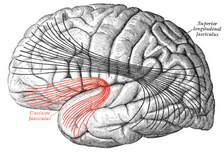
The uncinate fasciculus is a white matter association tract in the human brain that connects parts of the limbic system such as the temporal pole, anterior parahippocampus, and amygdala in the temporal lobe with inferior portions of the frontal lobe such as the orbitofrontal cortex. Its function is unknown though it is affected in several psychiatric conditions. It is one of the last white matter tracts to mature in the human brain.
The uncinate fasciculus is a hook-shaped bundle of axons that links anterior portions of the temporal lobe with the inferior frontal gyrus and the lower surfaces of the frontal lobe. It arises in the anterior temporal lobe and amygdala, in the temporal lobe curving in an upward pathway behind the external capsule inward of the insular cortex and continuing up into the posterior part of the orbital gyrus. It does not appear to have cell bodies in the hippocampus proper.
The average length of the uncinate fasciculus is 45 mm with a range 40–49 mm. Its volume in adults is 1425.9±138.6 mm3, being slightly larger in men, at 1504.3±150.4, than women 1378.5±107.4.
It has three parts: a ventral or frontal extension, an intermediary segment called the isthmus or insular segment and a temporal or dorsal segment.
The uncinate fasciculus is a bi-directional pathway between the temporal lobe and frontal lobe; it is traditionally considered to be part of the limbic system. It has been proposed that the uncinate fasciculus allows mnemonic representations stored in the temporal lobe to interact with and guide decision making in the frontal lobe.
Diffusion tensor imaging, a reconstruction model available from a diffusion MRI scan, shows a greater fractional anisotropy on the left side than on the right. The difference in this measure of anisotropy has been linked to the left hemispheric specialization for language. However, the use of electrical brain stimulation upon it fails to disrupt language, suggesting it might not be involved in language, though it is possible that this disruption failed to happen because it was functionally compensated by alternative pathways.
The uncinate fasciculus appears to have a role in some, but not all, types of learning and memory. Reversal learning, in which a stimulus-reward association is learned over many trials, then reversed such that the initial stimulus is no longer is associated with a reward, is associated with individual variation in the uncinate fasciculus. In addition, the ability to learn associations through trial and error, such as the pairing of a name with a face, correlates with uncinate fasciculus microstructure. This relates to early work showing that surgical damage to the uncinate fasciculus is reliably associated with deficits in proper name retrieval.
The uncinate fasciculus has the longest period of development in terms of fractional anisotropy as it alone amongst the major white fibre tracks continues to develop until the age of 30. Developmental disorders with core problems relating to memory retrieval, reward and valuation computation, and impulsive decision making may be linked to aberrations in uncinate fasciculus microstructure, e.g. temporal lobe epilepsy and conduct disorder.
Uncinate fasciculus
The uncinate fasciculus is a white matter association tract in the human brain that connects parts of the limbic system such as the temporal pole, anterior parahippocampus, and amygdala in the temporal lobe with inferior portions of the frontal lobe such as the orbitofrontal cortex. Its function is unknown though it is affected in several psychiatric conditions. It is one of the last white matter tracts to mature in the human brain.
The uncinate fasciculus is a hook-shaped bundle of axons that links anterior portions of the temporal lobe with the inferior frontal gyrus and the lower surfaces of the frontal lobe. It arises in the anterior temporal lobe and amygdala, in the temporal lobe curving in an upward pathway behind the external capsule inward of the insular cortex and continuing up into the posterior part of the orbital gyrus. It does not appear to have cell bodies in the hippocampus proper.
The average length of the uncinate fasciculus is 45 mm with a range 40–49 mm. Its volume in adults is 1425.9±138.6 mm3, being slightly larger in men, at 1504.3±150.4, than women 1378.5±107.4.
It has three parts: a ventral or frontal extension, an intermediary segment called the isthmus or insular segment and a temporal or dorsal segment.
The uncinate fasciculus is a bi-directional pathway between the temporal lobe and frontal lobe; it is traditionally considered to be part of the limbic system. It has been proposed that the uncinate fasciculus allows mnemonic representations stored in the temporal lobe to interact with and guide decision making in the frontal lobe.
Diffusion tensor imaging, a reconstruction model available from a diffusion MRI scan, shows a greater fractional anisotropy on the left side than on the right. The difference in this measure of anisotropy has been linked to the left hemispheric specialization for language. However, the use of electrical brain stimulation upon it fails to disrupt language, suggesting it might not be involved in language, though it is possible that this disruption failed to happen because it was functionally compensated by alternative pathways.
The uncinate fasciculus appears to have a role in some, but not all, types of learning and memory. Reversal learning, in which a stimulus-reward association is learned over many trials, then reversed such that the initial stimulus is no longer is associated with a reward, is associated with individual variation in the uncinate fasciculus. In addition, the ability to learn associations through trial and error, such as the pairing of a name with a face, correlates with uncinate fasciculus microstructure. This relates to early work showing that surgical damage to the uncinate fasciculus is reliably associated with deficits in proper name retrieval.
The uncinate fasciculus has the longest period of development in terms of fractional anisotropy as it alone amongst the major white fibre tracks continues to develop until the age of 30. Developmental disorders with core problems relating to memory retrieval, reward and valuation computation, and impulsive decision making may be linked to aberrations in uncinate fasciculus microstructure, e.g. temporal lobe epilepsy and conduct disorder.
Recent media
Recent media