Community hub
Recent from talks
Knowledge base stats:
Talk channels stats:
Members stats:
Acute intermittent porphyria
Acute intermittent porphyria (AIP) is a rare metabolic disorder affecting the production of heme resulting from a deficiency of the enzyme porphobilinogen deaminase. It is the most common of the acute porphyrias.
The clinical presentation of AIP is highly variable and non-specific. The patients are typically asymptomatic, with most gene carriers having no family history because the condition had remained latent for several generations. The syndrome marked by acute attacks affects only 10% of gene carriers. The mean age at diagnosis is 33 years old. Like other porphyrias, AIP is more likely to present in women. A distinguishing feature of AIP that separates it from other porphyrias is the absence of photosensitive cutaneous symptoms that occur in addition to acute attacks.
AIP is one of the four porphyrias that presents as an acute attack. 90% of affected individuals never experience an acute attack and are asymptomatic, while an estimated 5% of affected individuals experience repeat attacks. Attacks are most common in young adult women and are rare before puberty or after menopause. Severe acute attacks may require hospitalization. Patients usually experience symptoms in attacks that last from several hours to a few days. Between attacks, patients are asymptomatic.[citation needed]
The most frequent presenting symptoms are abdominal pain and tachycardia. The abdominal pain is typically severe, colicky, poorly localized, and often associated with pain in back and legs. Patients may also present with vomiting and constipation, but diarrhea is unusual. The onset of back and leg pain is characterized by severe pain and stiffness in back and thighs followed by loss of tendon reflexes and paralysis. Psychiatric symptoms are present, such as paranoid schizophrenia-like features but rarely psychosis and hallucinations. The acute attacks classically present with dark-red photosensitive urine (often called port-wine urine), but this is a nonspecific symptom. Physical examination often shows no abnormalities.
Hyponatremia is the most common electrolyte abnormality during acute attacks, occurring in 40% of patients and presenting as SIADH. Hypomagnesemia is also common. There are no pathognomonic signs or symptoms.[citation needed]
The most common identified triggers for acute attacks are medications, weight loss diets, and surgery. Many medications have been associated with AIP including antibiotics, hormonal contraceptives, seizure medications, anesthetics, and HIV treatment drugs.
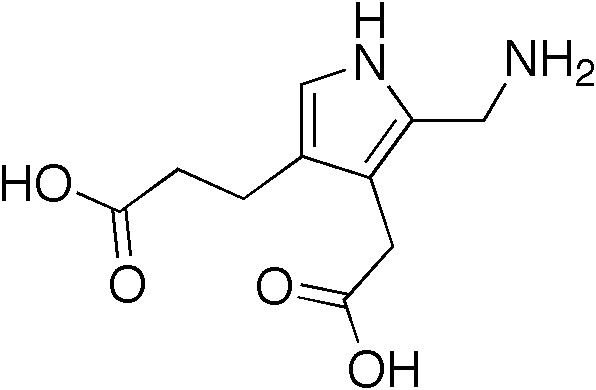
Porphyrias are caused by mutations in genes that encode enzymes in heme synthesis. In normal physiology, heme synthesis begins in the mitochondrion, proceeds into the cytoplasm, and finishes back in the mitochondrion. Heme is produced in all cells, but 80% of all heme is produced in erythropoietic cells in bone marrow and 15% in parenchymal cells in the liver, where turnover of hemoproteins is high. In AIP, over 100 mutations have been identified on the long arm of chromosome 11 at the HMBS gene, which codes for the cytoplasmic enzyme porphobilinogen deaminase. This deficiency prevents heme synthesis, which can not be completed and the metabolite porphobilinogen accumulates in the cytoplasm.
AIP is an autosomal dominant porphyria resulting in about 50% normal activity of the affected enzyme. The penetrance of AIP is incomplete with only 10% of gene carriers experiencing acute attacks suggesting role for other modifying genes or environment.
Hub AI
Acute intermittent porphyria AI simulator
(@Acute intermittent porphyria_simulator)
Acute intermittent porphyria
Acute intermittent porphyria (AIP) is a rare metabolic disorder affecting the production of heme resulting from a deficiency of the enzyme porphobilinogen deaminase. It is the most common of the acute porphyrias.
The clinical presentation of AIP is highly variable and non-specific. The patients are typically asymptomatic, with most gene carriers having no family history because the condition had remained latent for several generations. The syndrome marked by acute attacks affects only 10% of gene carriers. The mean age at diagnosis is 33 years old. Like other porphyrias, AIP is more likely to present in women. A distinguishing feature of AIP that separates it from other porphyrias is the absence of photosensitive cutaneous symptoms that occur in addition to acute attacks.
AIP is one of the four porphyrias that presents as an acute attack. 90% of affected individuals never experience an acute attack and are asymptomatic, while an estimated 5% of affected individuals experience repeat attacks. Attacks are most common in young adult women and are rare before puberty or after menopause. Severe acute attacks may require hospitalization. Patients usually experience symptoms in attacks that last from several hours to a few days. Between attacks, patients are asymptomatic.[citation needed]
The most frequent presenting symptoms are abdominal pain and tachycardia. The abdominal pain is typically severe, colicky, poorly localized, and often associated with pain in back and legs. Patients may also present with vomiting and constipation, but diarrhea is unusual. The onset of back and leg pain is characterized by severe pain and stiffness in back and thighs followed by loss of tendon reflexes and paralysis. Psychiatric symptoms are present, such as paranoid schizophrenia-like features but rarely psychosis and hallucinations. The acute attacks classically present with dark-red photosensitive urine (often called port-wine urine), but this is a nonspecific symptom. Physical examination often shows no abnormalities.
Hyponatremia is the most common electrolyte abnormality during acute attacks, occurring in 40% of patients and presenting as SIADH. Hypomagnesemia is also common. There are no pathognomonic signs or symptoms.[citation needed]
The most common identified triggers for acute attacks are medications, weight loss diets, and surgery. Many medications have been associated with AIP including antibiotics, hormonal contraceptives, seizure medications, anesthetics, and HIV treatment drugs.
Porphyrias are caused by mutations in genes that encode enzymes in heme synthesis. In normal physiology, heme synthesis begins in the mitochondrion, proceeds into the cytoplasm, and finishes back in the mitochondrion. Heme is produced in all cells, but 80% of all heme is produced in erythropoietic cells in bone marrow and 15% in parenchymal cells in the liver, where turnover of hemoproteins is high. In AIP, over 100 mutations have been identified on the long arm of chromosome 11 at the HMBS gene, which codes for the cytoplasmic enzyme porphobilinogen deaminase. This deficiency prevents heme synthesis, which can not be completed and the metabolite porphobilinogen accumulates in the cytoplasm.
AIP is an autosomal dominant porphyria resulting in about 50% normal activity of the affected enzyme. The penetrance of AIP is incomplete with only 10% of gene carriers experiencing acute attacks suggesting role for other modifying genes or environment.